
Rabies is a fatal worldwide viral disease and contributes to a great economic load in Egypt through the death of animals and control strategy costs. Due to Egypt’s privileged location and friction with neighboring countries and far countries from Europe and Asia throughout the ages, the current research goal is to evaluate the extent of their influence on nucleotide and amino acids variation of rabies virus isolates from the brain tissues of three dogs from 2017 to 2019 at the genetic level of the N and P genes from three governorates of Delta region in Egypt with earlier isolates, locally used vaccinal strains and other used strains internationally. Ten brain tissues from confirmed rabid cases in three governorates; Dakahleya (3), Alexandria (4); and Beheira (3) were diagnosed using viral cell culture and Direct Fluorescent Test (DFT) for the rabies virus. The samples were tested positive by DFT and isolated on baby hamster kidney cell culture. Of them, three confirmed rabies samples were picked to determine their N and P genes by sequencing further (at the level of nucleotide and amino acid). The N and P genes in this study isolates have a great similarity to previous isolates from Egypt as well as adjacent bordering countries. At the N and P genes level, the isolates bear little resemblance to the vaccinal strains (Evelyn-Rokitnicki-Abelseth, Pasteur, the China human rabies vaccine strain AG and others at this study) due to the presence of mutation sites. As the P gene plays a role in viral pathogenicity, the detected variance in nucleotide and amino acid identities of the P gene in our local circulating isolates over the years confirmed the anticipation of distinction in the pathogenicity of such isolates. All vaccinal strains have a genetic distance from field Dakahleya, Beheira, and Alexandria isolates (ranging from 88.2% to 88.9% for the China human rabies vaccine AG).Such results of nucleotide identity among Egyptian isolates over the years confirm the unity of their source. The application of strict quarantine measures on imported rabies vaccinated pets in addition to prospective studies on the pathogenicity of local circulating isolates regarding the N and P genes are strongly recommended in the control strategy.
Research Article · Volume 11, Issue 1, 17 December 2023 · Open Access
Virologic Detection and Molecular Characterization of Rabies Virus in Delta Region, Egypt
·
Affiliations & Notes ⌄
- 1Virology Department, Animal Health Research Institute (AHRI), Agricultural Research Centre (ARC), Giza, Egypt
- 2Mycoplasma Department, Animal Health Research Institute (AHRI), Agricultural Research Centre (ARC), Giza, Egypt. , *Correspondence | Morcos Ibrahim Yanni, Virology Department, Animal Health Research Institute (AHRI), Agricultural Research Centre (ARC), Giza, Egypt
- 3Email: morcosyanni70@gmail.com
Article Info ⌄
- Journal:
- Journal of Virological Sciences
- Published:
- 17 December 2023

From the article
- At the level of the P gene, the % of identity between the new Egyptian isolates and some compared vaccinal strains was found to range from 88.2 % to 89.8% for AG vaccine (Figure 9 and Table 3).
- Phylogenetic analysis of this study indicated clustering of Egyptian field rabies isolates at the N gene-level into comparable phylogenic clusters, showing close genetic distance (from 99% to 95.9%).
- In the current study, a phylogenetic analysis of the tested Egyptian field rabies isolates at the P gene revealed that the field Dakahleya isolate was genetically close to the majority of Egyptian isolates (from 98% to 95.9%).
- The field rabies Dakahleya isolates shared a genetic distance of 90.7 % or more with isolates from nearby countries (Israel, Tunisia, and Ethiopia) and a genetic distance of 89.5 % and less compared to isolates from far away countries (South Africa and Tanzania).
Selected from the article's own text.
Abstract
Introduction
The rabies virus (RABV) is a member of the Lyssavirus genus, which is part of the Rhabdoviridae family. Lyssaviruses have seven identified genotypes (G.T.) and unclassified G.T.s. Except for Antarctica and a few islands, the RABV GT1 isolates are distributed worldwide , Tordo et al. (1986) defined the viral genome as a non-segmented single-stranded negative-sense RNA of about 12 kb that encodes five genes: nucleoprotein (N), phosphoprotein (P), matrix protein (M), glycoprotein (G), and RNA polymerase (L). Because the N gene in the entire genome of the rabies virus is highly conserved and can be easily produced, it is commonly employed as a judging index for rabies virus genotyping and population variance .
There are seven recognized genotypes of lyssavirus defined on the basis of their genetic similarity: rabies virus (genotype 1), Lagos bat virus (genotype 2), Mokola (genotype 3), Duvenhage virus (genotype 4), European bat lyssavirus type 1 (genotype 5), European bat lyssavirus type 2 (genotype 6) and Australia bat lyssavirus (genotype 7). Additionally, four novel genotypes: Aravan virus, Khujand virus, Irkut virus, and West Caucasian were recovered from bats in Eurasia. Genotype 1 is responsible for classical rabies in terrestrial mammals throughout the world.
The molecular epidemiology of rabies in Africa was described into three phylogenetic lineages of RABV: Africa 1, 2, and 3 .
The Africa 1 and 2 lineages were isolated from dogs or humans bitten by rabid dogs, whereas the Africa 3 lineage was found associated with mongoose species, principally the yellow mongoose (Cynictis penicillata) from South Africa. The Africa 1 lineage was subdivided into two subgroups: 1a, restricted to North and West Africa, and 1b, limited to Southeast Africa. In general, the Africa 1 lineage was the most similar to current Eurasian RABV lineages, suggesting its recent introduction to Africa . The Africa 2 lineage includes wild-type strains that originated from several central and eastern African countries and is phylogenetically ancestral to the cluster that includes the Eurasian and Africa 1 RABVs. The Africa 3 lineage, of mongoose origin, is distant from all dog RABV variants.
Seven clades (I–VII) were identified in Israel and one (Africa 4) in Egypt. This novel clade, Africa 4 was placed ancestral to the cluster of RABV variants that were associated with the Middle East and the middle latitudes of Eurasia, and to Africa 1. This clade is part of the cosmopolitan canine RABV lineage that is believed to have originated in Europe and widely disseminated as a consequence of colonial activity during the 16th to 19th centuries . Molecular studies were done on Egyptian isolates from different localities as rabid horses from Cairo in 2014 , water buffalos from Menofeya in 2015 , and rabid cattle and fox from Elwadi Elgedid in 2021 . Imported bat from Egypt or Togo to France was diagnosed to be infected with LBV (Lagos bat virus) .
The N and P genes are involved in and facilitate the pathogenicity of RABV Gene . The rabies virus phosphoprotein P5 binding to the BECN1 (Beclin1) ring benefits RABV replication by inducing BECN1 signaling pathway-dependent incomplete autophagy, potentially targeting antiviral drugs against RABV .
Because of the P proteins ability to bind the signal transducer and activator of transcription (STATs) via the C-terminal domain (CTD) and residues 186–297 of RABV P protein ; ; , P-proteins are considered major interferon (IFN) antagonist of lyssaviruses ; . The average reported human rabies cases is around 50 cases per year, 70% of them didn’t receive post exposure prophylaxis, mostly in rural areas at the world rabies day celebration 2021 (https://www.fao. org/egypt/news/detail-events/en/c/1441644/).
P gene aids rabies virus replication in muscles by preventing interferon . In this current study, it was reviewed the national and regional available data to describe simply the epidemiology of rabies in Egypt focusing on the delta region to present the current state of the rabies control efforts and to determine the nucleotide variation of the N and P genes between the local Egyptian street isolates and the vaccinal stains.
Materials and Methods
Samples
Ten suspected rabid individuals brain tissues of dogs and cattle from the governorates of Alexandria, Dakahleya, and Beheira were euthanized after showing nervous signs (Table 1).
Table 1: Table showing the full data of examined samples at this study.
| S. | Locality | year | Species | Age | Clinical signs | gender |
| 1 | alex | 2019 | cattle | 1y | + | Not specified (n.s.) |
| 2 | alex | 2019 | dog | 8m | + | n.s. |
| 3 | alex | 2019 | dog | 1y | + | n.s. |
| 4 | alex | 2019 | dog | 2y | + | n.s. |
| 5 | dakahleya | 2018 | dog | 2y | + | n.s. |
| 6 | dakahleya | 2019 | cattle | 4y | + | n.s. |
| 7 | dakahleya | 2017 | cattle | 1y | + | n.s. |
| 8 | beheira | 2018 | cattle | 3y | + | n.s. |
| 9 | beheira | 2017 | cattle | 2y | + | n.s. |
| 10 | beheira | 2016 | cattle | 1y | + | n.s. |
Methods
The samples were sent to the Biosafety Level-2 Rabies Laboratory of the Animal Health Research Institute (AHRI), Giza, Egypt for rabies diagnosis using Fluorescent Antibody Technique (FAT). The samples were crushed and suspended in 10% MEM (Minimum Essential Medium) for virus isolation on BHK cells via rapid rabies antigen test and molecular research applying PCR and nucleotide sequencing of N and P genes.
Rapid rabies antigen test from China (Zrbio and batch number; 2018061212).
Fluorescence antibody technique (FAT)
Using a rabies polyclonal conjugate supplied by ARC-Onderstepoort Veterinary Institute Batch Number (N4-22), impressed smears from all evaluated cases were analyzed for the presence of rabies antigen (OIE, 2018).
Isolation in tissue culture
Supernatants from 10% crushed brain samples were seeded on monolayers of Baby Hamster Kidney (BHK-21/C-13) cells and monitored at 37°C/72h with 5% CO2. Three passages of 48 h each were performed according to the techniques specified by . FAT was used to verify the isolates. FAT was performed in a tissue culture chamber slide in which the isolates were fixed in 80% acetone, stained by the conjugate at 37◦/1h, washed 3 times for a duration of 10 min each using phosphate buffer saline and examined under the fluorescent microscope after applying a mounting buffer.
Extraction of nucleic acids
The QIAamp viral RNA mini kit batch number 1151234567 was used to extract RNA from three selected positive samples (Qiagen, Germany, GmbH). For 10 minutes, 140µl of the sample suspension was incubated at room temperature with 560µl of viral lysis buffer (AVL) and 5.6µl of carrier RNA. 560 µl of 100 percent ethanol was added to the lysate after incubation. Following the manufacturer’s instructions, the sample was washed and centrifuged. The nucleic acid was eluted using 60 µl of the kit’s elution buffer (QIAamp Viral RNA Mini Kits, 2013).
To amplify a 684-bp fragment of the N gene of RABV, the primer set (5′-ATGGATGCCGACAAGATTGT-3′) and (5′-CCCACT CTGATTGCC GAATA -3′), and to amplify a 882-bp fragment of P gene of RABV, the primer set (5′-TTGGTACCATCCCAAGTATGA-3′) and (5′-TCT TGCATGATTTT GCTTAGCTTGTC-3′) were designed based on . The oligonucleotide primers were obtained from Metabion Co., Germany.
PCR amplification
In a 25 µl reaction, 12.5 µl of Quantitect probe rt-PCR buffer (Qiagen, Gmbh), 1 µl of each primer at 20 pmol concentration, 0.25 µl of rt-enzyme, 4.25 ml of water and 6 µl of template were used. An applied biosystems 2720 thermal cycler was used to carry out the reaction. Reverse transcription was performed at 50°C for 30 minutes, followed by a 5-minute primary denaturation phase at 95°C, followed by 35 cycles of 94°C for 30 seconds (annealing: 50°C for 40 seconds for P and N genes) and 45 seconds at 72°C, final extension phase was performed at 72°C for 10 min, .
The PCR products were separated using 5V/cm gradients on a 1.5 percent agarose gel (Applichem, Germany, GmbH) in 1x TBE buffer at room temperature. 15 µl of the products were put into each gel slot for analysis. The sizes of the fragments were determined using the gene ruler 100bp ladder (Fermentas, Thermo, Germany). A gel documentation system (Biometra, Germany) was used to photograph the gel and the data was processed using computer software. The product of three positive PCR samples was sequenced using a big dye terminator V3.1 sequencing kit on a genetic analyzer 3500 (life technology). Bioedit and CLC’s Main Workbench 6 software (2022) are used for the analysis. Bioedit software version 6 based on Clustal W alignment tool has been used to create alignments for each gene containing representative sequences for the different related strains.
A BLAST® analysis (Basic Local Alignment Search Tool) was initially performed to establish sequence identity to GenBank accessions. Then, the N and P gene fragments were aligned using Bioedit 6 software , and the phylogenetic tree was created using Mega 7 software .
Results and Discussion
All ten tested brain tissue samples were positive for the presence of rabies antigen (Ag) using the rapid Ag test kit (Figure 1). Using the fluorescent antibody technique, impression smears, and the isolates on BHK cells tested positive for the presence of rabies Ag (Figure 2A). Positive and negative controls (Figure 2B, D) were included.
At the level of the N gene, the three recent and old Egyptian isolates in this study originated from the same clade with a high bootstrap (Table 2, Figure 5A). Amino acids analysis of Dakahleya 18 and Alex 19 N protein demonstrated their great resemblance with that of the old isolate CA 99 and their difference from that of the Beheira isolate due to the presence of mutation loci (Table 2, Figure 5B).
The % of N nucleotide identity here between Dakahleya isolate and many Egyptian isolates was 99% with (CA98, CA99, and Fay99), 99.1% with (Beheira18), 99.4% with (Alex19), 97.6 % with (Wadi19), and 95.9 % with (Wadi19) (egy79). Alex and Beheira isolates produced a strong correlation (Figure 6).
Table 2: The description of isolates from NCBI used for comparison in this study at the level of N gene.
| No | Access no | Host | Isolation year | Country | Variant | Abbre-viation | % of identity | ||
| BEH18 | ALEX19 | DAK18 | |||||||
| 1 | DQ837463.1 | DOG | 1999 | Egypt (Cairo) | CA99 | 99 | 99 | 98.7 | |
| 2 | DQ837462.1 | DOG | 1999 | Egypt (Fayoum) | Fay99 | 99 | 99 | 98.7 | |
| 3 | DQ837461.1 | DOG | 1998 | Egypt (Cairo) | CA98 | 99 | 99 | 98.7 | |
| 4 | MT036064 | FOX | 2017 | Egypt (Wadi) | Wadi17 | 97.3 | 97.6 | 97.3 | |
| 5 | MT036065 | CATTLE | 2018 | Egypt (Wadi) | Wadi18 | 97.3 | 97.6 | 97.3 | |
| 6 | MT036066 | CATTLE | 2019 | Egypt (Wadi) | Wadi19 | 97.6 | 97.9 | 97.6 | |
| 7 | U22627.1 | HUMAN | 1979 | Egypt | Egy79 | 95.6 | 95.9 | 95.9 | |
| 8 | KY12454.1 | DOG | 2014 | Chad | Chad14 | 89.2 | 89.2 | 88.9 | |
| 9 | EV514580 | 2006 | Mauritania | Mor06 | 90.4 | 99.4 | 90.1 | ||
| 10 | U22634.1 | DOG | 1987 | Cameron | Cam87 | 90.7 | 90.7 | 90.4 | |
| 11 | EF206707.1 | ERA | ERA | 92.4 | 92.4 | 92.1 | |||
| 12 | KM198893.1 | MOSCO-W3253 | MOSC-3253 | 92 | 92 | 92 | |||
| 13 | GV565704.1 | Flury HEP | Hep. | 92.6 | 92.5 | 92.3 | |||
| 14 | GV565703.1 | Flury LEP | Lep. | 92.9 | 92.9 | 92 | |||
| 15 | GQ412744.1 | AG | Chinese AG | 91.4 | 91.3 | 91.6 | |||
| 16 | GQP18139.1 | CVS.11 | CVS | 92.9 | 92.9 | 92.6 | |||
| 17 | JQ944709.1 | Rabies EPH Vac | ephvac | 93.5 | 93.5 | 93.2 | |||
| 18 | DQ837425.1 | cow | 1998 | Jordan | JOR98 | 92.9 | 92.9 | 92.5 | |
| 19 | DQ837423.1 | DONKEY | 1999 | Jordan | JOR99 | 92.9 | 92.5 | 92.3 | |
| 20 | DQ837465.1 | STONE- NARTIN | 1997 | Lebanon | LEB97 | 93 | 92.7 | 92.4 | |
| 21 | DQ837429.1 | DOG | 1996 | Israel | ESR96 | 92.7 | 92.7 | 92.4 | |
| 22 | DQ837466.1 | FOX | 2002 | Israel | ESR02 | 93 | 93 | 92.7 | |
| 23 | DQ837442.1 | FOX | 1998 | Israel | ESR98 | 93 | 92.7 | 92.4 | |
| 24 | MG011654.1 | HOMO-SAPIENS | 2016 | France | FR16 | 89.8 | 89.8 | 89.5 | |
| 25 | DQ837480.1 | DOG | 2000 | Turkey | TRK00 | 93.5 | 93.5 | 93.2 | |
| 26 | MT134104 | CATTLE | 2018 | Egypt (Beheira) | BEH18 | 100 | 99.4 | 99.1 | |
| 27 | MT134105 | DOG | 2019 | Egypt (Alex) | ALEX19 | 99.4 | 100 | 99,4 | |
| 28 | MT134106 | DOG | 2018 | Egypt (Dakahleya) | DAK18 | 99.1 | 99.4 | 100 | |
The recent Egyptian isolates, the non Egyptian isolates from Africa (Chad14, Moritanya2006, Cameron87) and neighboring countries (Lebanon97, Israel96, 98, 02 and Jordon 98) originated from a common ancestor (Table 2, Figure 5A) with the presence of mutation sites at their N Proteins (Figure 5B).
The degree of nucleotide identity between the Dakahleya isolate as well as certain non-Egyptian isolates at the N gene was reported to be 89.2% with Chad14, 90.4 % with Mauritania 2006, 90.7 % with Cameroon 87 and 92.7% to 93% with Chad, ISR96, ISR98, ISR02, LEB, JORD98 and JORD99) (Table 2).
At the level of the N gene, the % of identity between the Dakahleya isolate and some compared vaccinal strains was found to be 92 % for (EPHVAC and Moscow 3235 strain), 92.4 % for the ERA strain, 92.9 % for the LEP strain, 92.6 % for HEP and 91.4% for the Chinese AG strain. On the other hand, the % of identity between the Dakahleya isolate and some comparative vaccinal isolates were found to be 88.2% with EPHvac, 88.5% with (HEP and Moscow3235 strain), 88.7% with (LEP and ERA), and 88.9% with the Chinese AG strain. Alex and Beheira isolates showed similar figures (Table 2 and Figure 7).
In comparison to the Pasteur reference rabies virus strain partial N gene sequence (Genbank accession No. GU992320), all the current study Egyptian strains showed the following mutations: [(Thr3Ala), (Val21Ile), (His26Tyr), (Ser61Asn), (Phe80Leu), (Val90Leu), (Gly106Asp), (Asn119Ser), (Pro135Ser)], While strain EgyBEH18 showed another mutation (Lys5Arg.), while in comparison to the lastly published Egyptian strains, sequences were identical except for 2 mutations [(Val22Ala) and (Asn202Ser)] that were recorded in the Egyptian strains MT036064-MT036066.
At the level of the P gene, the % of identity of the Dakahleya isolate with (Behe18) was 98%, 97.9% with (Alex19), 95.7% with (Egy79), 88.7% with Wadi18, and 88.5 % with (Wadi19) (Table 3). The P genes of the old and recent isolates all originated from the same clade (Figure 8) with the presence of a mutation at the AA composition of their P proteins (Figure 9).
The % of nucleotide identity between the Dakahleya isolate and some non-Egyptian isolates at the P gene were 95.6 % with ESR50, 90.4 % with TUN88, 90.7 % with Ethio, 89.5 % with the South Africa06 and the Israeli isolate ESR01, 89.5 % with the South Africa02, 87.6 % with Tanzania and 85.5 % with South Africa 00. Alex isolates and Beheira isolate both yielded similar findings.
At the level of the P gene, the % of identity between the new Egyptian isolates and some compared vaccinal strains was found to range from 88.2 % to 89.8% for AG vaccine (Figure 9 and Table 3).
Table 3: The description of isolates from NCBI used for comparison in this study at the level of P gene.
| No | Access no | Host | Isolation year | Country | Abbre-viation | Variant | % of nucleotide identity | ||
| Beh18 | Alex19 | Dkh18 | |||||||
| 1 | MG458317.1 | DOG | 1999 | Egypt | CA99 | 97.6 | 97.4 | 97.6 | |
| 2 | MG458315.1 | DOG | 1998 | Egypt | CA98 | 97.6 | 97.4 | 97.6 | |
| 3 | MT036069 | CATTLE | 2019 | Egypt (Wadi) | Wadi19 | 95.5 | 95.4 | 88.5 | |
| 4 | MT036067 | FOX | 2017 | Egypt (Wadi) | Wadi17 | 95.7 | 95.8 | 88.9 | |
| 5 | MT036068 | CATTLE | 2018 | Egypt (Wadi) | Wadi18 | 95.7 | 95.6 | 88.7 | |
| 6 | KX148101.1 | MOUSE | 1979 | Egypt | Egy79 | 95.4 | 95.2 | 95.7 | |
| 7 | HQ266628 | DOG | 2006 | South Africa | Sa06 | 89.8 | 89.9 | 89.5 | |
| 8 | KT336436.1 | DOG | 2012 | South Africa | Sa12 | 89.4 | 89.7 | 89.3 | |
| 9 | MG458307 | CAT | 2000 | South Africa | Sa00 | 84.9 | 85.1 | 85.5 | |
| 10 | KY210275.1 | COW | 2013 | Tanzania | Tanz13 | 87.9 | 88 | 87.6 | |
| 11 | MG458306.1 | FOX | Chekoslovakia | chkosl | 89 | 89 | 89.3 | ||
| 12 | AF369331 | DOG | Ethiopia | eth | 90.4 | 90.6 | 90.7 | ||
| 13 | AF359322 | 1988 | Tunisia | Tunis98 | 90.5 | 90.6 | 90.4 | ||
| 14 | NC001542 | Pasteur | Pasteur | 89 | 89.2 | 89 | |||
| 15 | EF206715.1 | Rabifox (SAD) | SAD | 88.6 | 88.9 | 88.9 | |||
| 16 | JQ944709 | EPH VAC | 87.8 | 87.7 | 88.2 | ||||
| 17 | GQ412744.1 | AG | AG | 89.4 | 89.8 | 88.9 | |||
| 18 | EF206707.1 | ERA | ERA | 88.5 | 88.7 | 88.7 | |||
| 19 | KM198893.1 | MOSCO-W3253 | 88.5 | 88.4 | 88.5 | ||||
| 20 | GU565704.1 | FLURY HEP | HEP | 88.3 | 88.2 | 88.5 | |||
| 21 | GU565703.1 | FLURY LEP | LEP | 88.5 | 88.4 | 88.7 | |||
| 22 | AF369312 | DOG | 2001 | ISRAEL | Esr01 | 89.7 | 89.9 | 89.5 | |
| 23 | AF369311 | DOG | 2001 | ISRAEL | Esr01 | 89.5 | 88.7 | 89.5 | |
| 24 | AF369283 | FOX | 2001 | ISRAEL | Esr01 | 88.3 | 88.4 | 88.3 | |
| 25 | KF154998 | DOG | 1950 | ISRAEL | Esr50 | 95.4 | 95.2 | 95.6 | |
| 26 | MT134107 | CATTLE | 2018 | Egypt (Beheira) | Beh18 | 100 | 99.4 | 98 | |
| 27 | MT134108 | DOG | 2019 | Egypt (Alex) | Alex19 | 97.9 | 100 | 97.9 | |
| 28 | MT134109 | DOG | 2018 | Egypt (Dakahleya) | Dkh18 | 98 | 97.9 | 100 | |
Presence of AA divergence among the recent Egyptian isolates and the vaccinal strains in this study is due to the presence of mutation sites (Figures 8B, 10).
When compared to the Pasteur reference rabies virus strain full P gene sequence (Genbank accession No. NC_001542), all the current Egyptian strains showed the following mutations; [(Gly54Arg), (His57Gln), (Pro64Ser), (Pro66Leu), (Met69Val), (Ala70Thr), (Lys71Ala), (Ile108Met), (Ala130Thr), (Pro137Ser), (Leu153Pro), (Lys154Arg), (Thr158Pro), (Pro159Ser), (Ala171Val), (Thr177Val), (Leu223Phe), (Arg252His), (Ser280Ala) and (Ser284Asn)]. However, MT134109 showed 2 extra mutations [(Ser90Gly) and (Ser140Val)]. However, Ser140Leu was detected in MT134107 and MT134108.
As the rabies virus does not produce the appreciable cytopathic effect (CPE) in BHK-21 cells, the presence of virus antigen was confirmed in tissue culture by using the florescent antibody technique (FAT).
This research aimed to characterize rabies virus (RABV) strains that were circulating in Egypt. Phylogenetic analysis of this study indicated clustering of Egyptian field rabies isolates at the N gene-level into comparable phylogenic clusters, showing close genetic distance (from 99% to 95.9%). The tested field rabies Dakahleya isolate exhibited a genetic distance of 92.7% or more with isolates from nearby countries (Israel, Lebanon, and Jordan) and of 90.7% and less compared to isolates from remote nations (Chad, Mauritania, and Cameron). The genetic difference between the field Dakahleya isolate and the vaccinal strains in this study ranged from 91.4 % (Chinese AG strain) to 92.9 % (LEP vaccinal strain), including the ERA strain (The used strain in Egypt in veterinary rabies vaccines).
In the current study, a phylogenetic analysis of the tested Egyptian field rabies isolates at the P gene revealed that the field Dakahleya isolate was genetically close to the majority of Egyptian isolates (from 98% to 95.9%). It has a large genetic distance from El-Wadi governorate isolates, ranging from 88.5 % to 88.7 % (wadi19 and wadi18, respectively). The field rabies Dakahleya isolates shared a genetic distance of 90.7 % or more with isolates from nearby countries (Israel, Tunisia, and Ethiopia) and a genetic distance of 89.5 % and less compared to isolates from far away countries (South Africa and Tanzania). At the P gene level, all vaccinal strains in this investigation have a genetic distance from our field Dakahleya isolate ranging from 88.2 % to 88.9 % (for the Chinese AG vaccinal strain). Beheira and Alex isolates produced similar results.
It was notable that all Egyptian field isolates shared a high level of genetic similarity. The following previous studies supported these findings. Lineage 1 from Africa RABVs are quite similar to RABVs seen in Europe and Asia and they are assumed to have just arrived in Africa from Europe as a result of colonial activity from the 16th to 19th centuries . it is a partner of the cosmopolitan canine RABV lineage.
Isolates from Middle Eastern and European countries have a significant correlation, but they differ from isolates from Asian and African countries . The tight genetic similarity between isolates from European and Middle Eastern nations shows that the viruses circulating in foxes in both zones may have emerged from the same source . According to a previous study, the three Egyptian isolates (CA98, CA99, and FAY99) belonged to Africa 4, a novel clade with a high bootstrap value (99 %). It was reported to be ancestral to the cluster of RABV variants associated with the Middle East and Eurasia’s middle latitudes, as well as Africa 1 .
Dog trading and the porosity of the border areas that promote and facilitate rabies virus transmission according to Silberfein et al. (2006), Sunday et al. (2014) explain and support that finding.
Conclusions and Recommendations
The examined rabies Egyptian field isolates, most of the previous Egyptian isolates, isolates from nearby countries (Israel, Lebanon, and Jordan), and the vaccinal strains used in this study, particularly the Chinese AG strain and the LEP vaccinal strain, have a small genetic distance with each other and a significant genotypic variation with isolates from far countries to Egypt at the N gene level. Except for isolates from El-Wadi governorate, all the analyzed Egyptian field isolates and isolates from nearby countries had a close genetic distance at the P gene level (Israel, Tunisie, and Ethiopia). Isolates from far-flung places (South Africa and Tanzania) and vaccine strains employed in this study have a wide range of genetic distances. It could be concluded that dog trading and the porosity of the border areas have a great role in the spread of the disease in Egypt. Strict quarantine measures on the importation of unvaccinated animals, border follow-up, and control are recommended.
Acknowledgement
Authors thanks to the memebers of Virology Department, Animal Health Research Institute, Agriculture Research Centre.
Novelty Statement
Our results shoed the importance of dog trading and the porosity of borders on Rabies virus control.
Author’s Contribution
Bothe auhtors prepared the manuscript and planned the work. MY conducted the experiments and wrote the manuscript. EA was responsible of sampling and revision of the the manuscript.
Conflict of interest
The authorts have declared no conflictof interest.
Figures
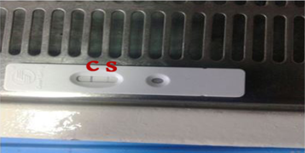
Positive brain sample using the rapid antigen test kit C control line /S positive sample line.
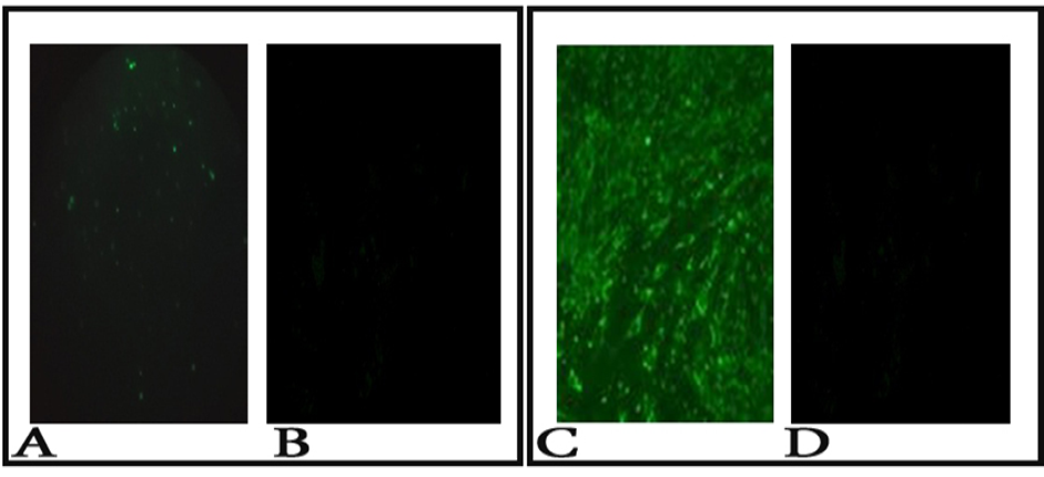
A: Positive fluorescence on brain impression smear 100x (dust like appearance of apple green color); B: No fluorescence at a negative control brain impression smear; C: Positive fluorescence sample on BHK cells100x (intracytoplasmic fluorescence); D: No fluorescence at a negative control BHK cells.
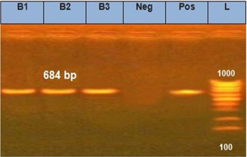
The electrophoretic pattern of PCR products (684 bp) to the brain samples (B1, B2, B3) for N gene.
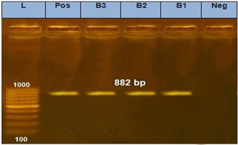
The electrophoretic pattern of PCR products (882 bp) to the brain samples (B1, B2, B3) for the P gene.
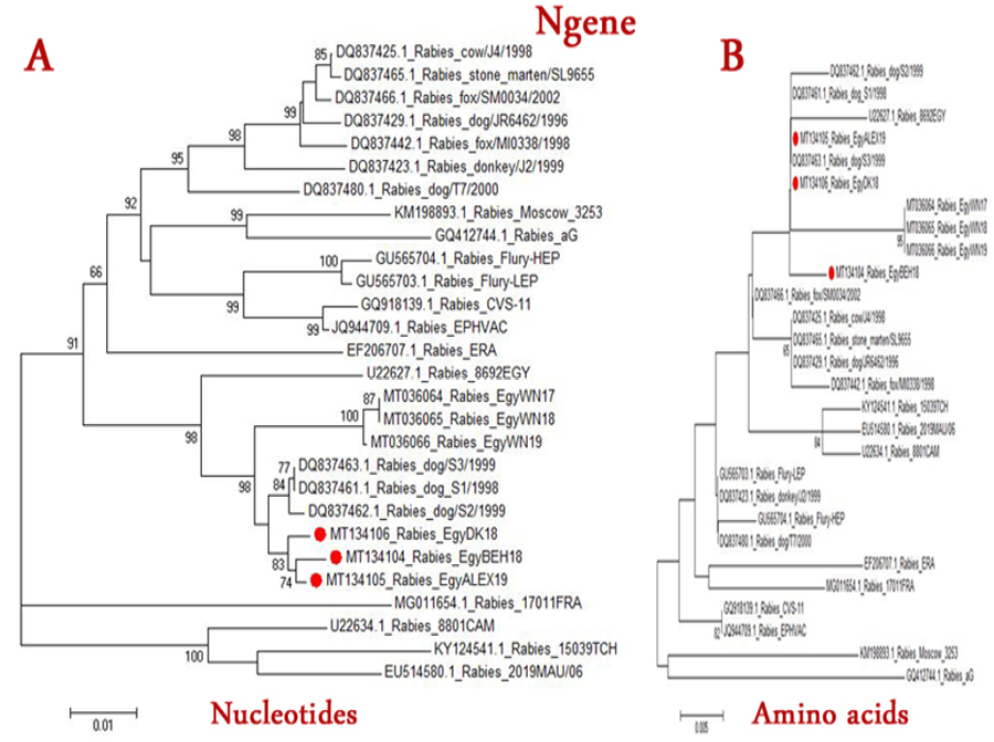
The Phylogenetic tree of Rabies viruses based on nucleotide (A) or Amino acid (B) sequence of N gene. Phylogenetic tree is based on maximum likelihood calculations (Mega 7 software) under the best fit model. Numbers at nodes represent measures of robustness based on 1000 bootstrap values. The sequences of selected publicly available strains were obtained from GenBank. Strains of current study are labeled with red circles.
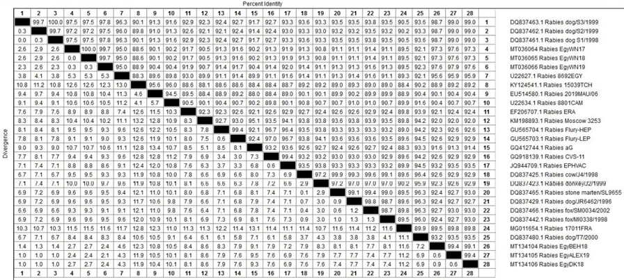
Figure illustrating the % of nucleotide identity of some Egyptian rabies isolates with other rabies virus isolates and strains at the level of the N gene.
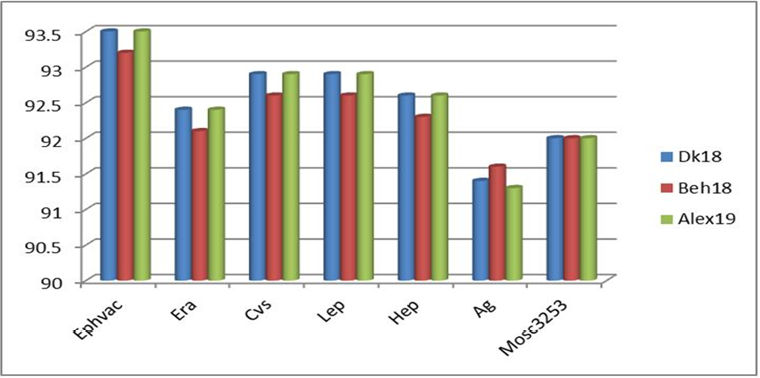
Comparing the nucleotide identity between each of the new Egyptian isolates and some vaccinal strains at the level of the N gene.
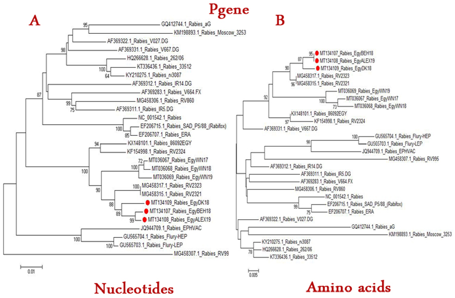
The phylogenetic tree of Rabies viruses based on nucleotide (A) or Amino acid (B) sequence of P gene. Phylogenetic tree is based on maximum likelihood calculations (Mega 7 software) under the best fit model. Numbers at nodes represent measures of robustness based on 1000 bootstrap value. The sequences of selected publicly available strains were obtained from GenBank. Strains of current study are labeled with red circle.
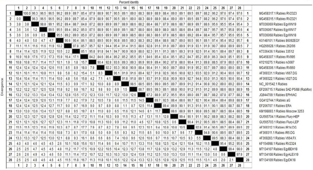
Figure illustrating the % of nucleotide identity of some Egyptian rabies isolates with other rabies virus isolates and strains at the level of P gene.
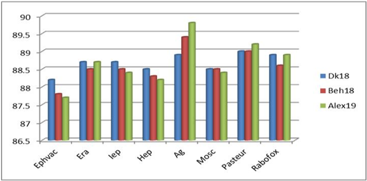
Chart comparing the nucleotide identity between each of the new Egyptian isolates and some vaccinal isolates at the level of the P gene.
References
- Altschul, S.F., Gish, W., Miller, W., Myers, E.W. and Lipmanl, D.J., 1990. Basic local alignment search tool. J. Mol. Biol., 215: 403-410. https://doi.org/10.1016/S0022-2836(05)80360-2[CrossRef]
- Assenberg, R., Delmas, O., Ren, J., Vidalain, V., Verma, A., Larrous, F., Graham, S., Tangy, F., Grimes, J. and Bourhy, H., 2010. Structure of the nucleoprotein binding domain of Mokola virus phosphoprotein. J. Virol., 84: 1089–1096. https://doi.org/10.1128/JVI.01520-09[CrossRef]
- Botvinkin, A.D., Poleschuk, E.M., Kuzmin, I.V., Borisova, T.I., Gazarian, S.V., Yager, P., Rupprecht, C.E.N., 2003. lyssavirus isolated from bat in Russia. Emerg. Infect. Dis., 9: 1623–1625. https://doi.org/10.3201/eid0912.030374[CrossRef]
- Brzozka, K., Finke, S. and Conzelmann, K.K., 2005. Identification of the rabies virus alpha/beta interferon antagonist: Phosphoprotein P interferes with phosphorylation of interferon regulatory factor 3. J. Virol., 79: 7673–7681. https://doi.org/10.1128/JVI.79.12.7673-7681.2005[CrossRef]
- CLC’s Main Workbench 6 Software, 2022. http//clc-genomics-workbench.software.informer.com/6.0/
- David, D., Hughes, G.J., Yakobson, B.A., Davidson, I., Hikmat, U.N., Orhan Aylan, Ivan, V. Kuzmin and Charles, E.R., 2007. Identification of novel canine rabies virus clades in the Middle East and North Africa. J. Gen. Virol., 88: 967–980. https://doi.org/10.1099/vir.0.82352-0[CrossRef]
- David, D., Yakobson, B., Smith, J.S. and Stram, Y., 2000. Molecular epidemiology of rabies virus isolates from israel and other middle- and near-eastern countries. Urnal Clin. Microbiol., 38(2): 755–762. https://doi.org/10.1128/JCM.38.2.755-762.2000[CrossRef]
- Dhaval H.V., Bharat, B.B., Rafyuddin, A.M. and Mayurdhvaj, K.J., 2017. Sequencing and sequence analysis of partial nucleoprotein (N) gene and phylogenetic analysis of rabies virus field isolates from Gujarat state, India. Virus Dis., 28(3): 320-327. https://doi.org/10.1007/s13337-017-0387-3[CrossRef]
- Hall, T., 1999. BioEdit: A user-friendly biological sequence alignment editor and analysis program for Windows 95/98/NT. Nucl. Acids Symp. Ser., 41: 95–98.
- Jessica, C., Marike, G., Kevin, R. and Wanda, M., 2021. Lagos bat virus, an under-reported rabies-related lyssavirus. Viruses, 13(4): 576. https://doi.org/10.3390/v13040576[CrossRef]
- Kanitz, F.A., Cargnelutti, J.F., Weiblen, R., Batista, H.B.C.R. and Flores, E.F., 2015. Virus isolation in cell culture for confirmatory diagnostic of rabies in bovine specimens. Cienc. Rural., Santa Maria. 45(12): 2193-2196. https://doi.org/10.1590/0103-8478cr20141690[CrossRef]
- Kissi, B., Tordo, N. and Bourhy, H., 1995. Genetic polymorphism in the rabies virus nucleoprotein gene. Virology, 209: 526–537. https://doi.org/10.1006/viro.1995.1285[CrossRef]
- Kumar, S., Stecher, G. and Tamura, K., 2016. MEGA7: Molecular Evol., evolutionary genetics analysis version 7.0 for bigger datasets. Mol. Biol., 33: 1870–1874. https://doi.org/10.1093/molbev/msw054[CrossRef]
- Kuzmin, I.V., Orciari, L.A., Arai, Y.T., Smith, J.S., Hanlon, C.A., Kameoka, Y. and Rupprecht, C.E., 2003. Bat lyssavirus (Aravan and Khujand) from Central Asia: phylogenetic relationships according to N, P and G gene sequence. Virus Res., 97: 65–79. https://doi.org/10.1016/S0168-1702(03)00217-X[CrossRef]
- Lieu, K.G., Brice, A. and Wiltzer, L., 2013. The rabies virus interferon antagonist p protein interacts with activated STAT3 and inhibits Gp130 receptor signaling. J. Virol., 87(14): 8261–8265. https://doi.org/10.1128/JVI.00989-13[CrossRef]
- Lui, J., Liao, M., Yan, Y., Yang, H., Wang, H. and Zhou, H., 2020. Rabies virus phosphoprotein P5 binding to BECN1 regulates self-replication by BECN1-mediated autophagy signaling pathway. Cell Commun. Signal, 18(1): 153. https://doi.org/10.1186/s12964-020-00644-4[CrossRef]
- Mavrakis, M., McCarthy, A.A., Roche, S., Blondel, D. and Ruigrok, R.W., 2004. Structure and function of the C-terminal domain of the polymerase cofactor of rabies virus. J. Mol. Biol., 343: 819–831. https://doi.org/10.1016/j.jmb.2004.08.071[CrossRef]
- Michael, S., Fouad, S.E.M., Zeinab, R.A., Ayman, S.E-H., Marcus, I.Y. and Saad, S.A.S., 2021. Molecular detection and phylogenetic analyses to evaluate the evolutionary pattern of N, P, and G gene sequences of some recent rabies virus (RABV) isolates from Elwadi- Elgedid Province in Egypt. Adv. Anim. Vet. Sci., 9(11): 1989-1994. https://doi.org/10.17582/journal.aavs/2021/9.11.1989.1994[Full text][CrossRef]
- Mohamed, E-T., Mohamed, E-B. and Mohamed, F.H., 2015. Identification and genetic characterization of rabies virus from Egyptian water buffaloes (Bubalus bubalis) bitten by a fox. Virus Dis., 26(3): 141–146. https://doi.org/10.1007/s13337-015-0263-y[CrossRef]
- Nadin-Davis, S.A., 2007. Molecular epidemiology. In: Jackson AC, Wunner W.H. (eds) Rabies, 2nd eds. Academic Press, San Diego, pp. 69–122. https://doi.org/10.1016/B978-012369366-2/50005-1[CrossRef]
- OIE Office International des Epizooties, 2018. Chapter 3.1.1.17. Rabies (infection) with rabies virus and other Lyssaviruses. In: OIE manual of diagnostic tests and vaccines for terrestrial animals. 8th ed. Paris, pp. 578-597.
- QIAamp Viral RNA Microkit, 2013. https://www.qiagen.com/us/products/diagnostics-and-clinical-research/sample-processing/qiaamp-viral-rna-kits
- Satoko, Y., Naoto, A.I., Ohka, A.A.S., Kaneda, C.S., Nakamura, D.E.H., Agari, D.E.T., Masaani, B.T., Nakagawa, A.K., Okada, A.K., Okadera, A.K., Mitake, A.H., Fujii, A.T., and Sugiyama, M., 2013. Involvement of the rabies virus phosphoprotein gene in Neuroinvasiveness. J. Virol., 87(22): 12327–12338. https://doi.org/10.1128/JVI.02132-13[CrossRef]
- Shoji, Y., Inoue, S., Nakamichi, K., Kurane, I., Sakai, T. and Morimoto, K., 2004. Generation and characterization of P gene-deficient rabies virus. Virology, 318(1): 295-305. https://doi.org/10.1016/j.virol.2003.10.001[CrossRef]
- Silberfein, M. and Conteh, A.H., 2006. Boundaries and conflict in the mano river region of west Africa (2006). Conf. Manage. Peace Sci., 23: 343–361. https://doi.org/10.1080/07388940600972685[CrossRef]
- Smith, J.S., Lillian, A.O., Pamela, Y.H., David, S. and Cynthia, K., 1992. Epidemiologic and historical relationships among 87 rabies virus isolate as determined by limited sequence analysis. J. Inf. Dis., 166(2): 296–307. https://doi.org/10.1093/infdis/166.2.296[CrossRef]
- Sunday, O.V.O., Okechukwu, O. and Okechukwu, R., 2014. Cross border crimes in West African Sub-Region: Implications for Nigeria’s national security and external relations. Glob. J. Hum. Soc. Sci. F Polit. Sci., 14: 45–57.
- Tordo, N., Poch, O., Ermine, A., Keith, G. and Rougeon, F., 1986. Walking along the rabies genome: Is the large G-L intergenic region a remnant gene? Proc. Natl. Acad. Sci. U.S.A., 83: 3914–3918. https://doi.org/10.1073/pnas.83.11.3914[CrossRef]
- Vidy, A., Chelbi-Alix, M. and Blondel, D., 2005. Rabies virus P protein interacts with STAT1 and inhibits interferon signal transduction pathways. J. Virol., 79: 14411–14420. https://doi.org/10.1128/JVI.79.22.14411-14420.2005[CrossRef]
- Wei, X.K., Zhong, Y.Z., Pan, Y., Li X.N., Liang, J.J. and Luo, T.R., 2018. The N and P genes facilitate pathogenicity of the rabies virus G gene. Vet. Med., 63: 561-570. https://doi.org/10.17221/63/2018-VETMED[CrossRef]
- Yanni, M.I, Hanaa, A.A., Lamyaa, A.A., Hanan, A.F., Ibrahim, E.M. and Aggour, M.G., 2014. Molecular studies on rabies isolates from horses in Cairo Egyptian J. Virol., 11(1): 117-127.
Services
Article usage statistics
Loading…