
This study aims at measuring expressions of β-catenin and senescence marker protein-30 (SMP30) after normal human skin fibroblasts (NHSFs) irradiated by different UVB doses as well as durations, thus unveiling mechanisms underlying HSF senescence induced by Ultraviolet B (UVB). NHSFs treated under different doses (100, 200, 300 mJ/cm2) of UVB with different durations (1, 2, 3 days) were case group, while those without UVB irradiation were control group. Expressions of gene/protein were obtained with real-time quantitative PCR (RT-qPCR) and western blot; cell proliferation inhibition rates (CPIR) of HSF were obtained by MTT, and HSF apoptosis was detected using Flow Cytometry (FCM). UVB irradiations significantly inhibit HSF growth, showing positive correlation with the increasing of CPIRs and apoptotic rates; The relative expression levels of β-catenin and Senescence marker protein-30 (SMP30) gene in UVB treated group were significantly lower than that of the control group; Gene expression levels also varied with different UVB doses and durations, indicating statistically significant dose- and time-dependent effects of UVB. UVB treatments inhibit cell growth and promote apoptosis in a dose-dependent and time-dependent manner, and high level as well as prolonged UVB irradiation lead to NHSFs senescence.
Research Article · Volume 52, Issue 6, 16 September 2020 · Open Access
UVB Irradiation Induces Human Skin Fibroblasts Senescence Through Regulation of Î’-Catenin and Smp30 Expression
· · · · ·
Affiliations & Notes ⌄
- 1Departments of Obstetrics and Gynecology, Taihe Hospital, Hubei University of Medicine, Shiyan, Renmin Southern 32, Hubei 442000, P.R. China 2Department of Medical offices, Taihe Hospital, Hubei University of Medicine, Shiyan, Renmin Southern 32, Hubei 442000, P.R. China 3Department of Laboratory Medicine, Taihe Hospital, Hubei University of Medicine, Shiyan, Renmin Southern 32, Hubei 442000, P.R. China 4Department of Anesthesiology, Ren-ming Hospital of Yun-xi, Shiyan, Hubei 442000, P.R. China
Article Info ⌄
- Journal:
- Pakistan Journal of Zoology
- Published:
- 16 September 2020

From the article
- The study showed that the NHSF CPIR and apoptosis rate increased gradually with increasing UVB dose and irradiation duration.
- This result suggests that the occurrence of damages, such as cell apoptosis, reduces survival rate of HSF given UVB irradiation under certain dose and duration.
- Moreover, the β-catenin and SMP30 gene expression in the case group decreased gradually with increasing UVB dose, indicating time- and dose-dependent impact of UVB induction.
- Hence, the decreased β-catenin gene expression in HSF induced by UVB oxidative stress may serve as a mechanism for normal HSF.
Selected from the article's own text.
Abstract
INTRODUCTION
Long-term exposure of human skin fibroblasts (HSF) to oxidative stress may induce alterations in cell senescence, morphology, G1 phase stagnation, and target gene expression ; . The long-term exposure of the skin may cause oxidative stress damage, which plays an important role in cell senescence and skin aging . UVB mainly acts on the shallow layer of the dermis and epidermis. Meanwhile, β-catenin is an effector protein that plays a key role in the Wnt signal pathway, particularly in regulating cell proliferation and apoptosis to protect against senescence . SMP30, a senescence marker protein that was first recognized in rat liver, exhibits decreased expression with increasing host age; this protein, together with β-catenin, may mediate and delay senescence . This study aims to simulate damage states after irradiation under different UVB doses as well as durations and determine HSF proliferation, apoptosis rate, gene expression differentiations to explore functions of β-catenin in UVB-induced skin damage to unveil the possible mechanism of oxidative-stress-induced photodamage.
MATERIALS AND METHODS
Cell culture and UVB treatment
Frozen and thawed cells were revived in DMEM culture medium containing 10% fetal calf serum. After fusion growth to 80%, the cells were subcultured with 1:1 trypsin and digested in EDTA. Two to four generations of well-grown cells were obtained for testing, and the remaining cells were preserved in liquid nitrogen. HSFs treated under different doses (100, 200, 300 mJ/cm2) of UVB as well as durations (1, 2, 3 days) were case group, and NHSFs without UVB irradiation were control group. The collected cells were culture for 24 h and used to determine NHSF proliferation, apoptosis rate and gene expression differentiation.
Detection of HSF proliferation
MTT method was used to detect HSF proliferation. About 2×10³ logarithmic phase cells (about 200 μl of the culture solution) were obtained and inoculated onto the culture plate. After single-layer adherent growth of cells for 24 h and culture plate coverage of about 50%-60%, the supernatant was discarded. The cells were irradiated with 100, 200, 300 mJ/cm2 UVB for 1, 2, 3 days from a distance of 30 cm. After irradiation, the culture was terminated, and the supernatant was discarded. DMSO was added to each hole for shaking and mixing, and absorbance A at 570 nm for each hole was recorded. CPIR was used to determine cell proliferation activity in the two groups, where CPIR= (AControl group-ATest group / AControl group)×100%. Each experiment was repeated 5 times.
Detection of HSF apoptosis
HSF apoptosis was detected using FCM. After irradiation, 1×10⁶ cells were collected and washed with PBS three times. The cells were added with 10 μl of annexin V-FITC and 5 μl of PI. The cells were subjected to lucifugal incubation for 30 min and mixed in 100 μl of binding buffer. Apoptosis rate was detected within 1 h.
RNA extraction and reverse transcription
Total RNA was extracted using TRIzol® Reagent (Invitrogen, USA) according to the manufacture’s protocol. The concentration and purity of total RNA was determined by measuring the absorption with Nano Drop-2000 (Thermo Fisher) at 260 nm and 280 nm. First-strand cDNA was prepared from total RNA using Promega reverse transcription system (Promega, USA) based on the manufacturer’s instructions. cDNA was used immediately or stored at -80°C until use.
Real-time PCR detected the expression of gene
The primers were used for the polymerase chain reactions (PCR) as follows: 5’-CAC TAC CAC AGC TCC TTC TC-3’ (forward) and 5’-GAG CAG CAT CAA ACT GTG TA-3’ (reverse) for β-catenin gene, 5’-CCG TGG ATG CCT TTG ACT AT-3’ (forward) and 5’-TCC AAA GCA GCA TGA AGT TG-3’ (reverse) for SMP30 gene, 5’-GAA GGT GAA GGT CGG AGT C-3’ (forward) and 5’-GAA GAT GGT GAT GGG ATT TC-3’ (reverse) for GAPDH gene. The primes produced 236, 233 and 226 samples for β-catenin, SMP30 and GAPDH, respectively. PCR amplification system: 25 mmol/L Mg2+ 2.4 μl, 2 μL each of 5’ and 3’ primer, 5 U/μl Taq 0.3 μl, 2 mmol/L dNTP 1.5 μl, 10×SYBR-Green I 1 μl, 10× Buffer 3 μl, cDNA 5 μl, with sterile water having volume of 30 μL. Reaction conditions: 95°C denaturation for 5 minutes; 94°C 30 seconds, 60°C 30 seconds, 72°C 1 min with 35 cycles; At the last cycle, dissociation curve analysis was performed and the reaction products were separated electrophoretically on a 2% agarose gel and stained with ethidium bromide for further confirmation of the PCR products. MRNA expression levels of β-catenin and SMP30 were measured as a relative ratio to GAPDH in each sample, and gene expression levels were normalized and determined by the 2-∆∆Ct method.
Western blot analysis
Total proteins were extracted using RIPA buffer and concentrations were determined by the BCA protein quantitative kit. In total 10 μg protein was loaded onto sodium dodecyl sulfate-polyacrylamide gel electrophoresis for electrophoresis and transferred to PVDF membranes. After transfer, Blots were blocked with 5% nonfat dry milk–TBS–0.1% Tween 20 for 2h at room temperature and then washed, then incubated overnight at 4°C with primary polyclonal antibodies against β-catenin and SMP30 according to the manufacturer’s instructions. Signals were detected after incubation for 2h at room temperature in horseradish peroxidase-conjugated secondary antibody. GAPDH acted as the reference protein for loading control.
statistical analysis
SPSS version 16.0 was employed to analyze the results, and P<0.05 was considered to indicate a statistically significant difference. The measured data were expressed as the mean ± standard deviation. A Student’s t-test, the variance analysis of repeated measurement data and Bilinear regression were used to compare groups.
RESULTS
Changes of every detection indicator with different UVB doses for different durations
At different irradiation time points, the comparison between two irradiation dose groups showed that the effect of time factors varies with the different dose of radiation. UVB irradiation with different UVB doses for different durations significantly inhibited NHSF growth, the CPIR and apoptotic rate increased gradually with increasing irradiation dose and duration. The relative expression level of the β-catenin and SMP30 gene in the case group were significantly lower than that in the control group after UVB treatment. Gene expression also varied with different UVB doses and durations, indicating the dose- and time-dependent effects of UVB, the difference of gene expression between different groups was statistically significant. According to the Sphericity Assumption, different analysis results were used (Tables I and II). The changes of every detection indicator with different UVB doses for different durations are shown in Table III and Figs. 1, 2.
Table I. Test of within-subjects effects (expression of gene).
| Source | Type III sum of squares | df | Mean square | F | Sig | ||
| β-catenin | day | Sphericity assumed | 0.249 | 2 | 0.125 | 28.638 | <0.001 |
| day*group | Sphericity assumed | 0.334 | 6 | 0.056 | 12.784 | <0.001 | |
| SMP30 | day | Sphericity assumed | 0.282 | 2 | 0.141 | 142.66 | <0.001 |
| day*group | Sphericity assumed | 0.293 | 6 | 0.049 | 49.287 | <0.001 |
Table II. Multivariate TestsC (CPIR and apoptosis rate).
| Effect | Value | F | Hypothesis df | Error df | Sig | ||
| CPIR | day | Pillai's trace | 0.961 | 183.791 | 2 | 15 | <0.001 |
| day*group | Pillai's trace | 0.68 | 2.748 | 6 | 32 | <0.001 | |
| Apoptosis rate | day | Pillai's trace | 0.975 | 297.075 | 2 | 15 | <0.001 |
| day*group | Pillai's trace | 1.215 | 8.257 | 6 | 32 | <0.001 |
Table III. The changes of every detection indicator with different UVB doses for different durations.
| UVB(mJ/cm2) | CPIR | Apoptosis rate | β-catenin mRNA | SMP30 mRNA | ||||||||
| 1d | 2d | 3d | 1d | 2d | 3d | 1d | 2d | 3d | 1d | 2d | 3d | |
| Control group | 0 | 0 | 0 | 1.96 ±0.41 | 2.81 ±0.57 | 4.01 ±0.38 | 1.14 ±0.14 | 1.27 ±0.11 | 1.29 ±0.11 | 1.08 ±0.10 | 1.18 ±0.10 | 1.20 ±0.10 |
| 100 | 4.12 ±0.76 | 6.02 ±1.08 | 8.38 ±1.27 | 6.52 ±1.51 | 10.06 ±1.50 | 12.06 ±1.17 | 0.96 ±0.10 | 0.82 ±0.08 | 0.71 ±0.09 | 0.94 ±0.08 | 0.82 ±0.08 | 0.69 ±0.10 |
| 200 | 7.14 ±0.84 | 9.14 ±1.44 | 11.42 ±1.51 | 9.61 ±1.14 | 12.24 ±1.19 | 14.32 ±0.86 | 0.82 ±0.08 | 0.70 ±0.08 | 0.59 ±0.07 | 0.80 ±0.11 | 0.66 ±0.08 | 0.53 ±0.09 |
| 300 | 9.52 ±0.53 | 12.37 ±0.96 | 14.64 ±1.05 | 12.06 ±1.17 | 14.18 ±1.16 | 16.02 ±1.32 | 0.68 ±0.14 | 0.53 ±0.11 | 0.39 ±0.07 | 0.63 ±0.12 | 0.48 ±0.08 | 0.36 ±0.07 |
Bilinear regression analysis of the relationship between t detection indicator and irradiation dose for different durations
The CPIR (apoptosis rate, gene expression) as the dependent variable and the different irradiation dose with different durations as independent variables for the regression analysis showed that in line with the linear relationship between the CPIR (apoptosis rate, gene expression) and irradiation dose for different durations. (Table IV and Fig. 3), P<0.05.
Table IV. Regression analysis of the influence factors of detection indicator.
| Model | B | Std. Error | Beta | t | Sig | |
| CPIR | constant | -6.42 | 0.681 | -9.42 | <0.001 | |
| group | 3.96 | 0.176 | 0.91 | 22.51 | <0.001 | |
| day | 1.71 | 0.241 | 0.29 | 7.09 | <0.001 | |
| Apoptosis rate | constant | -3.42 | 0.766 | -4.46 | <0.001 | |
| group | 3.6 | 0.198 | 0.863 | 18.21 | <0.001 | |
| day | 2.033 | 0.271 | 0.356 | 7.51 | <0.001 | |
| β-catenin | constant | 1.536 | 0.061 | 25.282 | <0.001 | |
| group | -0.221 | 0.016 | -0.86 | -14.114 | <0.001 | |
| day | -0.079 | 0.021 | -0.223 | -3.667 | <0.001 | |
| SMP30 | constant | 1.489 | 0.055 | 27.16 | <0.001 | |
| group | -0.215 | 0.014 | -0.868 | -15.23 | <0.001 | |
| day | -0.084 | 0.019 | -0.247 | -4.335 | <0.001 |
Gene/Protein expression in the control group and the experimental group
The expression level of β-catenin gene in the control group was 1.23 ±0.13, and in the experimental group was 0.70 ±0.18, the difference was statistically significant (t=10.68, P<0.001); The expression level of SMP30 gene in the control group was 1.15 ±0.11, and in the experimental group was 0.66 ±0.19, the difference was statistically significant (t=9.5, P<0.001). β-catenin and SMP30 expression levels are presented in Figure 4.
DISCUSSION
As an important core signal molecule in the Wnt pathway, β-catenin is located in chromosome 3p21, which plays a key role in occurrence and development of some tumors, regulating cell proliferation, apoptosis and anti-senescence ; . Senescence is a biological phenomenon that involves gradual degenerative changes in organs and progressive decline in physiological function caused by endogenous and exogenous factors with aging of a mature individual . In this process, the expression of related senescence genes, including mRNAs, within the cell mainly decreases at molecular level. Senescence may also occur in skin. In this study, HSF was treated with different UVB doses for different durations to determine β-catenin, SMP30 expression before and after simulating damage states and study the relationship between such changes and UVB irradiation. The potential mechanism underlying the effect of UVB irradiation on skin senescence was also explored.
The study showed that the NHSF CPIR and apoptosis rate increased gradually with increasing UVB dose and irradiation duration. This result suggests that the occurrence of damages, such as cell apoptosis, reduces survival rate of HSF given UVB irradiation under certain dose and duration. Wang reported that 300 mJ/cm2 UVB irradiation can induce NHSF to an optically damaged state; the treatment also leads to a series of oxidative stress damages, such as G1 phase stagnation, cell apoptosis, diminished SOD activity and increased release of oxidative stress damage products. Moreover, the β-catenin and SMP30 gene expression in the case group decreased gradually with increasing UVB dose, indicating time- and dose-dependent impact of UVB induction. After the damage was simulated, the gene expression significantly increased compared to that in the control group. This result is consistent with that presented by Tian et al. (2011). Furthermore, as the target gene regulating skin senescence, high β-catenin gene expression can augment HSF proliferation to a certain extent (Tian et al., 2015). Hence, the decreased β-catenin gene expression in HSF induced by UVB oxidative stress may serve as a mechanism for normal HSF.
As a senescence marker protein, SMP30 expression level decreased in mice when the oxidative stress level increased . Moreover, the high expression level of SMP30 inhibited the expression of senescence-related cell reactive oxygen species and β-galactosidase . However, after knocking out the SMP30 gene in mice, the pro-oxidant level increased with prolonged life of these mice . Upon SMP30 gene knockout, the mice could not synthesize sufficient Vc, resulting in diminished antioxidative stress ability, which accelerated free-radical-induced damage, and eventually hastened cell senescence . These studies showed that SMP30 serves as an antisenescence function in cells. When the skin is exposed to oxidative stress, such as UVB, oxygen radicals accumulate, thereby decreasing the expression of SMP30 that regulates skin senescence in NHSF. Moreover, different kinds of irreversible oxidative damage may be produced inside cells, leading to skin senescence.
Previous studies have shown that the photodamage induced by UVB is a path of skin senescence caused by the reduction of β-catenin and SMP30. Some studies have shown that FOXO3a can inhibit oxidation thereby delaying senescence through SOD2, catalase and related signaling pathways (Wnt pathway, PI3K/Akt pathway and so on) ; ; . This is also one of the key contents of our later research by cell transfer.
High UVB irradiation doses can cause DNA irreversible damage, cell necrosis, or even cancer, whereas low irradiation doses can cause oxidative-stress-induced photodamage and initiate cell aging ; . Oxidative stress damage plays an important role in the process of skin-cell premature senility. We have not discussed the effectiveness of controlling UVB damage as well as preventing oxidative-stress-induced skin senescence, but will be studied in future work. The presented results are helpful for further study in aesthetic medicine.
ACKNOWLEDGEMENTS
This study was funded by Taihe hospital projects (No.2016JZ26, No.2017JJXM044 and N0.2014JJXM019).
Disclosure of conflict of interest
The authors declare there is no conflict of interest.
Figures
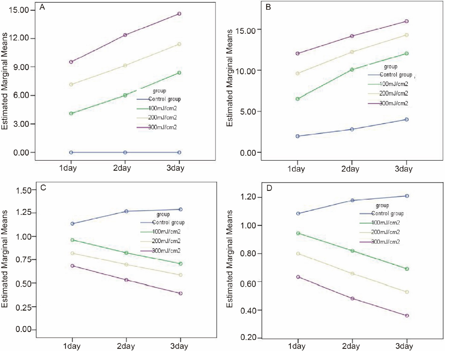
The changes of every detection indicator with different UVB doses for different durations. a, cell proliferation inhibition rate, CPIR; b, apoptosis rate; c, β-catenin mRNA expression; d, SMP30 mRNA expression.
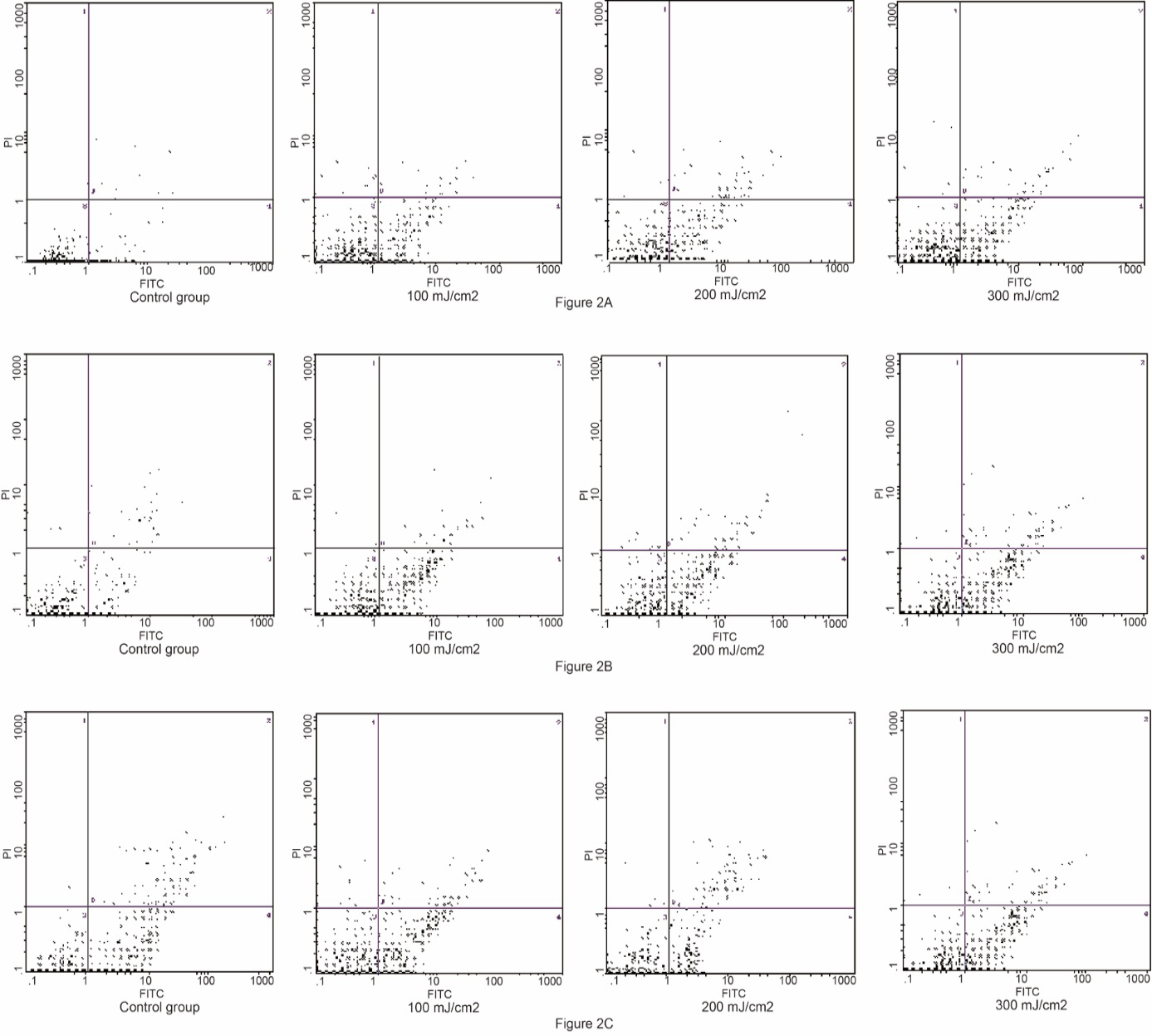
The changes of HSF apoptosis with different UVB doses for different durations. A, different doses UVB treatment 1 day; B, different doses UVB treatment 2 day; C, different doses UVB treatment 3 day.
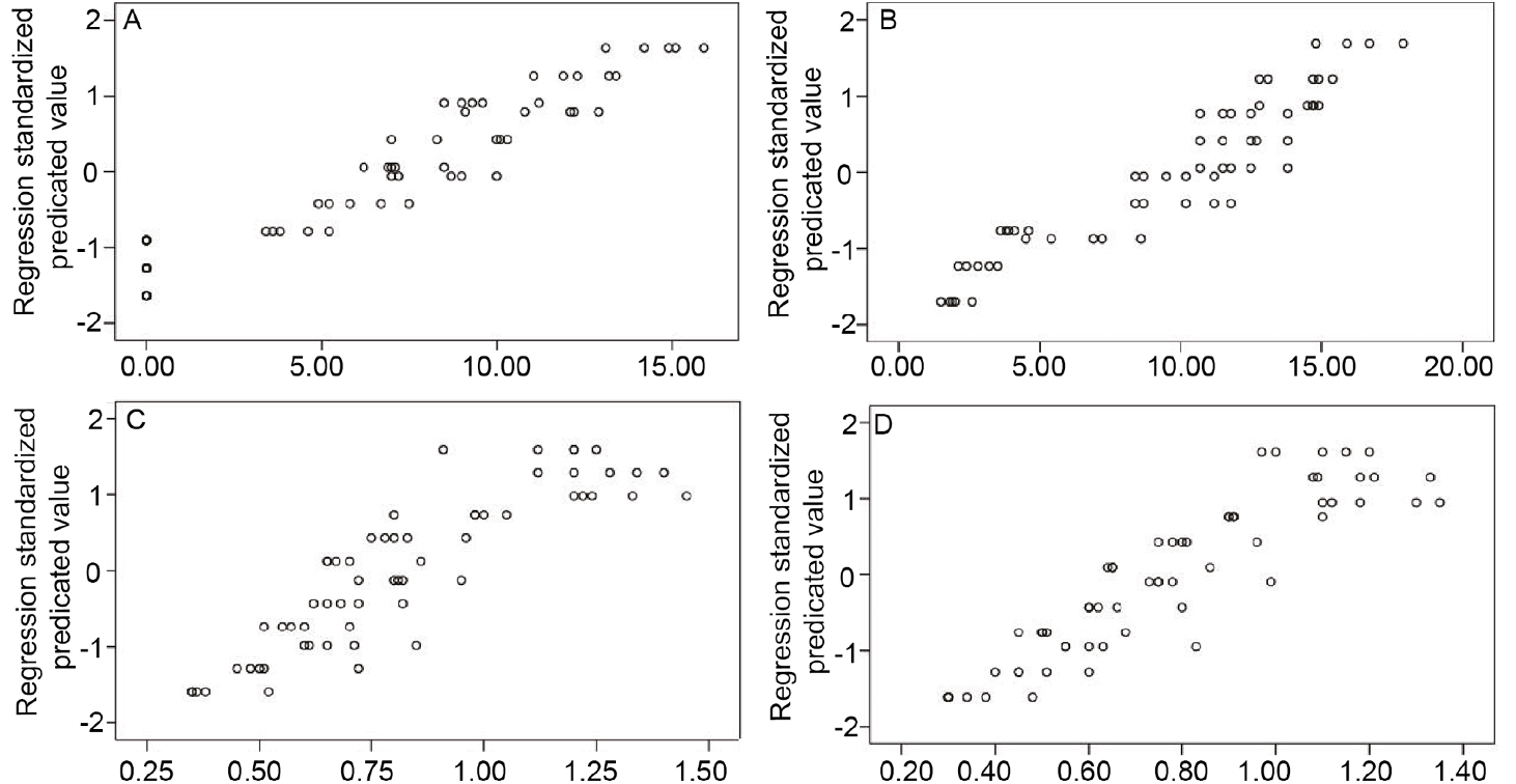
Analysis of the relationship between every detection indicator and different UVB doses for different durations by Bilinear regression. a, cell proliferation inhibition rate (CPIR); b, apoptosis rate; c, β-catenin; d, SMP30), P<0.05.
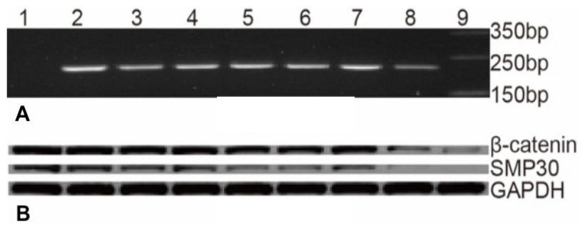
The expression ofβ-catenin and SMP30 mRNA /protein in every group. A, gene expression products by PCR: 1, control group; 2, GAPDH; 3~5, β-catenin; 6~8, SMP30; 9, Marker. B, protein expression products by Western Blot.
References
- Chen, P., Xu, S. and Qu, J., 2018. Lycopene protects keratinocytes against UVB radiation-induced carcinogenesis via negative regulation of FOXO3a through the mTORC2/AKT signaling pathway. J. Cell. Biochem., 1: 366-377. https://doi.org/10.1002/jcb.26189[CrossRef]
- Clavel, S., Siffroi-Fernandez, S., Coldefy, A.S., Boulukos, K., Pisani, D.F. and Dérijard, B., 2010. Regulation of the intracellular localization of Foxo3a by stress-activated protein kinase signaling pathways in skeletal muscle cells. Mol. Cell. Biol., 2: 470-480. https://doi.org/10.1128/MCB.00666-09[CrossRef]
- Hong, E.H., Lee, S.J., Kim, J.S., Lee, K.H., Um, H.D., Kim, J.H., Kim, S.J., Kim, J.I. and Hwang, S.G., 2010. Ionizing radiation induce cellular senescence of articular chondrocytes via negative regulation of SIRT1 by p38 kinase. J. biol. Chem., 285:1283-1295. https://doi.org/10.1074/jbc.M109.058628[CrossRef]
- Kagami, Y., Kondo, Y., Handa, S., Maruyama, N. and Ishigami, A., 2013. Senescence marker protein-30/gluconolactonase expression in the mouse ovary during gestation. Biol. Pharm. Bull., 36: 2005-2008. https://doi.org/10.1248/bpb.b13-00309[CrossRef]
- Mandinova, A., Lefort, K., Tommasi di Vignano, A., Stonely, W., Ostano, P., Chiorino, G., Iwaki, H., Nakanishi, J. and Dotto, G.P., 2008. The FoxO3a gene is a key negative target of canonical Notch signalling in the keratinocyte UVB response. EMBO J., 8: 1243-1254. https://doi.org/10.1038/emboj.2008.45[CrossRef]
- Mizukami, H., Saitoh, S. and Machii, H., 2013. Senescence marker protein-30 (SMP30) deficiency impairs myocardium-induced dilation of coronary arterioles associated with reactive oxygen species. Int. J. mol. Sci., 14: 9408-9423. https://doi.org/10.3390/ijms14059408[CrossRef]
- Pinnell, S.R., 2003. Cutaneous photodaamage, oxidative stress, and topical antioxidangt protection. J. Am. Acad. Dermatol., 48: 1-19. https://doi.org/10.1067/mjd.2003.16[CrossRef]
- Rass, K. and Reichrath, J., 2008. UVB damage and DNA repair in malignant melanoma and nonmelanoma skin cancer. Adv. exp. Med. Biol., l624: 162-178. https://doi.org/10.1007/978-0-387-77574-6_13[CrossRef]
- Sun, H., Liu, M., Wu, X., Yang, C., Zhang, Y., Xu, Z., Gao, K. and Wang, F., 2017. Overexpression of N-cadherin and β-catenin correlates with poor prognosis in patients with nasopharyngeal carcinoma. Oncol. Lett., 13: 1725-1730. https://doi.org/10.3892/ol.2017.5645[CrossRef]
- Tian, L.M., Peng, Y., Hu, R.Y., Zhang, P., Jiang, H.H., Xu, B., Tian, Q.J., Zhang, Z. and Wang P., 2016. Effect of senescence marker protein 30 on senescent phenotypes in hydrogen peroxide-induced senescence human skin fibroblasts. J. Xinxiang med. Univ., 33: 452-456.
- Tian, L.M., Xie, H.F., Li, J., Yang, T., Peng, Y. and Hu, W., 2015. Effects of β-cateninon the proliferative activity of and expressions of two apoptosis-related genes Bcl-2 and Bax by human skin fibroblasts induced by hydrogen peroxide. Chin. J. Dermatol., 48: 112-115.
- Tian, L.M., Xie, H.F., Li, J., Yang, T., Wang, W.Z. and Hu, Y.H., 2011. Expression of β-catenin in oxidative stress-induced premature senescence of hmuan skin flbroblast. Chin. J. Dermatol., 44: 259-262.
- Tian, L.M., Xie, H.F., Xiao, X., Yang, T., Hu, Y.H., Wang, W.Z., Liu, L.S., Chen, X. and Li, J., 2011. Study on the roles of β-catenin in hydrogen peroxide-induces senescence in human skin finro-blasts. Exp. Dermatol., 20: 836-838. https://doi.org/10.1111/j.1600-0625.2011.01324.x[CrossRef]
- Toussaint, O., Royer, V., Salmon, M. and Remacle, J., 2002. Stress-induced premature senescence and tissue ageing. Biochem. Pharmacol., 64: 1007-1009. https://doi.org/10.1016/S0006-2952(02)01170-X[CrossRef]
- Wang, Y.L., Wu, W. and Peng, G.P., 2008. Oxidative stress in human skin fibroblasts induced by UVB irradiation. Chin. J. Dermatol., 41:465-468.
- Woo, D.K. and Shadel, G.S., 2011. Mitochondrial sress signals revise an old aging theory. Cell, 144: 1-12. https://doi.org/10.1016/j.cell.2010.12.023[CrossRef]
- Yang, Y., Zhang, N., Zhu, J., Hong, X.T., Liu, H., Ou, Y.R., Su, F., Wang, R., Li, Y.M. and Wu, Q., 2017. Downregulated connexin32 promotes EMT through the Wnt/β-catenin pathway by targeting Snail expression in hepatocellular carcinoma. Int. J. Oncol., 50: 1977-1988. https://doi.org/10.3892/ijo.2017.3985[CrossRef]
- Zhang, Q.S. and Gu, H., 2008. Effects of UVB on the autophagy of human skin fibroblasts. China. J. Lepr. Skin. Dis., 24: 511-513.
- Zhang, W., Yan, S., Liu, M., Zhang, G., Yang, S., He, S., Bai, J., Quan, L., Zhu, H., Dong, Y. and Xu, N., 2010. beta-Catenin/TCF pathway plays a vital role in selenium induced-growth inhibition and apoptosis in esophageal squamous cell carcinoma (ESCC) cells. Cancer Lett., 296: 113-122. https://doi.org/10.1016/j.canlet.2010.04.001[CrossRef]
Services
Article usage statistics
Loading…