This study was carried out at the summer season 2015 in King Faisal University Research Station, Saudi Arabia. The field work continued for 10 weeks while, the laboratory work extended after that. Then the data analysis and prepare the manuscript exposed to some unexpected delay. One hundred twenty-eight sires from four different genetic background chicken lines were used for the study. Thirty-two sires of Cobb broiler breeder and 32 sires of Hisex layer breeder were obtained from commercial poultry projects at 35 weeks of age. Thirty-two sires of local Saudi Hajar1 line and 32 sires of local Saudi Hajar2 line were obtained from King Faisal University Research Station at the same age. Hajar1 and Hajar2 lines characterization were demonstrated by Ahmed and Alabbad (2014). From 1-day old, all birds were subjected to the same ND vaccination program. All birds were housed in an individual caging system, as one bird per cage, placed in a closed house system for 5 weeks prior to study. At 40 weeks of age, each line was divided into two groups: a control group and a treatment group, each of 16 sires in four replicates (4 birds/replicate). Water and commercial diet were provided ad libitum for all birds. Experimental birds were treated according to the mandatory roles of animal care and ethics guidelines set by the Deanship of Scientific Research, King Faisal University.
Cytokines-rich supernatant was generated according to the procedure of Ford et al. (2001), to produce IL-2 rich supernatant with slight modification. Nine Hajar1 cocks at 25 weeks of age were sacrificed, and then their spleens were removed and dissected individually under sterile conditions. The spleens were dissected and pressed through sterile mesh screens into 10 ml RPMI 1640 media (Sigma, USA), cell biology laboratory grade. The suspensions were centrifuged at 1000 rpm for 30 min in new 15 ml Falcon tubes. Splenocytes were collected and washed twice with 10 ml of RPMI 1640 media. Viable cells were distinguished using 0.05% trypan blue stain, counted, and adjusted to 10 million live cells per 1 ml of complete media consisting of RPMI 1640 media supplemented with penicillin, streptomycin (100 µg/ml), and 2% fetal calf serum. Five ml of the splenocytes suspension (50 million splenocytes) was transferred to each T-75 tissue culture flask containing 45 ml of complete media. Stimulation was carried out by the addition of Concanavalin A (ConA) (Sigma, USA, molecular grade) at 5 μg ConA per ml cells. After 24 hours of incubation in a 37°C humidified incubator with 5% CO2, the tissue culture fluid was harvested, centrifuged, and then the cytokine-rich supernatant was pooled in 15 ml tubes and stored at -20°C. Total protein was determined to ensure cytokine generation. The amount of protein generatedwhich refers to the amount of cytokines-derived proteins produced was calculated as the difference between total protein concentration in complete media and supernatant after splenocytes stimulation. The result was 160 mg/100 ml of media.
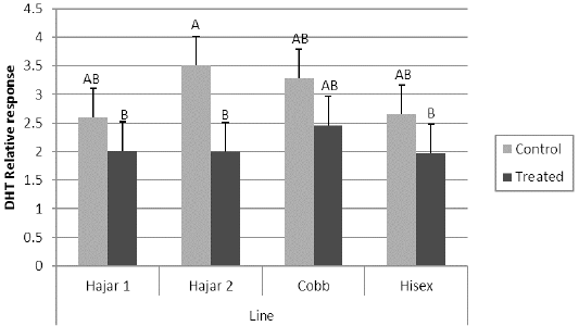
Parameters and data collection: At 40 weeks of age, zero time of the experimental period, blood samples were obtained from all birds to assess ND and SRBC zero time antibody titer. After sampling, only the treatment group birds in each line received 1 ml/kg body weight of the cytokine-rich supernatant via intramuscular injection. The control group birds were injected with 1 ml RPMI complete media containing the same concentration of ConA stimulant. Then all experimental birds were injected with 1 ml of 20% SRBC suspension via intramuscular route, to be used as an indicator for their humoral immunity. In addition, they all received a single dose of Lasota strain vaccine via eye drop route. Delayed hypersensitivity to BSA (Albumin bovine serum, lyophilized powder ≥96% Sigma, USA) as an indicator for cell-mediated immune response was achieved through the wattle test 20, where all birds at zero time of the experiment were subcutaneously injected with 4 mg BSA/bird. After 7 days of the experiment, all birds’ wattles thicknesses were recorded, and then the birds were reinjected with 1 mg of BSA in 0.1 ml saline intradermal. The delayed hypersensitivity test was calculated after 24 hours using micrometre as a relative response according to the following formula: Relative response = thickness of wattle after response (BSA response) / thickness of wattle before injection (control). Blood samples were collected from the wing veins of all birds at 3, 7, and 10 days post treatment for ND and SRBC titration. The SRBC antibody titers were assayed using micro agglutination as described by Nelson et al. (1995). A microtiter heamaglutination inhibition (HI) test (OIE, 2009), was used to assess the ND antibody titer.
Table 1: Effect of genetic line and Cytokine- rich supernatant treatment on Newcastle disease (ND) antibody titer.
* 0,3, 7 and 10 are zero day, 3days, 7days and 10 days of the experiment; ** T and C are the Treatment via cytokine -rich supernatant intramuscular injection, and control respectively; *** a, b, c, d values within a day with different superscript differ significantly (P ≤0. 05).
Table 2: Effect of genetic line and Cytokine- rich supernatant treatment on Sheep red blood cells (SRBC) antibody titer.
* 3, 7 and 10 are 3days, 7days and 10 days post SRBC injection; ** T and C are the Treatment via cytokine -rich supernatant intramuscular injection, and control respectively; *** a, b, c, d values within a day with different superscript differ significantly (P ≤0. 05).
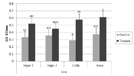
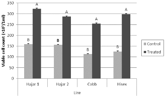
Mitogenic responses: Lymphocyte proliferation assays were performed. At 7 days of the experiment, 3 ml of peripheral blood was collected from all birds, and then peripheral blood lymphocytes of each sample were prepared. Lymphocytes were separated using Histopaque-1077 (sterile-filtered, density: 1.077 g/mL Sigma-Aldrich, USA), the number of viable cells was adjusted to 5 × 10⁶ cells/ml using the trypan-blue exclusion test. The cells were placed in 96-well flat-bottom tissue culture plates in three replicates at 100 µl/well and stimulated with phytohemagglutinine-p (PHA-p) (Phytohemagglutinin PHA-P, lyophilized powder, Cell biology grade Sigma-Aldrich, USA) (50 μg/ml) except for the negative-control wells. Two sets of plates were prepared for the following viable cell count and MTT assays. The plates were incubated at 41oC, as a suitable temperature for chicken cells, for 72 h in 5% CO2 in a humidified incubator. The MTT assay was applied to the first set of plates, according to Fernandez-Botran and Vetvicka (2001), a concentration of 5 mg/ml of MTT (98% Sigma-Aldrich, USA) was dissolved in RPMI-1640 media (Sigma, USA), and 10µl was added to each well. After incubation for 4 hr at 37oC with 5% CO2, 100 µl/well of acidic isopropanol solution was added to each well, mixed, and let stand for 10 min. The formed MTT formazan concentration was read on a micro ELISA reader and expressed as optical density (OD) at 570 nm wavelength. In addition, the second set of plates was used for the viable cell count using the trypan-blue exclusion test and expressed as cells/ml.
Statistical Analysis: The data were subjected to a two-way analysis of variance (ANOVA) for the effect of the genetic line and cytokine-rich supernatant treatment. Means were separated using Duncan’s multiple range tests. Data were analyzed using the general linear model procedure (SAS, 2005). JMP IN software version 5.1 (JMP IN 5.1, 2005) was used for statistical analysis. Statistical significance was considered as P ≤ 0.05 for all parameters.