
Cancer proteoforms are an innovation in identifying and understanding biomarkers and reaching for effective therapeutic targets. Proteoforms can be defined as individual species with different functions and been derived from a single gene using post-translational modifications and other mechanisms including splicing. Unlike traditional fixed protein isoforms, proteoforms describe the dynamic and functional aspect of proteins, which are of great importance in cancer research and clinical treatment. The discovery of specific diagnostic and prognostic cancer biomarkers by proteomic techniques has transformed the prevailing practices in oncology. The investigation of cancer proteoforms has shown that these structural modifications of proteins can contribute to the cancer development, metastasis and therapy. Proteoforms identification and discovery at present employ sophisticated proteomic instruments and high-definition mass spectrometers. They enable researches to select and describe proteoforms with a high level of specificity however, they are in difficulties with the issues of standardization and validation. The incorporation of proteoforms information into genomics and transcriptomes will in turn improve knowledge about cancer and provide input about biomarker advancements. In the therapeutic applications, proteoforms targeted approaches are appear as novel strategies. There are cognitive strategies where drugs and therapies got developed to interact with targeted proteoforms, which can be possibly helpful to overcome resistance issues and enhancing the necessities of treatment. Many of these advances are held to be creating a new generation of cancer treatments tailored to bring about a change in the manner of handling the disease.
Review · Volume 58, Issue 4, 21 June 2026 · Open Access
Unveiling Cancer Proteoforms: Insights into Biomarker Discovery and Therapeutic Strategies
· · · · · · · ·
Affiliations & Notes ⌄
- 1Department of Microbiology, Cholistan University of Veterinary and Animal Sciences, Bahawalpur, Pakistan 2Department of Biomedical and Diagnostic Sciences, University of Tennessee, Knoxville, USA 3Department of Parasitology, Cholistan University of Veterinary and Animal Sciences, Bahawalpur, Pakistan 4Department of Biotechnology, Baluchistan University of Information Technology, Engineering and Management Sciences, Quetta, Pakistan 5Veterinary Research Institute, Zarrar Shaheed Road, Lahore Cant
Article Info ⌄
- Journal:
- Pakistan Journal of Zoology
- Published:
- 21 June 2026

From the article
- Proteoforms, derived from genetic and post-translational alterations in cancer-associated proteins, provide intricate informative features about tumor biology, dynamics, and therapeutic prospects of heterogeneity and resistance.
- Recent molecular diagnostic techniques such as high-resolution mass spectrometry and single-cell proteomics are identifying innovative cancer-associated proteoforms that are revolutionizing cancer diagnostics and therapy.
- There are limitations like proteoforms complexity, integration of datasets, and biomarker confirmation and they have to be solved to bring the findings into clinical utility.
- Finally, the elucidation of cancer proteoforms can further improve cancer individualization as well as provide profound advancement in the field of oncology.
Selected from the article's own text.
Abstract
Introduction
Proteomics is the study of the proteome. The proteome encompasses the entire set of proteoforms present at a certain time in a cell, tissue, or individual in a given biological setting . Proteomics includes the assessment of global protein abundance, proteoforms levels, spatial conformations, chemical modifications, cellular localization, proteoforms functions, cofactors, and interacting partner networks . The dynamic shift in the research of cancer has resulted in a consideration of proteoforms which are multiple isotypes of a protein that are produced from the same gene through the action of different biological mechanisms . Proteoforms involve practically all the modifications, which can occur after translation or during transcription and can be seen as an exciting concept giving more dynamism to connections between protein structure and function in addition to isoforms . It is essential to consider the proteoforms as a key molecular player in the case of cancer and design new generation diagnostics and therapeutic approaches .
For this reason, proteoforms are different from protein isoforms since they are able to represent the complicated regulatory processes that occur in cells . With regards to cancer, then it can be observed that tumor cells have a distinct proteoforms variance compared to normal cells . This is an advantage since it offers the opportunity to investigate the tumor biology in addition to helping identify new biomarkers for the diagnosis, prediction of likely outcomes, and therapeutic targets . Certain prototypes linked to certain forms of cancer can act as biomarkers and provide a better picture of disease processes and tools for diagnostics and prognosis .
Proteoforms are not limited to discovery of biomarkers, they also provides the future possibilities for new therapeutic approaches . Since several cancer-associated proteoforms are synthetically lethal or significantly contribute to resistance mechanisms, scientists can design corresponding molecular inhibitors to disrupt these disease-promoting proteoforms selectively . This kind of approach is to maximize the treatment outcomes and minimize the side effects at the same time, and thus adhere to the concept of personalized medicine.
But the investigation of cancer proteoforms has its difficulties. Due to a high degree of proteoforms heterogeneity, conventional analytical approaches are insufficient to adequately identify proteoforms and their properties therefore, high-resolution mass spectrometry can be employed . Furthermore, proteoforms data need to be connected with other omics methods including genomics and transcriptomics for the better understanding of cancer processes .
With further development of the field, the diversification of cancer proteoforms can be considered a potent area of improvement for the diagnosis and treatment of cancer . Thus, it is possible to expand the knowledge of proteoforms regulation to make further steps in constructing more effective and targeted treatments, which directly affects patient’s health .
UNDERSTANDING CANCER PROTEOFORMS
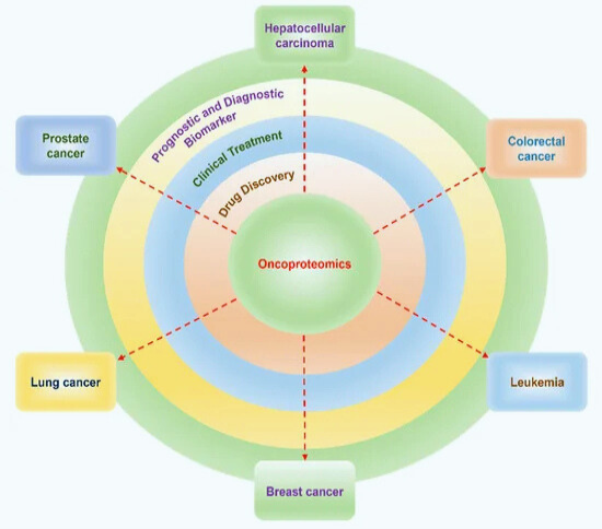
Cancer proteoforms identification show a great progress in the understanding and combating cancer . A proteoforms can be defined as a proteins isoform that is produced from a single gene utilizing different biological processes such as post translational modification, alternative splicing and proteolytic cleaving . Such differences are important for understanding of the mechanisms of cancer and for creation of the diagnostic and therapeutic tools as shown in Figure 1 . This section brings more detailed description of the cancer proteoforms their definition, their implication in the cancer and the difficulties that are associated with the analysis of the proteoforms .
Role of proteomics in the prognosis and diagnosis of cancer
In cancer, cells show quantitative and qualitative differences in proteoforms composition as compared to normal cells . These abnormalities can become altered expression of a protein, different modification of the protein or appearance of completely different protein fragment due to proteolysis . Understanding these variations is essential for several reasons.
Tumor progression
Proteoforms can affect process that are important for example, cell division, migration, and invasion . For instance, wrong folding of proteins due to errors in glycosylation leads to different cellular adhesion which is associated with metastasis .
Drug resistance
It is the variations with Proteoforms that may develop a resistance to the conventional therapies . Substances that cause changes in the effectiveness of treatment may include proteoforms that belong to certain enzymes in drug metabolism or signaling pathways .
Immune evasion
It is proposed that some proteoforms modify surface antigen or secrete immune-modulatory factor, thus enabling the tumor to escape immune system .
ROLE OF PROTEOFORMS IN CANCER PROGRESSION AND HETEROGENEITY
Cancer is a disease that has been associated with several proteins and in this case, proteoforms have been established to play a significant role in this disease . For instance, studies have bring into focus the fact that the existence of certain proteoforms of the prostate-specific antigen (PSA) enhances the ability to diagnose prostate cancer . Likewise, other altered proteoforms of metalloproteinase (MMPs) are associated with breast cancer development and metastasis . These examples show how knowledge of proteoforms can be useful in understanding the functions of malignant neoplasms and apply to the creation of more particular diagnostic tests and treatment strategies .
Cancer proteoforms are different molecular structures of proteins which are produced from a singular gene via distinct biological procedures . It is important for clinicians and researchers to learn about cancer proteoforms in order to compel some biomarkers for certain types of cancer as well as to initiate treatment processes . This elucidates the cancer specific proteoforms resulted out of post-translational modifications (PTM), alternative splicing (AS), and proteolytic cleavage (PC) and discourses regarding the utility of these identified proteoforms in the identification and eradication of cancer.
Post-translational modifications (PTMs)
The PTMs are co-translational modification that relates to modification that happens to the proteins after the translation process . They refer them as functionally, spatially and structurally altering a protein to a very great extent. Several types of PTMs are commonly observed in cancer proteoforms. Several PTMs are found to be significantly associated with cancer proteoforms and the following are some types of PTMs noted:
Phosphorylation
This modification involves the formation of covalent link between phosphate group and the amino acid residues which can be any one of the three residues including serine, threonine and tyrosine . Phosphorylation is associated with the processes that are relevant as regard to proteins and it also refers to the pathways, which can result in cancer development . This kind of disturbance is observed in several diseases for instance breast cancer, prostate cancer among others.
Glycosylation
Of all the post-translational modification of proteins, carbohydrate addition to proteins has been referred to as glycosylation which affects protein folding, stability and cell-surface interaction . Glycosylation is reported to be affected in a number of cancer types comprising ovarian and pancreatic carcinomas and the alterations help tumor cells to spread and evade immunity .
Ubiquitination
This involves the labeling of the proteins using a molecule called ubiquitin, this results into degradation of the protein . We also confirmed that defective ubiquitin system causes the enhanced protein degradation and signaling pathways which are associated with cancers .
Alternative splicing
Another interesting mechanism of gene expression regulation is this phenomenon called alternative splicing in which different groups of exons are joined to form different proteins from the same gene . In cancer, alternative splicing can generate proteoforms with altered functions . In cancer, AS can produce proteoforms which possesses either different function or even gain new properties:
Exon skipping
This type of an alternative splicing implies that some of the exons are skipped hence may produce low molecular weight or non-functional protein products . For instance, melanoma exon-skipping events lead to the formation of other proteins as biomarkers .
Intron retention
Due to presence of introns in mature mRNA, the proteoforms with the different functional capabilities are created . From a scientific perspective, intron retention has been related to the manufacture of proteins that install carcinoma and therapy resistances in cancers including glioblastoma.
Proteolytic cleavage
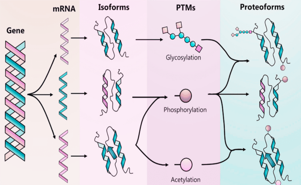
The others process is proteolysis, this is the enzymatic cleavage of proteins to smaller sections as shown in Figure 2. This process can generate proteoforms with distinct functions. This process can produce proteoforms with different activity levels , such as activation of proenzymes: Certain of the proenzymes are synthesized in compact form without an active core that has to be activated in order to become a functional molecule . In cancer, in the squamous cell carcinoma, activities such as the MMPs proenzymes which enhances tumor invasion and metastasis .
Oncofetal proteoforms
Some of these proteoforms are overexpressed in cancer tissues and are in fact classified as Oncofetal proteins because of their up regulation during fetal development . These proteins can serve as biomarkers for certain cancers. These proteins can be used as biomarkers of some types of cancer .
Alpha-fetoprotein (AFP)
AFP is a confirmed Oncofetal protein which is categorically overexpressed in hepatocellular carcinoma and in diseases associated with germ cell . It has been ascertained that in diagnosing and staging of these cancers, serum AFP is useful because its concentration is high within the blood.
Carcinoembryonic antigen (CEA)
The other Oncofetal protein known to have linkages with different types of cancer including the colorectal cancer is the CEA . Perhaps, the concentration of this cytokine in serum can be used for the evaluation of therapeutic outcomes and the existence of the disease relapse .
ROLE OF CANCER PROTEOFORMS IN BIOMARKER DISCOVERY
Proteoforms which are molecular variants of cancer proteins resulting from genetic and post translational modifications, are so vital in biomarker discovery and utility for early detection, diagnosis and prognostic marker of oncological diseases . Recent molecular markers are very selective in detecting cancer cells among normal cells and one cancer type from another . Different forms of PSA are more effective in diagnosing the prostate cancer than the total levels of PSA. In the same way, changes in glycosylation of the CA-125 proteoforms increases specificity as a biomarker that is useful in diagnosing and monitoring ovarian cancer .
PROTEOFORMS AND DISEASE PROGNOSIS
Proteoforms are rich in prognostic data since they indicate tumor malignancy and patient outcomes. There are Matrix metalloproteinase (MMPs) the variant being associated with cancer metastasis and short survival and in patients with increased MMP-2 and MMP-9 levels, the tumor is likely to be more invasive . Aberrant splicing isoforms in breast cancer confers resistance to therapy and prognosis of patient’s survival proteoforms variants assist with treatment .
Also, proteoforms are involved in assessment of treatment efficacy and identification of cancer relapses . Whereas lack of change in certain proteoforms suggests poor responses, increased or decrease in particular proteoforms is a better indication of patient response to treatment . Tumor-specific proteoforms in biological fluids are useful in early detection of recurrence enabling early intervention and therefore better prognosis .
TARGETING PROTEOFORMS FOR PRECISION THERAPY
Proteoforms present therapeutic agents that potential uniquely on targeting the changed molecular characteristic of carcinomas . Small molecules inhibitors can be rationally designed to selectively hit any pathogenic proteoforms. For instance, inhibitors selectively bind to certain proteoforms variants of kinases or other signaling proteins and impede cancer cell signaling and growth . Cancer vaccine can be designed to target specific proteoforms that are attained by the cancer cells. These vaccines intend to develop an immune response against the specific proteoforms related antigens, so as to enhance the immunological recognition of the cancer cells . Investigations about proteoforms specific antigens may provide the key for developing highly effective therapeutic vaccines.
ADVANCED PROTEOMICS TECHNOLOGIES
Scientific developments in the past years have further improved the identification, quantification, and characterization of cancer proteoforms, opening the field to new horizons and application in cancer studies . Consequently, high-resolution mass spectrometry offers the most feasible and powerful method to analyze proteoforms . Modernization in MS technologies include the trends like ultra-high-resolution mass spectrometers instrumentations that helped in better elucidation of proteoforms variants even in cases of subtle post-translational changes. These advancements enable the top-down proteome of 10 protein samples and enhanced definition of new cancer-specific proteoforms . Methods as label-free quantification and stable isotope labeling helped to analyze the alterations of the proteoforms abundance during different cancer stages and treatment . These quantitative methods offer information on dynamics and functions of proteoforms in connection with carcinogenesis as well as cancer treatment . Next-generation single-cell proteomics tools are developed to investigate proteoforms on the single-cell level . This approach is particularly useful to comprehend tumor heterogeneity and the proteoforms of a single cell in a complex and diverse tumor micro-environment. Single cell proteomics can help understand the role of different cell subtypes in cancer development and its treatment .
INTERACTION WITH GENOMICS AND TRANSCRIPTOMIC
The combination of proteomics with genomics and transcriptomic provides a systems-level understanding of the impact on proteoforms variation and function in cancer . Integration of these multi-omics methodologies unveils how genetic changes, specific splicing, and regulation impact proteoforms production and function and how it supports the creation of multi-targeted therapies . Proteogenomics strengthens this concept by integrating proteomic data with genomic and transcriptomic data to provide better identification of new proteoforms and their respective genes for providing a better understanding of molecular event in cancer .
EMERGING THERAPEUTIC STRATEGIES
New therapeutic approaches that are being derived from the concept of cancer proteoforms will seek to take advantage of these proteins . That is why one of the most promising strategies under investigation is the creation of drugs selectively acting on cancer-related proteoforms . With the utilization of these proteoforms specific inhibitors and antibodies, they dynamically selectively the abnormally translated proteoforms as to impair its function in preventing tumor growth, and thus enhance therapy selectivity and reduce side effects . Furthermore, therapeutic vaccines which are based on proteoforms specific antigens and gene editing approaches including CRISPR/Cas9 . These methods are the improvement of immune response to proteoforms which are unique to the tumor, as well as the elimination of mutations which lead to abnormal proteoforms production . These have been powerful strategies that have large potential for improvement of cancer care.
CONCLUSION
Proteoforms, derived from genetic and post-translational alterations in cancer-associated proteins, provide intricate informative features about tumor biology, dynamics, and therapeutic prospects of heterogeneity and resistance. Recent molecular diagnostic techniques such as high-resolution mass spectrometry and single-cell proteomics are identifying innovative cancer-associated proteoforms that are revolutionizing cancer diagnostics and therapy. There are limitations like proteoforms complexity, integration of datasets, and biomarker confirmation and they have to be solved to bring the findings into clinical utility. Future development will be also based on technological improvement, interdisciplinary collaboration and elimination of restrictions and moral issues. Finally, the elucidation of cancer proteoforms can further improve cancer individualization as well as provide profound advancement in the field of oncology.
Declarations
Acknowledgement
The authors acknowledge the contributions of researchers whose published work formed the basis of this review.
Funding
This review article did not receive any specific grant from funding agencies.
Generative AI and AI-assisted technology statement
The authors declare that no generative AI and AI assisted technology was used in the creation of this manuscript.
Statement of conflict of interest
The authors have declared no conflict of interest.
Figures
References
- Aebersold, R., Agar, J.N., Amster, I.J., Baker, M.S., Bertozzi, C.R., Boja, E.S., Costello, C.E., Cravatt, B.F., Fenselau, C. and Garcia, B.A., 2018. How many human proteoforms are there? Nat. Chem. Biol., 14: 206-214. https://doi.org/10.1038/nchembio.2576[CrossRef]
- Ardito, F., Giuliani, M., Perrone, D., Troiano, G. and Lo Muzio, L., 2017. The crucial role of protein phosphorylation in cell signaling and its use as targeted therapy. Int. J. mol. Med., 40: 271-280. https://doi.org/10.3892/ijmm.2017.3036[CrossRef]
- Aviña-Padilla, K., Ramírez-Rafael, J.A., Herrera-Oropeza, G.E., Muley, V.Y., Valdivia, D.I., Díaz-Valenzuela, E., García-García, A., Varela-Echavarría, A. and Hernández-Rosales, M., 2021. Evolutionary perspective and expression analysis of intronless genes highlight the conservation of their regulatory role. Front. Genet., 12: 654256. https://doi.org/10.3389/fgene.2021.654256[CrossRef]
- Bertoli, G., Cava, C. and Castiglioni, I., 2015. MicroRNAs: New biomarkers for diagnosis, prognosis, therapy prediction and therapeutic tools for breast cancer. Theranostics, 5: 1122. https://doi.org/10.7150/thno.11543[CrossRef]
- Bhatt, A.N., Mathur, R., Farooque, A., Verma, A. and Dwarakanath, B., 2010. Cancer biomarkers-current perspectives. Indian J. med. Res., 132: 129-149.
- Birzele, F., Csaba, G. and Zimmer, R., 2008. Alternative splicing and protein structure evolution. Nucl. Acids Res., 36: 550-558. https://doi.org/10.1093/nar/gkm1054[CrossRef]
- Brown, K.A., Melby, J.A., Roberts, D.S. and Ge, Y., 2020. Top-down proteomics: Challenges, innovations, and applications in basic and clinical research. Expert. Rev. Proteom., 17: 719-733. https://doi.org/10.1080/14789450.2020.1855982[CrossRef]
- Burger, A.M. and Seth, A.K., 2004. The ubiquitin-mediated protein degradation pathway in cancer: Therapeutic implications. Eur. J. Cancer, 40: 2217-2229. https://doi.org/10.1016/j.ejca.2004.07.006[CrossRef]
- Carbonara, K., Andonovski, M. and Coorssen, J.R., 2021. Proteomes are of proteoforms: Embracing the complexity. Proteomes, 9: 38. https://doi.org/10.3390/proteomes9030038[CrossRef]
- Carrieri, P.B., Provitera, V., Rosa, T.D., Tartaglia, G., Gorga, F. and Perrella, O., 1998. Profile of cerebrospinal fluid and serum cytokines in patients with relapsing-remitting multiple sclerosis. A correlation with clinical activity. Immunopharmacol. Immunotoxicol., 20: 373-382. https://doi.org/10.3109/08923979809034820[CrossRef]
- Chen, W., Geng, D., Chen, J., Han, X., Xie, Q., Guo, G., Chen, X., Zhang, W., Tang, S. and Zhong, X., 2024. Roles and mechanisms of aberrant alternative splicing in melanoma implications for targeted therapy and immunotherapy resistance. Cancer Cell Int., 24: 101. https://doi.org/10.1186/s12935-024-03280-x[CrossRef]
- Cufaro, M.C., Pieragostino, D., Lanuti, P., Rossi, C., Cicalini, I., Federici, L., De Laurenzi, V. and Del Boccio, P., 2019. Extracellular vesicles and their potential use in monitoring cancer progression and therapy: The contribution of proteomics. J. Oncol., 2019: 1639854. https://doi.org/10.1155/2019/1639854[CrossRef]
- De Strooper, B., 2010. Proteases and proteolysis in Alzheimer disease: A multifactorial view on the disease process. Physiol. Rev., 90: 465-494. https://doi.org/10.1152/physrev.00023.2009[CrossRef]
- Ercan, H., Resch, U., Hsu, F., Mitulovic, G., Bileck, A., Gerner, C., Yang, J.-W., Geiger, M., Miller, I. and Zellner, M., 2023. A practical and analytical comparative study of gel-based top-down and gel-free bottom-up proteomics including unbiased proteoform detection. Cells, 12: 747. https://doi.org/10.3390/cells12050747[CrossRef]
- Ferraro, S., Biganzoli, G., Bussetti, M., Castaldi, S., Biganzoli, E.M. and Plebani, M., 2023. Managing the impact of inter-method bias of prostate specific antigen assays on biopsy referral: The key to move towards precision health in prostate cancer management. Clin. Chem. Lab. Med., 61: 142-153. https://doi.org/10.1515/cclm-2022-0874[CrossRef]
- Forgrave, L.M., Wang, M., Yang, D. and DeMarco, M.L., 2022. Proteoforms and their expanding role in laboratory medicine. Pract. Lab. Med., 28: e00260. https://doi.org/10.1016/j.plabm.2021.e00260[CrossRef]
- Fusciello, M., Fontana, F., Tähtinen, S., Capasso, C., Feola, S., Martins, B., Chiaro, J., Peltonen, K., Ylösmäki, L. and Ylösmäki, E., 2019. Artificially cloaked viral nanovaccine for cancer immunotherapy. Nat. Commun., 10: 5747. https://doi.org/10.1038/s41467-019-13744-8[CrossRef]
- Gehring, N.H. and Roignant, J.Y., 2021. Anything but ordinary–emerging splicing mechanisms in eukaryotic gene regulation. Trends Genet., 37: 355-372. https://doi.org/10.1016/j.tig.2020.10.008[CrossRef]
- Gillette, T.G. and Hill, J.A., 2015. Readers, writers, and erasers: Chromatin as the whiteboard of heart disease. Circ. Res., 116: 1245-1253. https://doi.org/10.1161/CIRCRESAHA.116.303630[CrossRef]
- Goldstein, M.J. and Mitchell, E.P., 2005. Carcinoembryonic antigen in the staging and follow-up of patients with colorectal cancer. Cancer Investigat., 23: 338-351. https://doi.org/10.1081/CNV-200058878[CrossRef]
- He, K., Baniasad, M., Kwon, H., Caval, T., Xu, G., Lebrilla, C., Hommes, D.W. and Bertozzi, C., 2024. Decoding the glycoproteome: A new frontier for biomarker discovery in cancer. J. Hematol. Oncol., 17: 12. https://doi.org/10.1186/s13045-024-01532-x[CrossRef]
- Hershko, A. and Ciechanover, A., 1986. The ubiquitin pathway for the degradation of intracellular proteins. Prog. Nucl. Acid Res. Mol. Biol., 33: 19-56. https://doi.org/10.1016/S0079-6603(08)60019-7[CrossRef]
- Hunter, T., 2012. Why nature chose phosphate to modify proteins. Philos. Trans. R. Soc. B: Biol. Sci., 367: 2513-2516. https://doi.org/10.1098/rstb.2012.0013[CrossRef]
- Karimi, M.R., Karimi, A.H., Abolmaali, S., Sadeghi, M. and Schmitz, U., 2022. Prospects and challenges of cancer systems medicine: from genes to disease networks. Brief. Bioinf., 23: bbab343. https://doi.org/10.1093/bib/bbab343[CrossRef]
- Khan, A.R. and James, M.N., 1998. Molecular mechanisms for the conversion of zymogens to active proteolytic enzymes. Protein Sci., 7: 815-836. https://doi.org/10.1002/pro.5560070401[CrossRef]
- Kumar, D., Bansal, G., Narang, A., Basak, T., Abbas, T. and Dash, D., 2016. Integrating transcriptome and proteome profiling: Strategies and applications. Proteomics, 16: 2533-2544. https://doi.org/10.1002/pmic.201600140[CrossRef]
- Labib, M. and Kelley, S.O., 2020. Single-cell analysis targeting the proteome. Nat. Rev. Chem., 4: 143-158. https://doi.org/10.1038/s41570-020-0162-7[CrossRef]
- Li, J. and Zhan, X., 2021. Mass spectrometry-based proteomics analyses of post-translational modifications and proteoforms in human pituitary adenomas. Biochim. Biophys. Acta Proteins Proteom., 1869: 140584. https://doi.org/10.1016/j.bbapap.2020.140584[CrossRef]
- Lin, Y. and Lubman, D.M., 2023. The role of N-glycosylation in cancer. Acta Pharmaceut. Sin. B. https://doi.org/10.1016/j.apsb.2023.10.014[CrossRef]
- Lisitsa, A., Moshkovskii, S., Chernobrovkin, A., Ponomarenko, E. and Archakov, A., 2014. Profiling proteoforms: Promising follow-up of proteomics for biomarker discovery. Expert Rev. Proteom., 11: 121-129. https://doi.org/10.1586/14789450.2014.878652[CrossRef]
- Mäbert, K., Cojoc, M., Peitzsch, C., Kurth, I., Souchelnytskyi, S. and Dubrovska, A., 2014. Cancer biomarker discovery: Current status and future perspectives. Int. J. Radiat. Biol., 90: 659-677. https://doi.org/10.3109/09553002.2014.892229[CrossRef]
- Macklin, A., Khan, S. and Kislinger, T., 2020. Recent advances in mass spectrometry based clinical proteomics: Applications to cancer research. Clin. Proteom., 17: 17. https://doi.org/10.1186/s12014-020-09283-w[CrossRef]
- Malaney, P., Uversky, V. N. and Davé, V., 2017. PTEN proteoforms in biology and disease. Cell. Mol. Life Sci., 74: 2783-2794. https://doi.org/10.1007/s00018-017-2500-6[CrossRef]
- Maruvada, P., Wang, W., Wagner, P.D. and Srivastava, S., 2005. Biomarkers in molecular medicine: Cancer detection and diagnosis. Biotechniques, 38: 9-15. https://doi.org/10.2144/05384SU04[CrossRef]
- Meissner, F., Geddes-McAlister, J., Mann, M. and Bantscheff, M., 2022. The emerging role of mass spectrometry-based proteomics in drug discovery. Nat. Rev. Drug. Discov., 21: 637-654. https://doi.org/10.1038/s41573-022-00409-3[CrossRef]
- Metwali, E. and Pennington, S., 2024. Mass spectrometry-based proteomics for classification and treatment optimisation of triple negative breast cancer. J. Pers. Med., 14: 944. https://doi.org/10.3390/jpm14090944[CrossRef]
- Mijakovac, A., Miškec, K., Krištić, J., Vičić Bočkor, V., Tadić, V., Bošković, M., Lauc, G., Zoldoš, V. and Vojta, A., 2022. A transient expression system with stably integrated CRISPR-dCas9 fusions for regulation of genes involved in immunoglobulin G glycosylation. CRISPR J., 5: 237-253. https://doi.org/10.1089/crispr.2021.0089[CrossRef]
- Montero-Calle, A., Garranzo-Asensio, M., Torrente-Rodríguez, R.M., Ruiz-Valdepeñas Montiel, V., Poves, C., Dziaková, J., Sanz, R., Díaz del Arco, C., Pingarrón, J.M. and Fernández-Aceñero, M.J., 2023. p53 and p63 proteoforms derived from alternative splicing possess differential seroreactivity in colorectal cancer with distinct diagnostic ability from the canonical proteins. Cancers, 15: 2102. https://doi.org/10.3390/cancers15072102[CrossRef]
- Naryzhny, S. and Legina, O., 2019. Structural-functional diversity of p53 proteoforms. Biochem. Moscow Suppl. Ser. B, 13: 293–307. https://doi.org/10.1134/S199075081904005X[CrossRef]
- Ntai, I., LeDuc, R.D., Fellers, R.T., Erdmann-Gilmore, P., Davies, S.R., Rumsey, J., Early, B.P., Thomas, P.M., Li, S. and Compton, P.D., 2016. Integrated bottom-up and top-down proteomics of patient-derived breast tumor xenografts. Mol. Cell. Proteom., 15: 45-56. https://doi.org/10.1074/mcp.M114.047480[CrossRef]
- Petrosius, V. and Schoof, E.M., 2023. Recent advances in the field of single-cell proteomics. Trans. Oncol., 27: 101556. https://doi.org/10.1016/j.tranon.2022.101556[CrossRef]
- Piana, D., Iavarone, F., De Paolis, E., Daniele, G., Parisella, F., Minucci, A., Greco, V. and Urbani, A., 2024. Phenotyping tumor heterogeneity through proteogenomics: Study models and challenges. Int. J. mol. Sci., 25: 8830. https://doi.org/10.3390/ijms25168830[CrossRef]
- Pornchai, O., Rhys-Evans, P.H. and Eccles, S.A., 2001. Expression of matrix metalloproteinases and their inhibitors correlates with invasion and metastasis in squamous cell carcinoma of the head and neck. Arch. Otolaryngol. Head. Neck. Surg., 127: 813-820. https://doi.org/10.1097/CM9.0000000000000542[CrossRef]
- Qi, F., Li, Y., Yang, X., Wu, Y.P., Lin, L.J. and Liu, X.M., 2020. Significance of alternative splicing in cancer cells. Chin. med. J., 133: 221-228. https://doi.org/10.1097/CM9.0000000000000542[CrossRef]
- Raju, T.S., 2019. Co-and post-translational modifications of therapeutic antibodies and proteins. John Wiley and Sons. https://doi.org/10.1002/9781119053354[CrossRef]
- Rodrigues, J.G., Balmaña, M., Macedo, J.A., Poças, J., Fernandes, Â., de-Freitas-Junior, J.C.M., Pinho, S.S., Gomes, J., Magalhães, A. and Gomes, C., 2018. Glycosylation in cancer: Selected roles in tumour progression, immune modulation and metastasis. Cell. Immunol., 333: 46-57. https://doi.org/10.1016/j.cellimm.2018.03.007[CrossRef]
- Ruffinatti, F.A., Scarpellino, G., Chinigò, G., Visentin, L. and Munaron, L., 2023. The emerging concept of transportome: State of the art. Physiology, 38: 285-302. https://doi.org/10.1152/physiol.00010.2023[CrossRef]
- Schuster-Little, N., Madera, S. and Whelan, R., 2020. Developing a mass spectrometry–based assay for the ovarian cancer biomarker CA125 (MUC16) using suspension trapping (STrap). Anal. Bioanal. Chem., 412:6361–6370. https://doi.org/10.1007/s00216-020-02586-9[CrossRef]
- Shukla, H.D., 2017. Comprehensive analysis of cancer-proteogenome to identify biomarkers for the early diagnosis and prognosis of cancer. Proteomes, 5: 28. https://doi.org/10.3390/proteomes5040028[CrossRef]
- Sideras, K., Bots, S., Biermann, K., Sprengers, D., Polak, W., IJzermans, J., De Man, R., Pan, Q., Sleijfer, S. and Bruno, M., 2015. Tumour antigen expression in hepatocellular carcinoma in a low-endemic western area. Br. J. Cancer, 112: 1911–1920. https://doi.org/10.1038/bjc.2015.92[CrossRef]
- Su, J., Yang, L., Sun, Z. and Zhan, X., 2024. Personalized drug therapy: Innovative concept guided with proteoformics. Mol. Cell. Proteom., 23. https://doi.org/10.1016/j.mcpro.2024.100737[CrossRef]
- Su, M., Zhang, Z., Zhou, L., Han, C., Huang, C. and Nice, E.C., 2021. Proteomics, personalized medicine and cancer. Cancers, 13: 2512. https://doi.org/10.3390/cancers13112512[CrossRef]
- Uversky, V.N., 2016. p53 proteoforms and intrinsic disorder: An illustration of the protein structure–function continuum concept. Int. J. mol. Sci., 17: 1874. https://doi.org/10.3390/ijms17111874[CrossRef]
- Vainshelbaum, N.M., Giuliani, A., Salmina, K., Pjanova, D. and Erenpreisa, J., 2022. The transcriptome and proteome networks of malignant tumours reveal atavistic attractors of polyploidy-related asexual reproduction. Int. J. mol. Sci., 23: 14930. https://doi.org/10.3390/ijms232314930[CrossRef]
- van Schaick, G., Haselberg, R., Somsen, G.W., Wuhrer, M. and Domínguez-Vega, E., 2022. Studying protein structure and function by native separation–mass spectrometry. Nat. Rev. Chem., 6: 215–231. https://doi.org/10.1038/s41570-021-00353-7[CrossRef]
- Walsh, G. and Jefferis, R., 2006. Post-translational modifications in the context of therapeutic proteins. Nat. Biotechnol., 24: 1241–1252. https://doi.org/10.1038/nbt1252[CrossRef]
- Wu, L. and Qu, X., 2015. Cancer biomarker detection: Recent achievements and challenges. Chem. Soc. Rev., 44: 2963-2997. https://doi.org/10.1039/C4CS00370E[CrossRef]
- Zhan, X. and Su, J., 2024. Proteoform-based new perspectives for personalized drug therapy. https://doi.org/10.5772/intechopen.1007633[CrossRef]
- Zhang, B. and Kuster, B., 2019. Proteomics is not an island: Multi-omics integration is the key to understanding biological systems. Mol. Cell. Proteom., 18: S1-S4. https://doi.org/10.1074/mcp.E119.001693[CrossRef]
- Zhu, Z., Jiang, L. and Ding, X., 2023. Advancing breast cancer heterogeneity analysis: Insights from genomics, transcriptomics and proteomics at bulk and single-cell levels. Cancers, 15: 4164. https://doi.org/10.3390/cancers15164164[CrossRef]
Services
Article usage statistics
Loading…