
The fat content in yak milk is higher than that in dairy cows, but the molecular mechanisms responsible for milk fat synthesis in yaks are still unclear. This study examined the regulatory mechanism of milk fat synthesis in yak mammary epithelial cells (YMECs) by investigating the role of Ribosomal protein L8 (RPL8) in the mTORC1-SREBP1 signaling pathways. The results showed that over-expression or inhibition of RPL8 had a significant effect on triglyceride (TG) secretion, which also affected the sterol regulatory element-binding protein 1 (SREBP1) pathway. Similarly, the intervention of SREBP1 revealed that RPL8 promoted TG secretion through the SREBP1 pathway. Additionally, the study found that over-expression or inhibition of RPL8 regulated the signaling activity of the mammalian target of rapamycin complex 1 (mTORC1), which promoted the SREBP1 signaling pathway through mTORC1. Further examination by over-expressing or inhibiting SREBP1 or mTOR showed that mTOR promoted TG secretion through the mTORC1-SREBP1 signaling pathway. Moreover, YMECs treated with palmitic acid showed increased expression of RPL8, mTOR, and SREBP1, as well as increased TG secretion. Overall, the study’s findings provide insights into the underlying mechanisms of milk fat synthesis in yaks, and suggest that the RPL8 gene and mTORC1-SREBP1 signaling pathways could serve as potential genetic markers for milk fat synthesis in yak mammary glands.
Research Article · Volume 56, Issue 5, 28 July 2024 · Open Access
Uncovering the Role of Ribosomal Protein L8 in Milk Fat Synthesis Mechanisms in Yak Mammary Epithelial Cells
· · · · · · · · · ·
Affiliations & Notes ⌄
- 1State Key Laboratory of Hulless and Yak Germplasm Resources and Genetic Improvement, Lhasa, Tibet, 850000, China. 2Institute of Animal Husbandry and Veterinary, Tibet Autonomous Regional Academy of Agricultural Sciences, Lhasa, Tibet,850000, China. 3College of Life Sciences, Shihezi University, Shihezi, Xinjiang 832003, P.R. China. 4Rural Revitalization College, Hainan College of Vocation and Technique Haikou, Hainan, 570100, China. 5State Key Laboratory of Animal Nutrition, Beijing Engineering
Article Info ⌄
- Journal:
- Pakistan Journal of Zoology
- Published:
- 28 July 2024

From the article
- Our findings suggest that RPL8 is a positive regulator of milk fat synthesis, and its overexpression significantly promotes the secretion of TG in YMECs.
- Our results suggest that both RPL8 and mTOR act as upstream factors of SREBP1, promoting milk fat synthesis through the SREBP1 pathway.
- We have demonstrated that the expression of RPL8 is promoted by palmitic acid, which then activates the mTORC1-SREBP1 pathway to promote milk fat synthesis.
- Moreover, our study suggests that the RPL8 gene and mTORC1-SREBP1 signaling could serve as valuable markers for improving milk fat production in yak.
Selected from the article's own text.
Abstract
Introduction
Milk fat is a vital component of milk, containing a range of nutrients, including unsaturated fatty acids, phospholipids, linoleic acid, fat-soluble vitamins, and lipids ; . The milk fat content is a key indicator of milk quality, and it is regulated by genetic, hormonal, nutritional, and environmental factors in the mammary gland cells of milk-producing animals such as cows, yaks, and dairy goats ; ; .
The yak (Bos grunniens) is a distinctive breed of cattle that has its roots in the Qinghai-Tibet Plateau (Xin et al., 2020a, b). The breed and the environment in which it lives are critical factors that influence milk yield in both dairy yak and cow ; . Notably, studies have shown that yak milk contains higher levels of fat than cow milk . The synthesis of milk fat is a complex biological process that is regulated by various signaling pathways ; . While the mechanisms that control milk fat synthesis in cow and dairy goat mammary gland cells have been extensively researched , information on the genetic regulation of milk fat synthesis in dairy yaks is limited ; . The regulatory mechanisms, including the biological signaling pathways involved in milk fat synthesis in dairy yaks, require further investigation and validation. Two primary signaling pathways for milk fat synthesis in cow and dairy goat mammary gland cells are the peroxisome proliferator-activated receptor gamma (PPARγ) pathway ; and the sterol regulatory element-binding protein 1 (SREBP1) pathway ; .
Ribosomal protein L8 (RPL8) is an essential component of the 60S subunit of the ribosome, playing a crucial role in ribosome assembly and protein synthesis ; . Previous studies have suggested RPL8 as a potential candidate gene for the milk fat percentage trait in dairy cattle , 2014; . However, its biological signaling and regulatory role in milk fat synthesis in dairy Yak remain largely unknown and require further investigation.
Therefore, this study aims to explore the role of RPL8 and the underlying mechanism of milk fat synthesis in Yak mammary epithelial cells (YMECs). Our results reveal that RPL8 plays a critical role in promoting milk fat synthesis in YMECs through the mTORC1-SREBP1 pathway. These findings provide valuable insights into the mechanisms regulating milk fat synthesis in YMECs and broaden our understanding of the biological significance of RPL8 in this process.
Materials and Methods
Sample collection, Cell culture and identification
Fresh mammary gland tissues were obtained from local abattoir of Tibet, China. The mammary gland tissues were collected immediately after slaughter, and were stored in sterile saline solution at 37 ℃ containing 1% penicillin/streptomycin mixture, and transported to the laboratory in a thermos flask within 2 h. Yak mammary tissue was washed one time with 75% ethanol and then washed several times with DMEM/F12 medium (11320033, Gibco, California, USA) containing 100x penicillin-streptomycin-amphotericin B (C0224, Beyotime, Shanghai, China. The final concentration was 10 x). Then the tissue was cut into pieces (about 1mm3) in DMEM/F12 medium containing penicillin-streptomycin-amphotericin B and washed several times. The pieces were placed at the bottom of the cell culture dish pre-lined with collagen from rat tail Type I (MX0910, MKBio, Shanghai, China). The DMEM/F12 medium containing penicillin-streptomycin-amphotericin B was added to the cell culture dish until the pieces were flooded. The cell culture dish was cultured at 37°C in a atmosphere of 5% CO2. The medium was replaced after every 3 days. About a month later, the cells (mainly fibroblasts and mammary epithelial cells) that grow from the periphery of the pieces filled the bottom of the cell culture dish. The pieces were removed and the cells were digested with 0.25% trypsin and 0.02% ethylenediaminetetraacetic acid (EDTA) (C0201, Beyotime, China) at 37°C for 3 min.
The suspension (mainly fibroblasts) was discarded and the adherent cells (mainly mammary epithelial cells) were re-digested with 0.25% trypsin and 0.02% EDTA for 5 min . The adherent cells were gently blown and mixed evenly, and cultured with DMEM/F12 containing 10% FBS. After approximately 3-5 generations, purified YMECs were obtained. The expression of cytokeratin 18 (CK18, a marker of epithelial cell) and vimentin (a marker of fibroblast, used as a negative control) was measured by immunofluorescence (IF). The expression of β-casein (one of the representative components of milk protein) in YMECs was tested by western blotting (WB) and the secretion of triglyceride (TG) in YMECs was tested by triglyceride test kit (AKFA003M, boxbio, Beijing, China).
Western blotting
To collect the total protein from treated YMEC cells, Cell lysis buffer for Western and IP (P0013, Beyotime, Shanghai, China) containing 1 mM of phenylmethanesulfonyl fluoride (PMSF, ST506, Beyotime) was used. The protein concentration was measured using the enhanced BCA protein assay kit (P0010S, Beyotime). A 10% SDS-PAGE gel was used to separate approximately 30 μg of the total protein, which was then transferred onto a polyvinylidene fluoride (PVDF) membrane (FFP39, Beyotime). The membrane was blocked for 1.5 h at room temperature with TBSTx (ST675, Beyotime) containing 5% bovine serum albumin (BSA, ST025, Beyotime) and incubated overnight at 4°C with the primary antibody (diluted with TBSTx).
The membrane was washed three times (5 min at a time) with TBSTx and then incubated with HRP-conjugated secondary antibody (diluted with TBSTx) for 1 h at room temperature. The membrane was washed three times (10 min at a time) with TBSTx and then incubated with BeyoECL Plus (P0018S, Beyotime) for several sec. The chemical fluorescence of HRP was visualized with Tanon-5200 Chemiluminescent Imaging System (Tanon Science and Technology Co., Ltd, China).
The primary antibodies used in this research were as follows: β-actin (13E5) antibody (1: 1, 000, #4970, Cell Signaling Technology, Massachusetts, USA), beta casein antibody (1: 500, bs-0466R, Bioss, Beijing, China), PPAR gamma antibody (1:500, bs-0530R, Bioss), mTOR antibody (1: 1, 000, #2972, Cell Signaling Technology), p-mTOR antibody (1: 1, 000, #2971, Cell Signaling Technology), SREBP1 antibody (1: 1, 000, ab28481, Abcam, Cambridgeshire, UK), RPL8 antibody (1: 1, 000, ab155136, Abcam), FAS antibody (1: 1, 000, ab82419, Abcam), SCD1 antibody (1: 1, 000, ab236868, Abcam), ACC antibody (1: 1, 000, ab45174, Abcam), lamin B1 antibody (1: 1000, ab16048, Abcam, UK) and β-tubulin antibody (1: 1, 000; #2146, Cell Signaling Technology). The secondary antibodies were HRP-conjugated goat anti-rabbit IgG (H+L) (1: 2, 000, ZB-2301, ZSGB-BIO, Beijing, China) and HRP-conjugated goat anti-mouse IgG (H+L) (1: 2, 000, ZB-2305, ZSGB-BIO, Beijing, China).
Triglyceride secretion
YMECs were treated and cultured according to experimental requirements; the medium of each group of cells was collected. The triglyceride test kit (BC0625, Solarbio Science and Technology Co., Ltd, Beijing, China) was used to determine the content of triglyceride (TG) in the medium, following the manufacturer’s instructions.
Immunofluorescence
YMECs were planted into a 35 mm glass bottom dish (D35-20-1-N, in vitro Scientific, Hangzhou, China) and cultured with DMEM/F12 containing 10% FBS for 12 h and then treated with gene overexpression or silencing according to the experimental design. The treated cells were washed three times with PBS and fixed with ice-cold 4% paraformaldehyde at 4 °C for 15 min. The cells were washed three times with TBSTx (5 min at a time) and then incubated with primary antibodies (diluted with 5% BSA) for 1.5 h at room temperature. To observe the fluorescence of proteins, the cells were washed three times with TBSTx (5 min each time) and incubated with secondary antibodies conjugated to Alexa Fluor 488 and 647 for 1 hour at room temperature in the dark. After washing the cells three times with TBSTx (10 min each time), they were incubated with 2 μg/mL of 4, 6-diamidino-2-phenylindole (DAPI; C1005, Beyotime) for 15 min at room temperature in the dark, followed by three washes with TBSTx (10 min each time). Laser scanning confocal microscopy (LEICA, Germany) was used to observe the fluorescence of proteins. The primary antibodies used were vimentin antibody (1:500, ab92547, Abcam) and cytokeratin 18 antibody (1:500, ab7797, Abcam), while the secondary antibodies were goat anti-mouse IgG (H+L), Alexa Fluor 488 conjugated antibody (1:200, bs-40296G-AF488, Bioss) and goat anti-rabbit IgG HandL/Alexa Fluor 647 antibody (1:200, bs-0295G-AF647, Bioss).
Plasmid construction and gene over-expression
In this study, the over-expression plasmids of RPL8, mTOR and SREBP1 gene were constructed. The total RNA of YMECs was extracted with TRIZOL RNA isolation reagent (R0016, Beyotime) and the quality of RNA was tested by 2100 Bioanalyzer (Agilent, California, USA). The cDNA was synthesized with M-MLV reverse transcriptase (2641A, TaKaRa, Beijing, China). The PCR primers of RPL8, mTOR and SREBP1 gene were designed with Premier 5 and synthesized by Sangon Biotech Co., Ltd. The NCBI reference sequence of RPL8, mTOR and SREBP1 gene was XM_005911714.2, XM_005901305.2 and XM_014477492.1, respectively. The sequences of these PCR primers are shown in Table I. The gene was cloned and inserted into the eukaryotic expression plasmids pCMV-C-Myc (D2672, Beyotime). The over-expression plasmids of RPL8, mTOR and SREBP1 gene were named pCMV-RPL8-Myc, pCMV-mTOR-Myc and pCMV-SREBP1-Myc, respectively.
The over-expression plasmids of these genes were transfected into YMECs with Lipo6000™ transfection reagent (C0526, Beyotime) according to the protocol. In brief, YMECs were planted into 6 well plates and cultured to the confluence of cells was about 80% with DMEM/F12 medium containing FBS. The medium was changed to FBS-free DMEM/F12 medium. In order to transfect cells, a mixture of overexpression plasmids (2.5 μg/well) and Lipo6000™ transfection reagent (5 μl per well) was prepared by diluting them with 125 μl of FBS-free DMEM/F12 medium. These were then incubated separately for 5 min at room temperature before being mixed together and incubated for an additional 15 min at room temperature. The resulting mixture was added to 6-well plates containing the cells, and after 6 h, the medium was changed to DMEM/F12 with FBS, allowing the cells to be cultured for another 24 h. The blank control (B group) consisted of untransfected YMECs, while the empty plasmid control (EV group) consisted of YMECs transfected with the pCMV-C-Myc plasmid.
Table I. Sequences of primers utilized for construction of plasmid.
| Gene name | Primer sequence (5'-3') |
| RPL8 | F=CGGAATTCATGGGCCGCGTGATCCATGG (the EcoR I site is underlined) |
| R=GAAGATCTGTTCTCCTTCTCCTGCACAGTC (the Bgl II site is underlined) | |
| SREBP1 | F=AACTGCAGATGAAGGAGGTGGTGAGCTCC (the PstI site is underlined) |
| R=GCGTCGACGCTGGAGGTCACAGTGGTCCCAC (the Sal I site is underlined) | |
| mTOR | F=AACTGCAGATGCTTGGAACCGGCCCTGCTGC (the PstI site is underlined) |
| R=GCGTCGACCCAGAAAGGACACCAGCCAATG (the Sal I site is underlined) |
Small interfering RNA (siRNA) transfection
In this research, GenePharma Co., Ltd. (GenePharma, Shanghai, China) synthesized specific siRNAs targeting the RPL8, mTOR, and SREBP1 genes, as listed in Table II. The YMECs were transfected with these siRNAs, along with Lipo6000™ transfection reagent, using the same protocol as for gene overexpression. The dosage of siRNA and Lipo6000™ transfection reagent used per well was 100 pmol and 5 μl, respectively. YMECs that were not transfected were used as a blank control (B group), while those transfected with negative control siRNA were used as a negative control (NC group).
Table II. Sequences of small interfering RNAs (siRNAs).
| Gene name | Sequences of small interfering RNAs (5’-3’) | |
| RPL8 | Sense | AAAGAAACCAGUUUUAUUGAG |
| Antisense | CAAUAAAACUGGUUUCUUUUC | |
| SREBP1 | Sense | ACGAUCUUGUCAUUGAUGGCA |
| Antisense | CCAUCAAUGACAAGAUCGUUG | |
| mTOR | Sense | AUGACAUUCAGGAAUGUGGGC |
| Antisense | CCACAUUCCUGAAUGUCAUUC | |
| Negative control | Sense | UUCUCCGAACGUGUCACGUTT |
| Antisense | ACGUGACACGUUCGGAGAATT | |
Nuclear and cytoplasmic proteins extraction
The nuclear and cytoplasmic proteins were extracted using the nuclear and cytoplasmic protein extraction kit (P0027, Beyotime) according to the protocol. In brief, the treated cells were washed with PBS. The cells were scraped off with cell scraping (FSCP023, Beyotime), suspended with PBS and transferred to a clean centrifuge tube. The tube was centrifuged at room temperature, 800 g for 10 min, discarded the supernatant. The sediments (cells) were resuspended with cytoplasmic protein extraction reagent A (200 μl per two million cells) containing 1 mM of PMSF and vortexed to the cell precipitate completely dispersed. The cell suspensions were incubated on ice for 10 min. The cytoplasmic protein extraction reagent B was added (10 μl per two million cells), vortexed for 5 sec and incubated on ice for 1 min. Vortexed for 5 sec and centrifuged at 4 ℃, 12, 000 g for 5 min. The supernatant (the cytoplasmic proteins) was collected using a precooled, clean centrifuge tube and stored at -80 ℃. The sediments were resuspended with a nuclear protein extraction reagent (50 μl per two million cells) containing 1 mM of PMSF. The nuclear protein suspensions were vortexed for 20 sec and incubated on ice for 1 min. Repeat the vortex and incubation 30 times. The nuclear protein suspensions were centrifuged at 4 ℃, 12,000 g for 10 min. The supernatant (the cytoplasmic proteins) was collected using a precooled, clean centrifuge tube and stored at -80 ℃.
Statistical analysis
The statistical analysis of the data was conducted using a one-way ANOVA in Excel, and any differences with a p-value of less than 0.05 were deemed statistically significant. The results were expressed as the mean ± standard deviation (n=3) based on a sample size of three. A bar graph was created using GraphPad Prism 6 to visualize the data. Additionally, grayscale scanning of the WB band and the colocalization of IF were analyzed using ImageJ 2X software. It should be noted that all of the data presented in this study was derived from three independent experiments and then averaged.
RESULTS
YMECs
The YMECs were cultured and tested their morphology by immunofluorescence (IF) microscope (Fig. 1A). The expression of CK18 (a marker of an epithelial cell) was tested by IF. In every purified YMEC, the CK18 was expressed, and the vimentin (a marker of fibroblast, used as a negative control) was not expressed (Fig. 1B). The ability of milk synthesis of YMECs within 15 generations was evaluated. The expression of β-casein (one of the principal components of milk protein) was tested by WB. The β-casein was expressed in YMECs and it was stable within 5 to 15 generations (Fig. 1C, D). The secretion of TG (one of the principal components of milk fat) was tested using the TG test kit. The TG was secreted in YMECs and it was stable within 5 to 15 generations (Fig. 1E).
RPL8 regulates synthesis of milk fat in YMECs
The aim of this study was to investigate the impact of RPL8 expression on milk fat synthesis in YMECs. To achieve this, cells were subjected to varying levels of RPL8 expression (over-expression or silencing), and TG was assessed. The results showed that over-expression of RPL8 significantly increased the expression level of RPL8 (Fig. 2A-B) and the secretion of TG (Fig. 2C) in YMECs. Conversely, YMECs treated with RPL8 silencing showed a significant decrease in RPL8 expression (Fig. 2D, E) and TG secretion (Fig. 2F). This result suggested that RPL8 promoted milk fat synthesis in YMECs. We conducted further experimental trials to explore the biological mechanism through which RPL8 regulates milk fat synthesis in yak.
RPL8 activates the SREBP1 pathway in YMECs
The SREBP1 and PPARγ signaling pathways play a crucial role in regulating milk fat synthesis ; . To investigate the impact of RPL8 on these pathways, YMECs were subjected to RPL8 over-expression or silencing. Additionally, the expression of ACC, FAS, SCD1, SREBP1, and PPARγ, as well as the nuclear and cytoplasmic localization of SREBP1, were evaluated in YMECs after over-expression or silencing of RPL8. The results demonstrated that over-expression of RPL8 significantly increased the expression of ACC, FAS, SCD1, and SREBP1 (Fig. 3A, B), and promoted the nuclear localization (Fig. 3C, D) of SREBP1, while suppressing its cytoplasmic localization (Fig. 3C, E). Conversely, RPL8 silencing led to a significant decrease in the expression of ACC, FAS, SCD1, and SREBP1 (Fig. 3F, G), and inhibited the nuclear localization (Fig. 3H, I) of SREBP1, while enhancing its cytoplasmic localization (Fig. 3H, J). These findings suggest that RPL8 plays a crucial role in regulating milk fat synthesis through the SREBP1 and PPARγ signaling pathways. The expression of RPL8 (over-expression/silencing) did not affect expression of PPARγ in YMECs (Fig. 3A, B, F, G). These findings showed that RPL8 regulated SREBP1 signaling pathway not PPARγ pathway in YMECs.
RPL8 promotes milk fat synthesis through the SREBP1 pathway in YMECs
To determine if the SREBP1 pathway plays a role in RPL8-mediated promotion of milk fat synthesis, YMECs were co-treated with RPL8 over-expression or silencing and SREBP1 over-expression or silencing, and the expression of RPL8 and SREBP1, as well as TG secretion, were evaluated. The results showed that cells subjected to RPL8 over-expression displayed a significant increase in the expression of RPL8 and SREBP1, as well as TG secretion (Fig. 4A, C). However, the increase in SREBP1 expression and TG secretion was blocked by SREBP1 silencing (Fig. 4A, C). Conversely, cells subjected to RPL8 silencing displayed a significant decrease in the expression of RPL8 and SREBP1, as well as TG secretion (Fig. 4D, F). However, the decrease in SREBP1 expression and TG secretion was restored by SREBP1 over-expression in YMECs (Fig. 4D, F). Interestingly, the expression of RPL8 remained unchanged after SREBP1 silencing or over-expression (Fig. 4A, D, E). Therefore, it can be concluded that SREBP1 is involved in the regulation of RPL8-mediated milk fat synthesis and acts as a downstream factor of RPL8. This confirms that RPL8 promotes milk fat synthesis through the SREBP1 pathway in YMECs.
RPL8 activates the mTORC1 pathway in YMECs
Previous studies have suggested that the mTORC1 pathway plays a role in regulating milk fat synthesis and is one of the upstream pathways of SREBP1 . To investigate the impact of RPL8 on the mTORC1 pathway, YMECs were subjected to RPL8 overexpression or silencing, and the expression of mTOR and p-mTOR was evaluated. The results showed that the expression of both mTOR and p-mTOR (Fig. 5A, B), as well as the rate of p-mTOR/mTOR (Fig. 5C), was significantly increased in cells treated with RPL8 overexpression. Conversely, cells treated with RPL8 silencing displayed a significant decrease in the expression of both mTOR and p-mTOR (Fig. 5D, E), as well as the rate of p-mTOR/mTOR (Fig. 5F). These findings suggest that RPL8 activates the mTORC1 pathway in YMECs.
RPL8 activates the SREBP1 pathway via mTORC1 in YMECs
In order to investigate the potential involvement of mTORC1 in regulating the activation of the SREBP1 pathway by RPL8, YMECs were subjected to co-treatment with either RPL8 overexpression or silencing, as well as mTOR silencing or overexpression. The expression of SREBP1, mTOR, and RPL8, as well as the nuclear localization of SREBP1, were all assessed. The results demonstrated that overexpression of RPL8 led to a significant increase in the expression of SREBP1, mTOR, and RPL8 (Fig. 6A, B), as well as the nuclear localization of SREBP1 (Fig. 6C, D), accompanied by a significant decrease in the cytoplasmic localization of SREBP1 (Fig. 6C, E). These effects were found to be blocked by mTOR silencing. Conversely, silencing of RPL8 resulted in a significant decrease in the expression of SREBP1, mTOR, and RPL8 (Fig. 6F, G), which could be restored by mTOR overexpression. These findings suggest that mTORC1 functions as a downstream factor of RPL8, and is involved in regulating the SREBP1 pathway. Specifically, RPL8 appears to activate the SREBP1 pathway through mTORC1 in YMECs.
mTORC1 regulates synthesis of milk via SREBP1 pathway in YMECs
In order to assess the impact of mTORC1 on milk fat synthesis in YMECs, we conducted tests on cells that were either treated with mTOR overexpression or silencing, and measured the secretion of TG. The findings revealed a significant increase in the expression of mTOR (Fig. 7A, B) and secretion of TG (Fig. 7C) in cells subjected to mTOR overexpression. Conversely, mTOR silencing resulted in a significant decrease in the expression of mTOR (Fig. 7D, E) and secretion of TG (Fig. 7F).
To investigate whether the SREBP1 pathway played a role in regulating mTORC1 in milk fat synthesis in YMECs, we treated the cells with either mTOR overexpression or mTOR overexpression/SREBP1 silencing and measured the secretion of TG. The results demonstrated that the expression of mTOR and SREBP1 (Fig. 7G, H) and secretion of TG (Fig. 7I) were significantly increased in cells treated with mTOR overexpression. However, SREBP1 silencing blocked the increase in the expression of SREBP1 and secretion of TG (Fig. 7G-I), while the expression of mTOR remained unchanged in YMECs treated with SREBP1 silencing (Fig. 7G-I).
These findings indicate that mTORC1 promotes milk fat synthesis and that SREBP1 is involved in regulating mTORC1 in the context of milk fat synthesis. Furthermore, mTORC1 promotes milk fat synthesis through the SREBP1 pathway in YMECs.
Palmitic acid promotes the expression of RPL8, mTOR and SREBP1 and the secretion of TG in YMECs
Research has indicated that palmitic acid (PA) has unique and specific effects in lactating dairy cows, consistently increasing the concentration and yield of milk fat . In order to assess the impact of PA on the expression of RPL8, mTOR, and SREBP1, as well as the secretion of TG, YMECs were treated with PA at concentrations of 0, 50, and 100 μM, and the expression of these proteins and secretion of TG were measured. The findings revealed a significant increase in the expression of RPL8, mTOR, and SREBP1 (Fig. 8A, B) and secretion of TG (Fig. 8C) in cells treated with 50 and 100 μM of PA, indicating that PA promotes the expression of RPL8, mTOR, and SREBP1, as well as TG secretion in YMECs.
Discussion
The primary function of mammalian mammary epithelial cells is lactation, which involves the synthesis of milk protein and the secretion of milk fat . In this study, we isolated and cultured in vitro primary YMECs and confirmed their epithelial cell identity through the expression of the marker CK18 , b; . To assess the lactation function of the YMECs, we tested the expression of β-casein and the secretion of triacylglycerol (TG), the main components of milk protein and milk fat, respectively , b; . We found that the YMECs exhibited positive expression of CK18, β-casein, and secretion of TG, indicating successful purification and culturing of YMECs with lactation function. These cells can be used in subsequent gene function experiments.
RPL8 is a component of the 60S subunit of the ribosome and plays a role in protein synthesis ; . Previous studies have shown a strong association between RPL8 and milk fat percentage in dairy cattle through genome-wide association analysis , 2014). In lactating cows, the expression of genes related to milk fat synthesis is affected by RPL8, and it is considered a promising candidate gene for milk fat percentage trait in dairy cattle ; . In our study, we overexpressed and silenced RPL8 to investigate its regulatory role in milk fat synthesis in YMECs. Our findings suggest that RPL8 is a positive regulator of milk fat synthesis, and its overexpression significantly promotes the secretion of TG in YMECs.
Milk fat synthesis in mammalian mammary glands is regulated by several key genes and pathways, including the SREBP1 and PPARγ pathways . SREBP1 is a transcription factor that promotes fatty acid and triglyceride biosynthesis by activating genes encoding these enzymes . PPARs and their pathways are known to play a regulatory role in adipocyte differentiation and adipose metabolism (Bolsoni-Lopes et al., 2015). In our study, we investigated the effect of RPL8 on both the SREBP1 and PPARγ pathways. Our results indicate that RPL8 specifically activates the SREBP1 pathway, but not the PPARγ pathway, to promote milk fat synthesis in YMECs.
The SREBP1 pathway is activated by several upstream factors or pathways, such as insulin and growth factor through the PI3K-AKT-mTORC1 pathway, and cholesterol through LXR ; . In our study, we investigated the effect of RPL8 and mTOR on the activation of the SREBP1 pathway and milk synthesis. Our results suggest that both RPL8 and mTOR act as upstream factors of SREBP1, promoting milk fat synthesis through the SREBP1 pathway. Additionally, mTOR acts as a downstream factor of RPL8 and regulates milk fat synthesis through the mTORC1-SREBP1 pathway in YMECs.
Palmitic acid is a 16-carbon saturated fatty acid, which is the most common saturated fatty acid found in plants, animals, and many microorganisms ; . Recent studies have revealed that palmitic acid has unique and specific functions in lactating dairy cows beyond its ubiquitous energy source ; . Palmitic acid supplementation has been shown to consistently increase the concentration and yield of milk fat in Holstein cows .
In our study, we investigated the effect of palmitic acid on milk fat synthesis in YMECs. We observed that the addition of palmitic acid significantly regulated milk fat synthesis, as evidenced by an increase in the secretion of TG, and also elevated the expression level of genes involved in milk fat synthesis, such as RPL8, mTOR, and SREBP1. Our findings indicate that palmitic acid promotes milk fat synthesis through the RPL8-mTORC1-SREBP1 pathway in YMECs.
Conclusions
In conclusion, our study highlights the crucial role of RPL8 as a positive regulator of milk fat synthesis in YMECs. We have demonstrated that the expression of RPL8 is promoted by palmitic acid, which then activates the mTORC1-SREBP1 pathway to promote milk fat synthesis. These findings enhance our understanding of the molecular mechanisms underlying the regulation of milk fat synthesis in YMECs. Moreover, our study suggests that the RPL8 gene and mTORC1-SREBP1 signaling could serve as valuable markers for improving milk fat production in yak.
Acknowledgement
We acknowledge the financial support provided by Central Government Guided Local Science and Technology Development Fund Project (XZ202101YD0017C) and State Key Laboratory of Hulless and Yak Germplasm Resources and Genetic Improvement Project (XZNKY-2021-C-014-Z04).
Funding
The present study was supported by Central Government Guided Local Science and Technology Development Fund Project (XZ202101YD0017C) and State Key Laboratory of Hulless and Yak Germplasm Resources and Genetic Improvement Project (XZNKY-2021-C-014-Z04). The authors highly acknowledge their financial support.
Ethical statement and IRB approval
The animal study was reviewed and approved by the Animal Welfare Committee of the Ethics Committee of Dalian University. In brief, we confirmed that the ethical policies of the journal, as noted on the journal’s author guidelines page, have been adhered to and the appropriate ethical review committee approval has been received. All of the experimental procedures applied in this study were conducted according to the principles of state key laboratory of hulless and yak germplasm resources and genetic improvement (Lhasa, China) and Tibet Autonomous Regional Academy of Agricultural Sciences (Lhasa, China) Animal Care and Use Committee, which approved the study protocols.
Informed consent
The manuscript is an original work and has not been submitted. The study complies with current ethical consideration. We confirmed that all the listed authors have participated actively in the study, and have seen and approved the submitted manuscript. The authors do not have any possible conflicts of interest.
Date availability statement
All the data is available in the submitted manuscript.
Statement of conflict of interest
The authors have declared no conflict of interest.
Figures
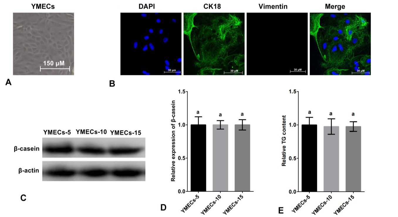
Identification of purified YMECs. A: Cell morphology (100×). B: Expression of CK18 in YMECs (400×). Cell nucleus were dyed with DAPI (blue), CK18 was visualized with Alexa Fluor 488 conjugated antibody (green) and vimentin (a marker of fibroblast, used as negative control) was visualized with Alexa Fluor 647 conjugated antibody red. C-D: Expression of β-casein in YMECs within 15 generations. E: Secretion of TG of YMECs within 15 generations. YMECs-5, 10 or 15: YMECs were 5, 10 or 15 generations, respectively.
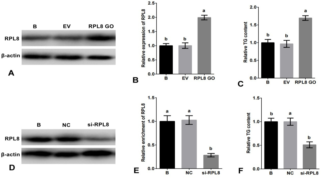
Role of RPL8 in promoting milk fat synthesis in YMECs. In A-B, the expression of RPL8 was increased in YMECs subjected to RPL8 over-expression. Similarly, C shows increased TG secretion following RPL8 over-expression in YMECs. On the other hand, D-E demonstrates a decrease in RPL8 expression in YMECs treated with RPL8 silencing. Furthermore, F depicts a decrease in TG secretion in YMECs subjected to RPL8 silencing. In B and E, the expression of RPL8 in the “B” group was set to “1”, while in C and F, the secretion of TG in the “B” group was also set to “1”. Cells were divided into different groups, including those that were not transfected B, those that were transfected with the empty vector (EV), those that were transfected with the RPL8 over-expression vector (RPL8 GO), those that were transfected with the negative control siRNA (NC), and those that were transfected with the RPL8 siRNA (Si-RPL8). In the bar charts, different superscript letters indicate significant differences (p<0.05), while the same letters indicate no significant difference (p>0.05).
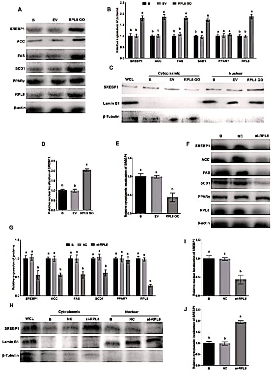
Activation of SREBP1 pathway by RPL8 in YMECs. In A-B, the expression of RPL8, ACC, FAS, SCD1, SREBP1, and PPARγ was evaluated in YMECs subjected to RPL8 overexpression. The nuclear and cytoplasmic localization of SREBP1 was also assessed in cells treated with RPL8 overexpression, as depicted in C-E. The results showed that the expression of these proteins, as well as the nuclear localization of SREBP1, was significantly increased in cells with RPL8 overexpression. Conversely, F-G displays the expression of RPL8, ACC, FAS, SCD1, SREBP1, and PPARγ in YMECs treated with RPL8 silencing. Similarly, H-J demonstrates the nuclear and cytoplasmic localization of SREBP1 in cells treated with RPL8 silencing. The results showed that the expression of these proteins, as well as the nuclear localization of SREBP1, was significantly decreased in cells with RPL8 silencing. In B and G, the expression of proteins in the “B” group was set to “1”. In D and I, the nuclear localization of SREBP1 in the “B” group was set to “1”, while in E and J, the cytoplasmic localization of SREBP1 in the “B” group was also set to “1”. Cells were divided into different groups, including those that were not transfected B, those that were transfected with the empty vector (EV), those that were transfected with the RPL8 overexpression vector (RPL8 GO), those that were treated with whole cell lysate (WCL), those that were transfected with the negative control siRNA (NC), and those that were transfected with the RPL8 siRNA (Si-RPL8). In the bar charts, different superscript letters indicate significant differences (p<0.05), while the same letters represent no significant difference (p>0.05).
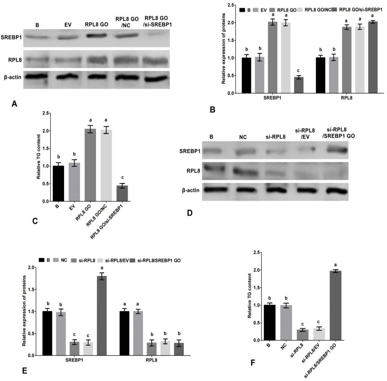
Promotion of milk fat synthesis by RPL8 through the SREBP1 pathway in YMECs. A-B The expression of RPL8 and SREBP1 in YMECs treated with RPL8 overexpression or RPL8 overexpression/SREBP1 silencing. C, The secretion of TG in YMECs treated with RPL8 overexpression or RPL8 overexpression/SREBP1 silencing. D-E, The expression of RPL8 and SREBP1 in YMECs treated with RPL8 silencing or RPL8 silencing/SREBP1 overexpression. F, the secretion of TG in YMECs treated with RPL8 silencing or RPL8 silencing/SREBP1 overexpression. In B and E, the expression of RPL8 and SREBP1 in “B” group was set to “1”. In C and F, the secretion of TG in “B” group was set to “1”. B: cells were no transfected. EV: cells were transfected with the empty vector. RPL8 GO: cells were transfected with RPL8 overexpression vector. NC: cells were transfected with the negative control siRNA. si-RPL8: cells were transfected with the RPL8 siRNA. RPL8 GO/ si-SREBP1: cells were co-transfected with the RPL8 overexpression vector and SREBP1 siRNA. In the bar charts, different superscript letters indicate significant differences (p<0.05), while the same letters represent no significant difference (p<0.05).
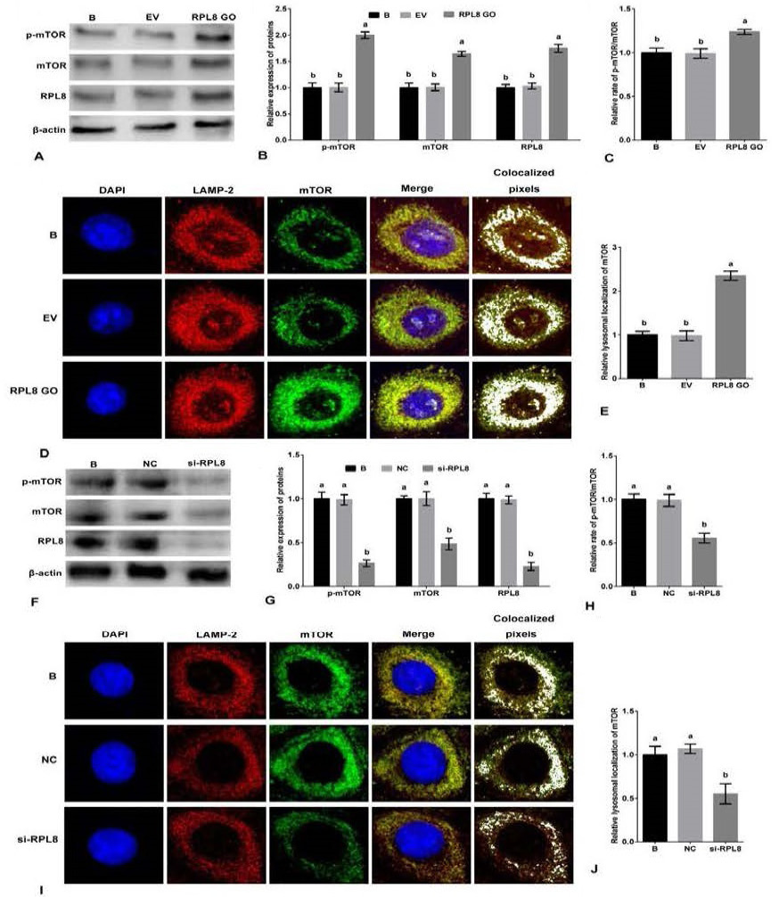
Activation of mTORC1 pathway by RPL8 in YMECs. A-B, the expression of mTOR and p-mTOR in YMECs treated with RPL8 overexpression. C, the rate of p-mTOR/mTOR in YMECs treated with RPL8 overexpression. D-E, the lysosomal localization of mTOR in YMECs treated with RPL8 overexpression. F-G, the expression of mTOR and p-mTOR in YMECs treated with RPL8 silencing. H, the rate of pmTOR/mTOR in YMECs treated with RPL8 silencing. I-J, the lysosomal localization of mTOR in YMECs treated with RPL8 silencing. In B and G, the expression of mTOR and p-mTOR in “B” group was set to “1”. In C and H, the rate of p-mTOR/mTOR in “B” group was set to “1”. In D and I, the colocalization between LAMP2 (green) and mTOR (red) was visualized and quantified using the ImageJ colocalization finder plugin (white). B: cells were no transfected. EV: cells were transfected with the empty vector. RPL8 GO: cells were transfected with RPL8 overexpression vector. NC: cells were transfected with the negative control siRNA. si-RPL8: cells were transfected with the RPL8 siRNA. In the bar charts, different superscript letters indicate significant differences (p<0.05), while the same letters represent no significant difference (p<0.05).
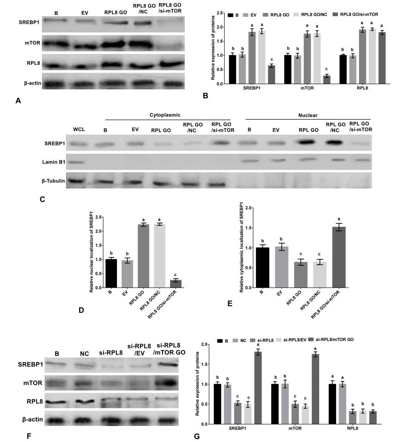
RPL8 activates the SREBP1 pathway through mTORC1 in YMECs. A-B, the expression of SREBP1, mTOR and RPL8 in YMECs treated with RPL8 overexpression or RPL8 overexpression/mTOR silencing. C-E, the nuclear localization (C-D) and cytoplasmic localization (C and E) of SREBP1 in cells treated with RPL8 overexpression or RPL8 overexpression/mTOR silencing. F-G, the expression of SREBP1, mTOR and RPL8 in YMECs treated with RPL8 silencing or RPL8 silencing/mTOR overexpression. Protein expression in the “B” group was standardized to “1” in B and G, while SREBP1 localization in the nucleus and cytoplasm of the “B” group was standardized to “1” in D and E, respectively. The experimental groups were categorized as WCL (whole cell lysate), B (non-transfected cells), EV (cells transfected with empty vector), RPL8 GO (cells transfected with RPL8 overexpression vector), NC (cells transfected with negative control siRNA), Si-RPL8 (cells transfected with RPL8 siRNA), and RPL8 GO/si-mTOR (cells co-transfected with RPL8 overexpression vector and mTOR siRNA). The bar charts display superscript letters indicating significant differences (p<0.05) between different experimental groups, while identical letters indicate no significant difference (p<0.05).
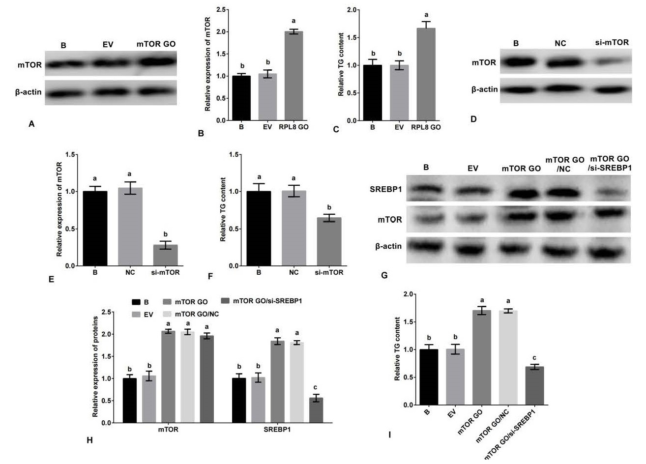
mTORC1 promotes milk fat synthesis in YMECs through the SREBP1 pathway. Specifically, the expression of mTOR in YMECs treated with mTOR overexpression is shown in A-B, while C displays the secretion of TG in YMECs treated with mTOR overexpression. The expression of mTOR in YMECs treated with mTOR silencing is depicted in D-E, and F shows the secretion of TG in YMECs treated with mTOR silencing. The expression of mTOR and SREBP1 in YMECs treated with mTOR overexpression or mTOR overexpression/SREBP1 silencing is shown in G-H, while I displays the secretion of TG in YMECs treated with mTOR overexpression or mTOR overexpression/SREBP1 silencing. In B, E, and H, the expression of mTOR or/and SREBP1 in the “B” group was set to “1”. In C, F, and I, the secretion of TG in the “B” group was set to “1”. The experimental groups were categorized as B (non-transfected cells), EV (cells transfected with empty vector), mTOR GO (cells transfected with mTOR overexpression vector), NC (cells transfected with negative control siRNA), si-mTOR (cells transfected with mTOR siRNA), and mTOR GO/si-SREBP1 (cells co-transfected with mTOR overexpression vector and SREBP1 siRNA). The bar charts display superscript letters indicating significant differences (p<0.05) between different experimental groups, while identical letters indicate no significant difference (p>0.05).
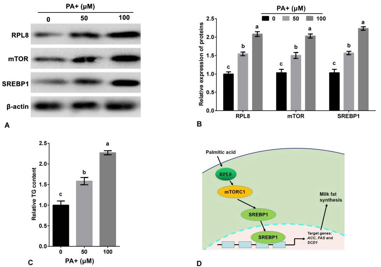
Palmitic acid promotes the expression of RPL8 and secretion of TG in YMECs. The expression of RPL8, mTOR, and SREBP1 in cells treated with 0, 50, and 100 μM of PA is shown in A-B, while C displays the secretion of TG in cells treated with the same concentrations of PA. D Shows a schematic model of how RPL8 promotes milk fat synthesis via the mTORC1-SREBP1 pathway in YMECs. In B, the expression of RPL8, mTOR, and SREBP1 in cells treated with 0 μM of PA was set to “1”. In C, the secretion of TG in cells treated with 0 μM of PA was set to “1”. PA refers to palmitic acid. The bar charts display superscript lowercase letters indicating significant differences (p<0.05) between different experimental groups, while identical letters indicate no significant difference (p>0.05).
References
- Bionaz, M. and Loor, J.J., 2008. Gene networks driving bovine milk fat synthesis during the lactation cycle. BMC Genomics, 9: 366. https://doi.org/10.1186/1471-2164-9-366[CrossRef]
- Bolsoni-Lopes, A., Deshaies, Y. and Festuccia, W.T., 2015. Regulation of brown adipose tissue recruitment, metabolism and thermogenic function by peroxisome proliferator-activated receptor γ. Temperature (Austin), 19: 476-482. https://doi.org/10.1080/23328940.2015.1011564[CrossRef]
- Chan, Y.L. and Wool, I.G., 1992. The primary structure of rat ribosomal protein L8. Biochem. biophys. Res. Commun., 185: 539-547. https://doi.org/10.1016/0006-291X(92)91658-D[CrossRef]
- Che, L., Xu, M., Gao, K., Zhu, C., Wang, L., Yang, X., Wen, X., Xiao, H., Jiang, Z. and Wu, D., 2019. Valine increases milk fat synthesis in mammary gland of gilts through stimulating AKT/MTOR/SREBP1 pathway. Biol. Reprod., 101: 126-137. https://doi.org/10.1093/biolre/ioz065[CrossRef]
- Contarini, G. and Povolo, M., 2013. Phospholipids in milk fat: Composition, biological and technological significance, and analytical strategies. Int. J. mol. Sci., 14: 2808-2831. https://doi.org/10.3390/ijms14022808[CrossRef]
- He, Q., Luo, J., Wu, J., Li, Z., Yao, W., Zang, S. and Niu, H., 2021. ELOVL6 promoter binding sites directly targeted by sterol regulatory element binding protein 1 in fatty acid synthesis of goat mammary epithelial cells. J. Dairy Sci., 104: 6253-6266. https://doi.org/10.3168/jds.2020-19292[CrossRef]
- Jiang, L., Liu, J., Sun, D., Ma, P., Ding, X., Yu, Y. and Zhang, Q., 2010. Genome wide association studies for milk production traits in Chinese Holstein population. PLoS One, 5: e13661. https://doi.org/10.1371/journal.pone.0013661[CrossRef]
- Jiang, L., Liu, X., Yang, J., Wang, H., Jiang, J., Liu, L., H,e S., Ding, X., Liu, J. and Zhang, Q., 2014. Targeted resequencing of GWAS loci reveals novel genetic variants for milk production traits. BMC Genomics, 15: 1105. https://doi.org/10.1186/1471-2164-15-1105[CrossRef]
- Jiang, N., Hu, L., Liu, C., Gao, X. and Zheng, S., 2015. 60S ribosomal protein L35 regulates β-casein translational elongation and secretion in bovine mammary epithelial cells. Arch. Biochem. Biophys., 583: 130-139. https://doi.org/10.1016/j.abb.2015.08.006[CrossRef]
- Jiang, N., Wang, Y., Yu, Z., Hu, L., Liu, C., Gao, X. and Zheng, S., 2015. WISP3 (CCN6) regulates milk protein synthesis and cell growth through mTOR signaling in dairy cow mammary epithelial cells. DNA Cell Biol., 34: 524-533. https://doi.org/10.1089/dna.2015.2829[CrossRef]
- Jiao, P., Yuan, Y., Zhang, M., Sun, Y., Wei, C., Xie, X., Zhang, Y., Wang, S., Chen, Z. and Wang, X., 2020. PRL/microRNA-183/IRS1 pathway regulates milk fat metabolism in cow mammary epithelial cells. Genes (Basel), 11: 196. https://doi.org/10.3390/genes11020196[CrossRef]
- Lee, J.N., Wang, Y., Xu, Y.O., Li, Y.C., Tian, F. and Jiang, M.F., 2017. Characterisation of gene expression related to milk fat synthesis in the mammary tissue of lactating yaks. J. Dairy Sci., 84: 283-288. https://doi.org/10.1017/S0022029917000413[CrossRef]
- Li, N., Zhao, F., Wei, C., Liang, M., Zhang, N., Wang, C., Li, Q.Z. and Gao, X.J., 2014. Function of SREBP1 in the milk fat synthesis of dairy cow mammary epithelial cells. Int. J. mol. Sci., 15: 16998-7013. https://doi.org/10.3390/ijms150916998[CrossRef]
- Li, X., Li, P., Wang, L., Zhang, M. and Gao, X., 2019. Lysine enhances the stimulation of fatty acids on milk fat synthesis via the GPRC6A-PI3K-FABP5 signaling in bovine mammary epithelial cells. J. Agric. Fd. Chem., 67: 7005-7015. https://doi.org/10.1021/acs.jafc.9b02160[CrossRef]
- Liu, L., Lin, Y., Liu, L., Wang, L., Bian, Y., Gao, X. and Li, Q., 2016. Regulation of peroxisome proliferator-activated receptor gamma on milk fat synthesis in dairy cow mammary epithelial cells. In Vitro Cell. Dev. Biol. Anim., 52: 1044-1059. https://doi.org/10.1007/s11626-016-0059-4[CrossRef]
- Liu, X., Yang, J., Zhang, Q. and Jiang, L., 2017. Regulation of DNA methylation on EEF1D and RPL8 expression in cattle. Genetica, 145: 387-395. https://doi.org/10.1007/s10709-017-9974-x[CrossRef]
- Loften, J.R., Linn, J.G., Drackley, J.K., Jenkins, T.C., Soderholm, C.G. and Kertz, A.F., 2014. Invited review: Palmitic and stearic acid metabolism in lactating dairy cows. J. Dairy Sci., 97: 4661-4674. https://doi.org/10.3168/jds.2014-7919[CrossRef]
- Lou, Q., Cao, S., Xu, W., Zhang, Y., Qin, Z. and Wei, W., 2014. Molecular characterization and mRNA expression of ribosomal protein L8 in Rana nigromaculata during development and under exposure to hormones. J. environ. Sci. (China), 26: 2331-2339. https://doi.org/10.1016/j.jes.2014.09.018[CrossRef]
- Luo, C.C., Yin, D.Y., Gao, X.J., Li, Q.Z. and Zhang, L., 2013. Goat mammary gland expression of Cecropin B to inhibit bacterial pathogens causing mastitis. Anim. Biotechnol., 24: 66-78. https://doi.org/10.1080/10495398.2012.745417[CrossRef]
- Luo, C., Qi, H., Huang, X., Li, M., Zhang, L., Lin, Y. and Gao, X., 2019. GlyRS is a new mediator of amino acid-induced milk synthesis in bovine mammary epithelial cells. J. cell. Physiol., 234: 2973-2983. https://doi.org/10.1002/jcp.27115[CrossRef]
- Luo, C., Zhao, S., Dai, W., Zheng, N. and Wang, J., 2018. Proteomic analysis of lysosomal membrane proteins in bovine mammary epithelial cells illuminates potential novel lysosome functions in lactation. J. Agric. Fd. Chem., 66: 13041-13049. https://doi.org/10.1021/acs.jafc.8b04508[CrossRef]
- Ma, L. and Corl, B.A., 2012. Transcriptional regulation of lipid synthesis in bovine mammary epithelial cells by sterol regulatory element binding protein-1. J. Dairy Sci., 95: 3743-3755. https://doi.org/10.3168/jds.2011-5083[CrossRef]
- Mohan, M.S., O’Callaghan, T.F., Kelly, P. and Hogan, S.A., 2021. Milk fat: Opportunities, challenges and innovation. Crit. Rev. Fd. Sci. Nutr., 61: 2411-2443. https://doi.org/10.1080/10408398.2020.1778631[CrossRef]
- Mu, T., Hu, H., Ma, Y., Feng, X., Zhang, J. and Gu, Y., 2021. Regulation of key genes for milk fat synthesis in ruminants. Front. Nutr., 8: 765147. https://doi.org/10.3389/fnut.2021.765147[CrossRef]
- Oppi-Williams, C., Suagee, J.K. and Corl, B.A., 2013. Regulation of lipid synthesis by liver X receptor α and sterol regulatory element-binding protein 1 in mammary epithelial cells. J. Dairy Sci., 96: 112-121. https://doi.org/10.3168/jds.2011-5276[CrossRef]
- Osorio, J.S., Lohakare, J. and Bionaz, M., 2016. Biosynthesis of milk fat, protein, and lactose: Roles of transcriptional and posttranscriptional regulation. Physiol. Genomics, 48: 231-256. https://doi.org/10.1152/physiolgenomics.00016.2015[CrossRef]
- Piantoni, P., Lock, A.L. and Allen, M.S., 2013. Palmitic acid increased yields of milk and milk fat and nutrient digestibility across production level of lactating cows. J. Dairy Sci., 96: 7143-7154. https://doi.org/10.3168/jds.2013-6680[CrossRef]
- Ren, Q.R., Zhang, H., Guo, H.Y., Jiang, L., Tian, M. and Ren, F.Z., 2014. Detection of cow milk adulteration in yak milk by ELISA. J. Dairy Sci., 97: 6000-6006. https://doi.org/10.3168/jds.2014-8127[CrossRef]
- Rico, J.E., Mathews, A.T., Lovett, J., Haughey, N.J. and McFadden, J.W., 2016. Palmitic acid feeding increases ceramide supply in association with increased milk yield, circulating nonesterified fatty acids, and adipose tissue responsiveness to a glucose challenge. J. Dairy Sci., 99: 8817-8830. https://doi.org/10.3168/jds.2016-11296[CrossRef]
- Ricoult, S.J., Yecies, J.L., Ben-Sahra, I. and Manning, B.D., 2016. Oncogenic PI3K and K-Ras stimulate de novo lipid synthesis through mTORC1 and SREBP. Oncogene, 35: 1250-1260. https://doi.org/10.1038/onc.2015.179[CrossRef]
- Sears, A., Gonzalez, O., Alberto, A., Young, A., de Souza, J., Relling, A. and Batistel, F., 2020. Effect of feeding a palmitic acid-enriched supplement on production responses and nitrogen metabolism of mid-lactating Holstein and Jersey cows. J. Dairy Sci., 103: 8898-8909. https://doi.org/10.3168/jds.2020-18232[CrossRef]
- Wang, D., Chen, Z., Zhuang, X., Luo, J., Chen, T., Xi, Q., Zhang, Y. and Sun, J., 2020. Identification of circRNA-associated-ceRNA networks involved in milk fat metabolism under heat stress. Int. J. mol. Sci., 21: 4162. https://doi.org/10.3390/ijms21114162[CrossRef]
- Western, M.M., de Souza, J. and Lock, A.L., 2020. Effects of commercially available palmitic and stearic acid supplements on nutrient digestibility and production responses of lactating dairy cows. J. Dairy Sci., 103: 5131-5142. https://doi.org/10.3168/jds.2019-17242[CrossRef]
- Wu, J., He, S., Yu, Z., Lan, D., Xiong, X. and Li, Z., 2020. Transcriptomic study of yak mammary gland tissue during lactation. Anim. Biotechnol., 22: 1-8.
- Xin, J.W., Chai, Z.X., Zhang, C.F., Yang, Y.M., Zhang, Q., Zhu, Y., Cao, H.W., Yang, J.C., Zhong, J.C. and Ji, Q.M., 2020b. Transcriptome analysis identified long non-coding RNAs involved in the adaption of yak to high-altitude environments. R. Soc. Open Sci., 7: 200625. https://doi.org/10.1098/rsos.200625[CrossRef]
- Xin, J.W., Chai, Z.X., Zhang, C.F., Zhang, Q., Zhu, Y., Cao, H.W., Ji, C.Y., Chen, X.Y., Jiang, H., Zhong, J.C. and Ji, Q.M., 2020a. Signature of high altitude adaptation in the gluteus proteome of the yak. J. exp. Zool. B Mol. Dev. Evol., 334: 362-372. https://doi.org/10.1002/jez.b.22995[CrossRef]
- Xu, H.F., Luo, J., Zhang, X.Y., Li, J. and Bionaz, M., 2019. Activation of liver X receptor promotes fatty acid synthesis in goat mammary epithelial cells via modulation of SREBP1 expression. J. Dairy Sci., 102: 3544-3555. https://doi.org/10.3168/jds.2018-15538[CrossRef]
- Zheng, X.R., Jiang, L., Ning, C., Hu, Z.Z., Zhou, L., Yu, Y., Zhang, S.L. and Liu, J.F., 2019. A novel mutation in the promoter region of RPL8 regulates milk fat traits in dairy cattle by binding transcription factor Pax6. Biochim. biophys. Acta Mol. Cell Biol. Lipids, 1864: 158528. https://doi.org/10.1016/j.bbalip.2019.158528[CrossRef]
- Zhou, F., Ouyang, Y. and Miao, Y., 2021. Peroxisome proliferator-activated receptor gamma regulates genes involved in milk fat synthesis in mammary epithelial cells of water buffalo. J. Anim. Sci., 92: e13537. https://doi.org/10.1111/asj.13537[CrossRef]
Cited by 1 article
In journals published by ResearchersLinks.
Services
Article usage statistics
Loading…