
In order to explore the mechanism underlying chemosensation in the tea looper, Scopula subpunctaria (Lepidoptera: Geometridae), the types, morphology, quantity and distribution of sensilla on the antenna were observed by scanning electron microscopy. The results showed that females and males antennae were filiform, bipectinate, respectively, which be divided into three segments, scape, pedical and flagellum that consisted numerous individual flagellomeres, respectively. There were eight kinds and twelve types of sensilla on the antennae, including 4 subtypes of sensilla trichodae, 2 subtypes of sensilla basiconica, sensilla chaetica, Böhm bristles, sensilla squamiformia, sensilla auricillica, sensilla coeloconica, and sensilla styloconica. No differences were found about the morphology, abundance, and distribution of most sensillum. However, the distributions and abundances of ST III, sensilla coeloconica and sensilla styloconica dimensions showed significant sexual dimorphism between male and female adults. In addition, the possible functions of sensilla were analyzed and discussed according to their morphology, distribution, and the previously reported sensilla function. These results could help to understand the chemical communication of S. subpunctaria and their host, and provide a scientific basis for the development of a novel biological control strategy of S. subpunctaria.
Research Article · Volume 56, Issue 4, 13 June 2024 · Open Access
Ultrastructure of Antennal Sensilla of Scopula subpunctaria (Herrich-Schaeffer) (Lepidoptera: Geometridae)
· · · · · · · ·
Affiliations & Notes ⌄
- 1School of Agricultural Sciences, Xinyang Agriculture and Forestry University, Xinyang 46400, China 2Xinyang Academy of Ecological Research, Xinyang, 464000, China. 3Department of Plant Medicals, Andong National University, Andong 36729, Republic of Korea * Corresponding author: shubao.301@163.com
Article Info ⌄
- Journal:
- Pakistan Journal of Zoology
- Published:
- 13 June 2024

From the article
- In conclusion, we observed sexual dimorphism in the antennae of females (filiform) and males (bipectinate) in S. subpunctaria.
- Eight types of sensilla were observed using SEM, including sensilla trichodea, chaetica, basiconica, coeloconica, squamiformia, and styloconica and Böhm bristles.
- No differences were found about the morphology, abundance, and distribution of most sensillum, except the distributions and abundances of ST III, sensilla coeloconica and sensilla styloconica dimensions between males and females adults.
- Whereas, further studies on electrophysiology, behaviors, and molecular mechanism, are still needed to verify the functions of those sensilla described in present study and hopefully design an effective semiochemical control methods in S. subpunctaria.
Selected from the article's own text.
Abstract
INTRODUCTION
Insect antennae, as the main olfactory and sensory structures, display multiple types of sensilla that vary in structure and functions on its surface ; , which has extremely critical role in detecting complex physical and chemical stimuli from the surrounding environment. The reception of specific stimuli is mediated by membrane receptors expressed on dedicated recipient neurons of the sensillum, which change them into electrical signals. These electrical signals are then transmitted to the central nervous system and further regulate and trigger various biological behaviors including forage, mating, habitat selection, predator evasion and oviposition decisions .
Each antenna sensillum is an independent sensory unit and displays various morphological characters which typically related to the perception of various signals ; . Generally, antenna sensilla are termed as Böhm bristles (BB), trichodea, basiconica, chaetica, styloconica, coeloconica, auricillica, and squamiformia according to their structures and appearances . The different sensilla types might be strongly associated with morph-specific functions. For example, the sensilla trichodea, basiconica, auricillica, and coeloconica with numerous pores on the cuticular surface are proposed to have olfactory function. The sensilla charactered one or a few pores at the tip have gustatory function, e.g. sensilla chaetica. The sensilla styloconica, squamiformia and Böhm bristles without any surface pores are considered as the mechanosensilla .
In the past few decades, several studies on the morphology, structure, as well as distribution of antennal sensilla of various insects have been carried out in different orders, such as Orthoptera , Coleoptera , Diptera , Hymenoptera , Hemiptera , and Lepidoptera ; . Furthermore, olfaction-based behavioral manipulation has been frequently used for the monitoring and prevention of pest and also developed a novel, effective and environmentally friendly strategies . Therefore, studies of the structures of antennal sensilla in the insects are important to help elucidate the olfactory mechanisms in insects.
Scopula subpunctaria (Lepidoptera: Geometridae), one of the most important pest of tea plantations, causes serious economic losses, which are widely distributed in many tea producing areas in China ; ; ; . This tea looper generally feed on hypodermis, mesophyll, margin of leaves, and even all the leaves under the larvae stages, resulting in the decline of quality and yield of tea. It is difficult to be identified because of the same body color of the larvae and tea plant , which is also challenge for control. Ma et al. (2019) and Geng et al. (2021) studies on S. subpunctaria had mainly focused on the reproductive behavior, the biosynthesis and transportation of sex pheromone components , and the population dynamics between S. subpunctaria and natural enemies . However, compared to the above-mentioned studies, the ultrastructure of antennae sensilla has been insufficient in S. subpunctaria.
Hence, the external morphology, types, distribution and of antennal sensilla between female and male of S. subpunctaria were observed by scanning electron microscopy (SEM). Moreover, the possible structural functions of sensilla were analyzed and speculated according to their morphological characters and the previously reported sensilla function. Our results elucidate the chemical communication mechanism of the olfactory and chemosensory in S. subpunctaria, which provide an essential basis for further study on behavioral mechanisms for this pest.
MATERIALS AND METHODS
Insects rearing
Larve of S. subpunctaria were collected from Maan Mountain, Xinyang, Henan, China (32°10′50″N, 113°50′17″E) in August 2021, and reared in a cage (45 cm×45 cm×45 cm) at 25±1℃, 60±5% relative humidity, with a photoperiod regime of 16:8 h (L: D) in the laboratory with fresh tea leaves. At 1-2 d post-emergence, fresh female and male S. subpunctaria adults were used as the specimen for SEM.
Scanning electron microscopy (SEM)
Antennae of female and male moths (n=20) of S. subpunctaria were selected for SEM observations. The antennae were quickly cut off from the head with a scalpel under a dissecting microscope (Olympus, szx-16, Tokyo, Japan), cleaned in an ultrasonic cleaner for 10 s, and fixed in 2.5% glutaraldehyde overnight at 4℃. Then, the samples were washed 10 min in phosphate-buffered saline (PBS; 0.1 M, pH 7.2) for three times, then dehydrated through a graded ethanol series (30%, 60%, 80%, 90%, and 95%) for 15 min at each gradation, using 100% ethanol three times (for 15 min each time). After natural drying, the antennae were mounted on SEM sample holders using double-sided adhesive tape, coated with gold film in a JFC-1600 ion sputter (JEOL, Tokyo, Japan). Finally, the samples were examined using a JSM-7900F scanning electron microscope (JEOL, Tokyo, Japan) at an accelerating voltage of 10 kV.
Terminology and data analysis
The terminology for naming and classification of the sensilla is based mainly on the their external morphology as described by Schneider (1964) and Zacharuk (1985) and Lepidoptera antenna structure ; ; .
Data analysis
The length and basal diameter of antennal sensilla were measured using Sigma Scan Pro Measurement System 5.0. All data were reported as means±SE. Any significant differences between the sexes were assessed using a two sample t-test in SAS statistical software 9.2 (SAS Institute Inc., Cary, NC, USA), with thresholds set at a P<0.05.
RESULTS
Antenna morphology of S. subpunctaria
The antenna of S. subpunctaria consisted of scape (Sc), pedicel (Pe), and flagellum (Fl) that was divided into numerous individual flagellomeres. The antennae of males and females of S. subpunctaria showed marked sexual dimorphism. Females were filiform antennae, thread-like, covered in arranged scales (S) and sensilla. While males were bipectinate types, feather-like, which had a central filament with lateral branches covered with overlapping scales and sensilla (Fig. 1). The average antenna length of females was significantly longer than that of males (female=14.24±0.73 mm; male=8.15±0.19 mm, t=13.99, P=0.0002) (Fig. 1A, B).
The scape, the first segment of the antenna, is elongated and cylindrical shape with significantly difference on average length between sexes (t=5.67, P=0.0048, females=256.74±2.60 μm; males=240.25±4.31 μm) (Table I). The pedicel, barrel-shaped, is the second segment. The average length in females is also significantly different from that of males (t=4.27, P=0.0129, females=201.44±1.06 μm, males=189.77±4.57 μm) (Table I). The flagellum, the largest segment of antenna, contain numerous individual flagellomeres. The antennal flagellomeres of both sexes are barrel or cylindrical in shape (Fig. 1C-D). The average length of the flagellomeres in females are dramatically and significantly greater than that in males (t=63.00, P<0.0001, female=102.58±0.93 μm; male=79.29±0.86 μm) (Table I). The width of flagellomeres gradually decrease from proximal to the distal end of antennae. Most sensilla are distributed on the ventral and lateral surfaces of the antennal flagellomeres.
Table I. Length of antenna of Scopula subpunctaria female and male adults.
| Antenal location | Length (μm) | |
| Female | Male | |
| Scape | 256.74±2.60** | 240.25±4.31 |
| Pedicel | 201.44±1.06* | 189.77±4.57 |
| Flagellomere | 99.89±1.32** | 49.45±0.42 |
| Total length | 14240±731.23** | 8146±186.10 |
Data are presented as means±SE, n=5. * and ** indicate significant difference between male and female in length at 0.05 and 0.01 level using the t-test, respectively.
Table II. Size and distribution of antennal sensilla on Scopula subpunctaria female and male adults.
| Sensilla type | Length (μm) | Basal width (μm) | Tip | Cuticular surface | Sex |
| Sensilla trichodea (St) | |||||
| St I | 97.19±1.00 | 9.30±0.58 | Sharp | Helical deep grooves | ♀ and ♂ |
| St II | 124.88±0.42 | 11.69±1.26 | Sharp | Rough and wrinkle | ♀ and ♂ |
| St III | 158.90±2.74 | 7.82±0.09 | Sharp | Horizontal deep grooves | ♂ |
| St IV | 80.70±1.54 | 5.55±0.16 | Sharp | Longitudinal grooves | ♀ and ♂ |
| Sensilla trichodea (Sb) | |||||
| Sb I | 28.29±0.40 | 2.46±0.11 | Blunt | Smooth | ♀ and ♂ |
| Sb II | 34.48±0.79 | 5.44±0.06 | Blunt | Corrugated | ♀ and ♂ |
| Sensilla chaetica (Sch) | 148.02±4.49 | 6.28±1.38 | Subacute | Grooved | ♀ and ♂ |
| Böhm bristles (BB) | 5.98±0.28 | 1.52±0.08 | Sharp | Smooth | ♀ and ♂ |
| Sensilla squamiformia (Ssq) | 108.09±5.12 | 7.10±0.09 | Sharp | Longitudinal grooves | ♀ and ♂ |
| Sensilla auricillica (Sau) | 45.09±0.69 | 5.13±0.11 | Blunt | Multiple pores | ♀ and ♂ |
| Sensilla coeloconica (Sco) | 11.01±0.59** | - | Blunt | Deep longitudinal grooved | ♀ |
| 4.92±0.43 | - | Blunt | ♂ | ||
| Sensilla styloconica (Sst) | 34.88±0.33** | 9.23±0.06 | Cone-shaped structure | Deep longitudinal grooved | ♀ |
| 41.47±0.64 | 15.92±0.10 | ♂ |
Sensilla on S. subpunctaria antennae
Eight different morphological types of antennal sensilla were identified in both male and female moths of S. subpunctaria (Table II), including four subtypes of sensilla trichodea (St I, St II, St III, and St IV), two subtypes of sensilla basiconica (Sb I and Sb II), one type of sensilla chaetica (Sch), Böhm bristles (BB), sensilla squamiformia (Ssq), sensilla auricillica (Sau), sensilla coeloconicum (Sco), and sensilla styloconica (Sst). We found there were some differences on the distributions and abundances of ST III, sensilla coeloconica and sensilla styloconica dimensions between males and females (Table II).
Sensilla trichodea (St)
Sensilla trichodea are the most abundant sensilla, and widely located on the ventral sides of the antennae flagellum of S. subpunctaria. They present significantly higher proportions on total number in males than females (Fig. 2A-B). Sensilla trichodea is slender, hair-like structure characterized by gradual thinning from the base to the top and had cuplike fossa at the base. These sensilla incline forward to the end of the antenna and form different angles with the attachment site. They are slightly curved at the apex and had significant differences on the surface.
Sensilla trichodea are classified into four subtypes (St I, St II, St III, and St IV) based on shape, size, distribution, and surface texture. St I and St II distributed mainly on the ventral and lateral surfaces of the flagellomeres and derived from a small conical basal socket (Fig. 2C). Their basal parts are almost vertical to the antennal surface and taper toward the end of antenna. St I characterized with helical deep grooves on the surface gradually marked toward the apex, while St II with rough and wrinkle surface (Fig. 2D). The average length and basal width of St I are 97.19±1.00 µm and 9.30±0.58 µm, respectively. St II were significantly longer and wider than those of St I (length: 124.88±0.42 µm, basal width: 11.69±1.26 µm) (tlength=-44.18, P<0.0001; tbasal width= -2.99, P= 0.0402) (Table II). St III distributed uniformly on the central filament and the lateral branches of the flagellomeres on male bipectinate antennae, and characterized as the longest of the subtypes (158.90±2.74 µm) (Table II). However, the average basal width of St III is 7.82±0.09 µm, which is significantly shorter than that of St II (t=-5.31, P=0.0330). One third of the tip of the shaft is curved about a 60 angle relative to the antennal surface. The wall of cuticular surface has parallel horizontal deep grooves and screw-thread patterns along its structure up to the apical tip (Fig. 2E-F). In contrast, St IV has longitudinal grooves on cuticular surface with an average length and basal width of 80.70±1.54 µm and 5.55±0.16 µm, respectively. They are inserted tightly into a small cuticular socket at a 45-60° angle to the surface of the antenna, and are randomly dispersed on the antennal flagellomeres on sexes (Fig. 2G-I).
Sensilla basiconica (Sb)
Sensilla basiconica (Sb) are sparsely presented on the ventral and dorsal surfaces of both males and females antennae flagellum and are primarily interspersed between sensilla trichoidea. These sensilla are short, rod-like structure, strong and robust, which look similar to sensilla trichodea in appearance but with corrugated surface texture and smaller size (Fig. 3). The blunt tipped shaft of sensilla basiconica incline forward on the antenna surface and toward the distal tip. They are inserted into a wide, slightly round circular sockets on the antennal surface (Fig. 3A).
Two subtypes (Sb I and Sb II) in sensilla basiconica of S. subpunctaria are identified according to their morphological characteristics, size and precise wall texture. The peg-like Sb I is short, thin and robust with a triangular shape at the apical extremity (Fig. 3B). They are 28.29 ±0.40 μm long with a base width of 2.46± 0.11 μm (Table II). Sb II is characterized by corrugated surface texture, and taper from the base to the apex with blunt setae (Fig. 3C). The size of Sb II (average length=30.89±3.12 µm; average basal width=2.23±0.26 µm) is significantly greater than those of Sb I (tlength=12.06, P=0.0003; tbasal width=-40.72, P<0.0001) (Table II), whereas the number did not show any difference in both males and females.
Sensilla chaetica (Sch)
Sensilla chaetica is present in the apical tip of the dorsal part of the antennae flagellum in both sexes (Fig. 3D). Compared to sensilla trichodea, sensilla chaetic are long, spiny-shaped and pointed, characterized by grooved surfaces and blunt tip. The shafts emerge from a round collar-like flexible socket at the antennal surface. The average length of sensilla chaetic is approximately 148.02±5.12 µm with average basal width of 6.28±1.38 µm (Table II), respectively. The basal width of the sensilla decrease from base towards the tip. Further, no significant difference was observed between male and female adults.
Böhm bristles (Bb)
Böhm bristles cluster only at the dorsal base of the scape and pedicel of the antennae of both males and females covering by numerous scales (Fig. 1C), but none does not exist on the flagellum. These sensilla are short thorns-shape with a smooth nonporous surface and sharp tip, and emerge directly from the broad socket on the base (Fig. 4A). They resemble sensilla chaetica in appearance, which are positioned almost perpendicular to the antennal surface. But the average length and basal width of Böhm bristles (average length=5.98±0.28 µm; average basal width=1.52±0.08 µm) are significantly smaller than sensilla chaetica (tlength=54.64, P<0.0003; tbasal width=6.05, P=0.0258) (Table II). Distinction of Böhm bristles between the sexes could not be found.
Sensilla squamiformia (Ssq)
Sensilla squamiformia are elongated and shuttle-like with a flexible socket at the base, deep longitudinal grooves at the surface, which were slightly curved toward the apex of the flagellum (Fig. 4B). They taper from the base to the apex with sharp setae and are irregularly found in each flagellomere. This type of sensilla is 108.09±5.12 μm in average length and 7.10±0.09 μm in average basal width (Table II), which was similar between the male and female antennae.
Sensilla auricillica (Sau)
Sensilla auricillica are short and elongated with slightly curved or blunt tips, which mainly scattered at the ventral and lateral parts of each flagellomeres (Fig. 4C). These sensilla have the appearance of ear or leaf shaped arose from round socket, covered with numerous open pores on the entire cuticular wall of the sensilla (Fig. 4D). They curve forward to form the apex of flagellum and situated almost parallel to the antennal surface. The mean length of this type is 45.09±0.69 μm, and are the mean basal width is 5.13±0.11 μm (Table II). No difference between sexes is observed in the number and shape of sensilla auricillica.
Sensilla coeloconica (Sco)
Sensilla coeloconica are exclusively located on the ventral surface of flagellomeres on both male and female in small quantities, either individually or in clusters (Fig. 5A-B). Most of which occur near the distal edge of antennae with no fixed distribution pattern. Such sensilla are inserted into the ellipsoid-shaped cavity wall with a protruding sensory peg in the center. The center of a protruding sensory peg (cp) is characterized by longitudinal groove dotted with numerous pores is surrounded by 10-16 finger-like cuticular spines (cs) with many ridges on the surface, curving toward the center, which forms a chrysanthemum-like shape (Fig. 5C-D). These finger-like spines are varied in length and size and taper gradually from the base to the tip, which are longer than the center sensory peg. The average length of center pegs in males (4.92±0.43 μm) is significantly shorter than that of females (11.01±0.59 μm) (t=−14.36, P=0.0001; Table I) and there is no sexual dimorphism on the number and shape.
Sensilla styloconica (Sst)
Sensilla styloconica are regularly arranged on the ventral surface of the terminal segment of each flagellomeres of the antennae with less dense, only one sensillum on each flagellomere. Such sensilla are scout upright on the whole with tip facing towards the distal end of the antennae and typically parallel or the angle between which and the antennal surface is about 45° (Fig. 6A). These sensilla resemble feeding-bottle shape with a reticulated inflexible base. They are covered with longitudinal cuticular ridges on the surface, with one, two or three cone-shaped structure in the apical region (Fig. 6B-D). These types of sensilla appear to be aporous and wat swollen slightly at the base. No significant differences are found in the location and shape of sensilla styloconica between the male and female except size. The size in males (average length=41.47±0.64 µm; average basal width=15.92±0.10 µm) is significantly different to that of females (average length=34.88±0.33 µm; average basal width=9.23±0.06 µm) (t=15.79, P<0.0001) (Table II).
DISCUSSION
Gross morphology of antennae
The antennae of insects have a variety of sensilla that play an important role in detecting the various stimuli from the environment . The present study was focused on the antennal sensilla which are most likely involved in chemical communication of S. subpunctaria. A significant sexual dimorphism was observed in the antennae of S. subpunctaria with female adults having filiform antennae with only a filamentous central stem, whereas male adults having bipectinate antennae with several lateral branches on each flagellomere. The characteristic of antenna dimorphism also has been described for Automeris liberia of Saturniidae and Erannis ankeraria of the same genus . This distinction demonstrated that the bipectinate antenna has a larger area covered sensory and olfactory sensilla than the filiform antennae , which facilitate locating the female for mating for male.
However, the female antennae possesses a greater number of flagellomeres than male antennae. The average antennal length of females is significantly longer than that of males, which is different from other species reported previously ; ; ; . The structure of antenna in both sexes consist of three segments, scape, pedicel and flagellum. The joint region of both scape and pedicel are covered with overlapping scales, which have been reported in several Lepidopteran species belonging to different families: Castniidae , Gelechiidae , Tortricidae , Crambidae , and Noctuidae . Generally, scales occurred along with sensilla on the antennae, which could protect the sensilla from mechanical damages or favor the ability of the insect to detect volatile stimuli or filter contaminating microparticles present in the air .
Different types of antennae sensilla
In the present study, eight morphologically distinct types of sensilla were identified on the antennae of both sexes of S. subpunctaria, including sensilla trichodea, basiconica, chaetica, squamiformia, auricillica, coeloconica, and styloconica and Böhm bristles by SEM. These types of sensilla resembled to those reported from other Lepidoptera species, such as A. liberia (Saturniidae) , Diaphania angustalis (Crambidae) , and Earias vittella (Nolidae) . The results of the antennae sensilla ultrastructure of S. subpunctaria showed that the external antennal sensilla morphological characteristics were almost identical in both sexes, but the distributions and abundances of ST III, sensilla coeloconica and sensilla styloconica dimensions showed significant sexual dimorphism between males and females adults. The functions of different sensilla were analyzed and speculated according to their morphology, distribution, and the previously reported sensilla function in our study.
Sensilla trichodea
Sensilla trichodea, as the most widespread and numerous sensilla in this study, is similar in shape to other Geometridae insects, such as Ectropis obliqua and E. ankeraria . In addition, there are more flourished sensilla trichodea in males than females moths, which was compatible with reports of E. obliqua , Monema flavescens and D. angustalis . According to their shape, size, and distribution, sensilla trichodea could be sorted into different subtypes. For example, two subtypes were found on Conogethes punctiferalis and E. ankeraria , three subtypes in Glenea cantor and A. Liberia , four subtypes in Eocanthecona furcellata (Hemiptera: Pentatomidae) . In this study, four subtypes of sensilla trichodea were distinguished. St I, St II, and St IV distributed mainly on the ventral and lateral surfaces of the flagellomeres in both sexes. While St III characterized by parallel horizontal deep grooves, was the longest of the subtypes and chiefly found in the lateral branches of male bipectinate antennae, which might be classically associated to the perception of sex pheromones.
Previous study have been validated that proteins and receptors of odoriferous molecules are expressed in sensilla trichodea using immunohistochemistry and in situ hybridization, indicating their participation in pheromone detection or plant volatiles or both . For example, in E. obliqua, EoblGOBP2 localized in sensilla trichodea is sensitive to tea leaf volatiles (E)-2-hexenal, methyl salicylate, and acetophenone . In consideration of the similarities of sensilla trichodea between S. subpunctaria and E. obliqua, we assume that sensilla trichodea in S. subpunctaria are olfactory sensilla, which have potential function of semiochemical detection.
Sensilla basiconica
The sensilla basiconica identified in S. subpunctaria are also common in other Lepidoptera insects ; ; , which is generally considered as olfactory receptors to sense sex pheromones and plant volatiles ; . In S. subpunctaria, two subtypes of sensilla basiconica were identified. Similar to our findings, different morphological subtypes of sensilla basiconica had been reported in other Lepidoptera insects ; ; , suggesting they could be responsible for different odorant detection, which needed more evidence from electrophysiology and behavior tests. The surfaces of sensilla basiconica are covered with many pore-like structures, allowing the entry of odorous substances, which suggest they possessed olfactory chemoreception function . It is previously reported that olfactory receptor gene is also expressed in sensilla basiconica. For example, a specific olfactory receptor gene (OR35) found in sensilla basiconica of locust antenna was reported to be responsible for the detection of aggregation pheromone . In Spodoptera litura, SlituOR12 expressed in the sensilla basiconica, exclusively tuned to the important plant volatile cis-3-hexenyl acetate . Other studies suggested sensilla basiconica responded to contact and taste .
Sensilla chaetica
Sensilla chaetica observed in S. subpunctaria has also been reported in other insects ; ; . The morphological characteristics were simillar to sensilla trichodea, but these sensilla were straight and pointed with a broad base. Moreover, the significant variations in the size and structure of these sensilla were not observed in the present study, which was similar to those of Spilonota albicana , E. ankeraria and Eutectona machaeralis . In S. subpunctaria, sensilla chaetica are located in the intermediate and apical regions of the antennae in both sexes. Generally, moths always use the apex of antennae to touch the odor source, indirectly proving that sensilla chaetica might have a mechanoreceptive function as shown previously . This behavior is also attributed to the increased lengths of sensilla chaetica at the distal section of antenna , which is validated in our present work. The average length of sensilla chaetica (148.02±5.12 µm) measured is longer than other sensilla, excepted the largest St III. Besides, the length of sensilla chaetic is also used as a taxonomic character in light microscopy analysis for differentiation within and between species especially in Diptera .
The olfactory and gustatory function of sensilla chaetica also has been reported in Paysandisia archon , Helicoverpa armigera and Spodoptera littoralis . In this study, we observed that the surface structure of the sensilla chaetica was similar to that of E. ankeraria without pores , suggesting that they might only sense mechanical stimulation but not chemical sensation. Accordingly, sensilla chaetica of antennae in S. subpunctaria are hypothesized to function in perceiving mechanical stimuli based on their distribution, structure and length.
Bohm bristles
Böhm bristles found in our study are short thorns-shape with a smooth nonporous surface clustered at the dorsal base of the scape and pedicel in both sexes of S. subpunctaria as in other Lepidoptera like A. liberia , E. machaeralis , and E. vittella with the same location and structure. Studies have indicated that these sensilla were considered as the mechanoreceptors with a proprioceptive function ; and as wind velocity receptors , which could mediate antennal position and movements and was response to sensory stimulus. Recent behavioral study in Daphnis nerii has been confirmed when Böhm bristles were ablated from the basal segments of antennae, moths were unable to bring their antennae into flight position, causing frequent collisions with the flapping wing . Whereas, some researchers interpreted them as membrane receptors to segment membranes for the scape-pedicel joint .
Sensilla squamiformia
In S. subpunctaria, sensilla squamiformia are elongated and shuttle-like with a flexible socket at the base, and deep longitudinal grooves at the surface. Similar type of sensilla were reported in the same genus insect E. obliqua and Gelechiidae Tuta absoluta . The distributions of these sensilla on flagellum in S. subpunctaria were identical to those of S. littoralis . According to previous studies, they were inferred to detect host plant odors or were probably responsible for acting as mechanoreceptive function . Consequently, further studies are necessary to elucidate its function in S. subpunctaria.
Sensilla auricillica
The external morphology and distribution of sensilla auricillica in S. subpunctaria are consistent with those described in other moth species such as Grapholitha molesta and E. furcellata . The ear shapes of sensilla auricillica exhibited numerous pores on their cuticular surface, which was similar to E. vittella , suggesting that these sensilla might be related to olfactory function detecting and perceiving various chemical compounds such as sex pheromones and plant volatiles in moth (Saeda, 2015; . As concerns sensilla auricillica are multiporous sensilla, we assumed sensilla auricillica on antennae of S. subpunctaria might be related to identification of sexual communication and host.
Sensilla coeloconica
Sensilla coeloconica found in antennae of both sexes of S. subpunctaria could be easily distinguished from the other sensilla due to their unique morphology with the longitudinal grooved peg surrounded by a number of finger-like previously reported in other moths ; ; and other insect orders ; . The presence of the longitudinal grooves on their surfaces allowed odorant molecules into the innervated lumen of the sensilla. The cuticular finger-like structure was suggested to protect the grooved peg from physical damage not olfactory function because the dendrites were absence in the internal structure . However, the grooves or finger-like structure were not detectable in some previous research ; . In Lepidopterans, sensilla coeloconica had been usually associated with olfactory receptors for sex pheromones or plant odors as demonstrated in S. littoralis ; and Drosophila . Based on the above information, this kind of sensilla in S. subpunctaria was supposed to as olfactory sensilla, but more electrophysiological and behavioral evidence were necessary.
Sensilla styloconica
Sensilla styloconica observed on the antennae of S. subpunctaria resembled feeding-bottle shape with one, two or three cone-shaped apically. They regularly arranged on the ventral surface of the terminal segment of flagellomere with less dense, similar to those observed in Plodia interpunctella , C. punctiferalis (Crambidae) and S. littoralis . This sensilla could be classified into different subtypes based on the presence or absence of apical structures , whereas only one type was identified in S. subpunctaria beacause of cone-shaped structure apically discarding its number. Aporous sensilla styloconica has been demonstrated to be temperature and humidity receptors , but sensilla styloconica of S. subpunctaria were covered with longitudinal cuticular ridges on the surface, which could help transmit external stimuli to the sensory cells. Thus we infer that sensilla styloconica of S. subpunctaria function as chemoreceptors, which further needs to be clarified.
CONCLUSION
In conclusion, we observed sexual dimorphism in the antennae of females (filiform) and males (bipectinate) in S. subpunctaria. Eight types of sensilla were observed using SEM, including sensilla trichodea, chaetica, basiconica, coeloconica, squamiformia, and styloconica and Böhm bristles. No differences were found about the morphology, abundance, and distribution of most sensillum, except the distributions and abundances of ST III, sensilla coeloconica and sensilla styloconica dimensions between males and females adults. The potential functions of different sensillum were speculated by comparing with other previous studies. These findings provided ultrastructural evidence which might help gain a better understanding of the chemical communication between S. subpunctaria and their host. Whereas, further studies on electrophysiology, behaviors, and molecular mechanism, are still needed to verify the functions of those sensilla described in present study and hopefully design an effective semiochemical control methods in S. subpunctaria.
Funding
This study was supported by the Key Project for University Excellent Young Talents of Henan Province (2020GGJS260), Key Scientific Research Projects of Universities in Henan Province (24B210012), the Project of Science and Technology Innovation Team (XNKJTD-007 and KJCXTD-202001), Special funds for Henan Provinces Scientific and Technological Development Guided by the Central Government (Z20221341063), Xinyang Academy of Ecological Research Open Foundation (2023XYQN08, 2023XYMS11).
Ethical statement
There are no researches conducted on animals or humans.
Statement of conflict of interest
The authors have declared no conflict of interest.
Figures
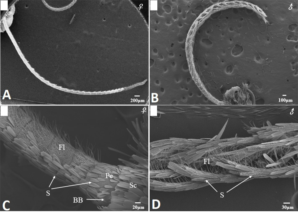
Scanning electron micrographs of the antennae in Scopula subpunctaria. (A) female filiform antenna; (B) male bipectinate antenna; (C) segments of flagellum (Fl), scape (Sc), pedicel (Pe) and Böhm bristles (BB) of female antenna; (D) segments of flagellum of male antenna.
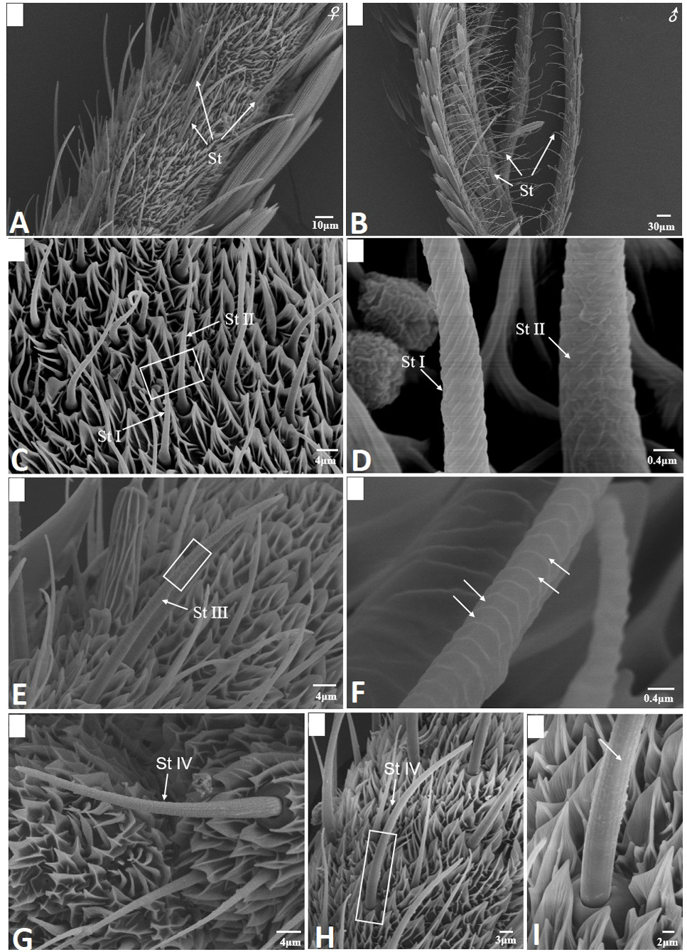
Scanning electron micrographs of sensilla trichodea in Scopula subpunctaria. (A) sensilla trichodea (St) on female antennae; (B) sensilla trichodea on male antennae; (C) St I and St II; (D) partial enlargement of St I and St II, showing helical deep grooves and wrinkle surface, respectively; (E) St III; (F) partial enlargement of St III, showing parallel horizontal deep grooves (arrows); (G-H) St IV; (I) partial enlargement on boxes of figure 2H, showing longitudinal grooves (arrows).
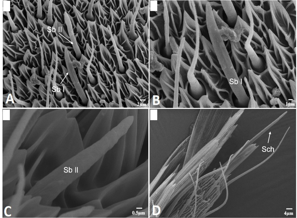
Scanning electron micrographs of sensilla basiconica and sensilla chaetica in Scopula subpunctaria. (A) sensilla basiconica I (Sb I) and sensilla basiconica II (Sb II); (B) sensilla basiconica I (Sb I); (C) sensilla basiconica II (Sb II); (D) sensilla chaetica (Sch).
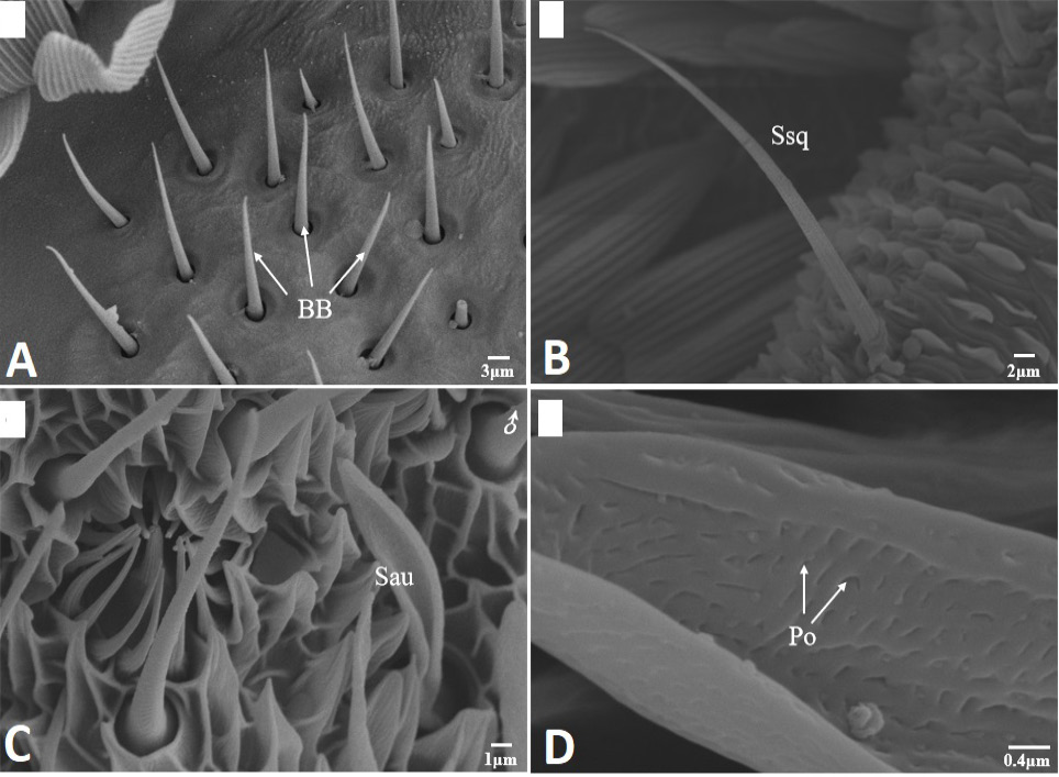
Scanning electron micrographs of Böhm bristles, sensilla squamiformia and sensilla auricillica in Scopula subpunctaria. (A) Böhm bristles (BB); (B) sensilla squamiformia (Ssq); (C) sensilla auricillica (Sau); (D) close-up of sensilla auricillica, showing Pore (Po).
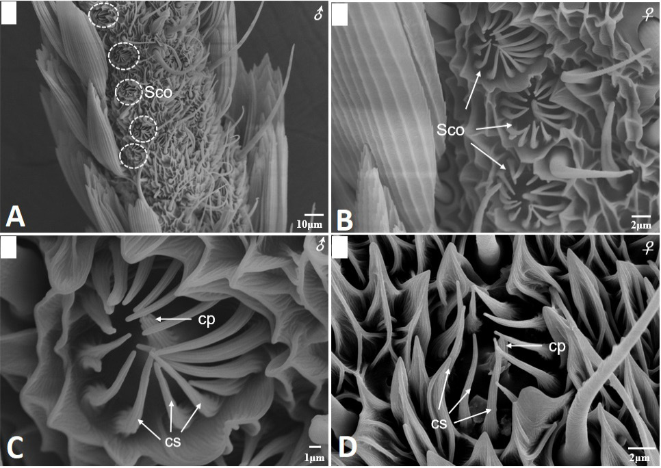
Scanning electron micrographs of sensilla coeloconica in Scopula subpunctaria. (A) sensilla coeloconica on male antennae flagellum; (B) sensilla coeloconica on female antennae flagellum; (C) higher magnification of sensilla coeloconica showing central peg (cp) surrounded by 16 cuticular spines (cs, three white arrows) on male antennae flagellum; (D) higher magnification of sensilla coeloconica showing central peg (cp) surrounded by 10 cuticular spines (cs, three white arrows) on female antennae flagellum.
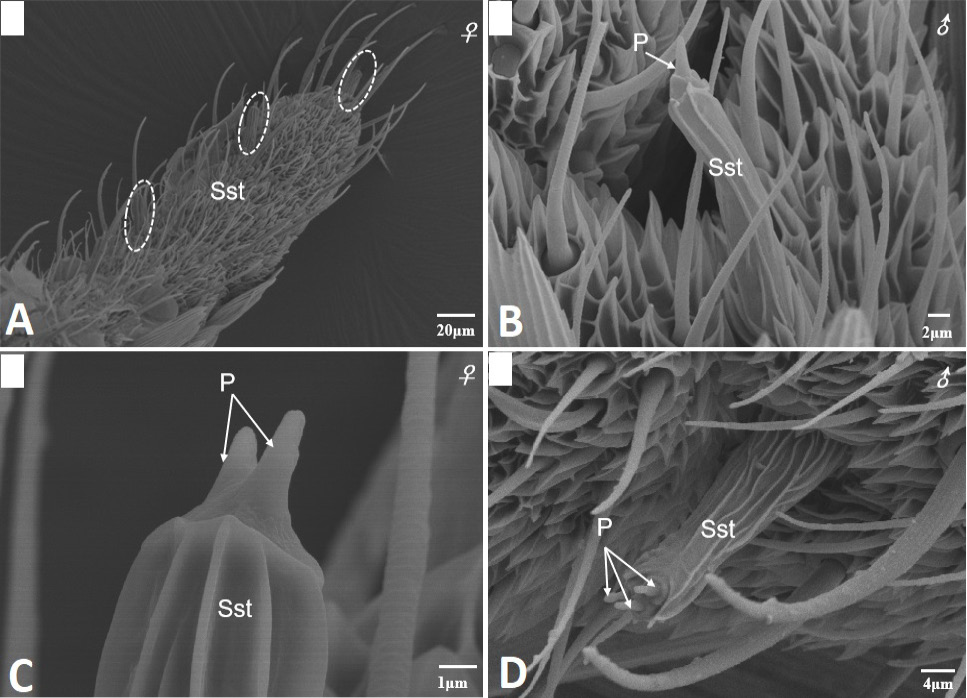
Scanning electron micrographs of sensilla styloconica on the female and male antennae of Scopula subpunctaria. (A) sensilla styloconica located on the ventral surface of the terminal segment of flagellum (in white circle); (B) sensilla styloconica found on male antennae showing single cone-shaped structure at the apex (white arrows); (C) sensilla styloconica found on female antennae showing two cone-shaped structure at the apex (white arrows); (D) sensilla styloconica found on male antennae showing three cone-shaped structure at the apex (white arrows).
References
- Bahia, A.C., Barletta, A.B.F., Pinto, L.C., Orfanó, A.S., Nacif-Pimenta, R., Volfova, V., Petr. V., Secundino N.F.C., Fernandes Freitas.de F., and Pimenta, P.F.P., 2021. Morphological characterization of the antennal sensilla of the afrotropical sand fly, Phlebotomus duboscqi (Diptera: Psychodidae). J. med. Ent., 58: 634-645. https://doi.org/10.1093/jme/tjaa247[CrossRef]
- Bawin, T., Collard, F., Backer De, L., Barthélémy, B., Compère, P., Francis, F. and Verheggen, J., 2017. Structure and distribution of the sensilla on the antennae of Tuta absoluta (Lepidoptera: Gelechiidae). Micron, 96: 16-28. https://doi.org/10.1016/j.micron.2017.01.008[CrossRef]
- Binyameen, M., Anderson, P., Ignell, R., Seada, M.A., Hansson, B.S. and Schlyter, F., 2012. Spatial organization of antennal olfactory sensory neurons in the female Spodoptera littoralis moth: Differences in sensitivity and temporal characteristics. Chem. Senses, 37: 613-629. https://doi.org/10.1093/chemse/bjs043[CrossRef]
- Castrejon-Gomez, V.R., Valdez-Carrasco, J., Cibrian-Tovar, J., Camino-Lavin, M. and Osorio, R.O., 1999. Morphology and distribution of the sense organs on the antennae of Copitarsia consueta (Lepidotera: noctuidae). Fla. Entomol., 82: 546-555. https://doi.org/10.2307/3496472[CrossRef]
- Cuperus, P.L., 1983. Distribution of antennal sense organs in male and female ermine moth, Yponomeuta vigintipunctatus (Retzius) (Lepidoptera: Yponomeutidae). Int. J. Insect Morphol., 12: 59-66. https://doi.org/10.1016/0020-7322(83)90035-1[CrossRef]
- da Silva, K.B., da Silva, C.B., Lisboa Ribeiro Júnior, K.A., de Freitas, J.M.D., de Freitas, J.D., Sanchez Chia, G., Salles Tinoco, R., da Costa, J., Fonseca Goulart, H. and Goulart Santana, A.E., 2019. Morphology and distribution of antennal sensilla of Automeris liberia (Lepidoptera: Saturniidae). Micron, 123: 102682. https://doi.org/10.1016/j.micron.2019.102682[CrossRef]
- Dong, Z.S., Yang, Y.B., Dou, F.G., Zhang, Y.J., Huang, H.X., Zheng, X.L., Wang, X.Y. and Wen, L., 2020. Observations on the ultrastructure of antennal sensilla of adult Glenea cantor (Cerambycidae: Lamiinae). J. Insect Sci., 20: 7. https://doi.org/10.1093/jisesa/ieaa013[CrossRef]
- Dyer, L.J. and Seabrook, W.D., 1978. Evidence for the presence of acceptor sites for different terpenes on one receptor cell in male Monochamus notatus (drury) (Coleoptera: Cerambycidae). J. Chem. Ecol., 4: 523-529. https://doi.org/10.1007/BF00988916[CrossRef]
- Fernandes, F.de F., Barletta, A.B.F., Orfanó, A.S., Pinto, L.C., Nacif-Pimenta, R., Miranda, J.C., Secundino, N.F.C., Bahia, A.C. and Pimenta, P.F.P., 2020. Ultrastructure of the antennae and sensilla of Nyssomyia intermedia (Diptera: Psychodidae), vector of American cutaneous leishmaniasis. J. med. Ent., 57: 1722-1734. https://doi.org/10.1093/jme/tjaa124[CrossRef]
- Geng, S.B., Hou, H.L., Wang, G.J., Jung, C., Yin, J. and Qiao, L., 2021. Temperature-dependent oviposition model of Scopula subpunctaria (Lepidoptera: Geometridae). J. Asia-Pac. Ent., 24: 948-953. https://doi.org/10.1016/j.aspen.2021.08.004[CrossRef]
- Guo, X., Yu, Q., Chen, D., Wei, J., Yang, P., Yu, J., Wang, X. and Kang, L., 2020. 4-Vinylanisole is an aggregation pheromone in locusts. Nature, 584: 584-588. https://doi.org/10.1038/s41586-020-2610-4[CrossRef]
- Hallberg, E., Hansson, B.S., and Steinbrecht, R.A., 1994. Morphological characteristics of antennal sensilla in the European cornborer Ostrinia nubilalis (Lepidoptera: Pyralidae). Tissue Cell, 26: 489-502. https://doi.org/10.1016/0040-8166(94)90002-7[CrossRef]
- Jeong, S.A., Kim, J., Byun, B.K., Oh, H.W. and Park, K.C., 2020. Morphological and ultrastructural characterization of olfactory sensilla in Drosophila suzukii: scanning and transmission electron microscopy. J. Asia-Pac. Ent., 23: 1165-1180. https://doi.org/10.1016/j.aspen.2020.06.009[CrossRef]
- Jiang, N.J., Tang, R., Guo, H., Ning, C., Li, J.C., Wu, H. and Wang, C.Z., 2020. Olfactory coding of intra- and interspecific pheromonal messages by the male Mythimna separata in North China. Insect Biochem. mol. Biol. 125: 103439. https://doi.org/10.1016/j.ibmb.2020.103439[CrossRef]
- Jiang, X.J., Ning, C., Guo, H., Jia, Y.Y., Huang, L.Q., Qu, M.J. and Wang, C.Z., 2015. A gustatory receptor tuned to D-fructose in antennal sensilla chaetica of Helicoverpa armigera. Insect Biochem. mol. Biol., 60: 39-46. https://doi.org/10.1016/j.ibmb.2015.03.002[CrossRef]
- Kelling, F.J., Biancaniello, G. and den Otter, C.J., 2002. Electrophysiological characterization of olfactory cell types in the antennae and palps of the housefly. J. Insect Physiol., 48: 997-1008. https://doi.org/10.1016/S0022-1910(02)00187-7[CrossRef]
- Koh, Y.H., Park, K.C. and Boo, K.S., 1995. Antennal sensilla in adult, Helicoverpa assulta (Lepidoptera: Noctuidae). Annls entomol. Soc. Am., 88: 519-530. https://doi.org/10.1093/aesa/88.4.519[CrossRef]
- Krishnan, A., Prabhakar, S., Sudarsan, S. and Sane, S.P., 2012. The neural mechanisms of antennal positioning in flying moths. J. exp. Biol., 215: 3096-3105. https://doi.org/10.1242/jeb.071704[CrossRef]
- Lan, L.J., Wang, S.K., Hu, K.Y., Ma, T. and Wen, X.J., 2020. Ultrastructure of antennal morphology and sensilla of teak skeletonizer, Eutectona machaeralis Walker (Lepidoptera: Crambidae). Microsc. Microanal., 26: 1274-1282. https://doi.org/10.1017/S1431927620024599[CrossRef]
- Li, Y., Liu, F., Du, X., Li, Z. and Wu, J., 2018. Ultrastructure of antennal sensilla of three fruit borers (Lepidoptera: Crambidae or Tortricidae). PLoS One, 13: e0205604. https://doi.org/10.1371/journal.pone.0205604[CrossRef]
- Liu, F., Li, F., Zhang, S., Kong, X. and Zhang, Z., 2019. Ultrastructure of antennal sensilla of Erannis ankeraria Staudinger (Lepidoptera: Geometridae). Micros. Res. Tech., 82: 1903-1910. https://doi.org/10.1002/jemt.23358[CrossRef]
- Liu, S., Rao, X., Li, M., Feng, M., He, M. and Li, S., 2015. Identification of candidate chemosensory genes in the antennal transcriptome of Tenebrio molitor (Coleoptera: Tenebrionidae). Comp. Biochem. Physiol. Part D Genom. Proteom., 13: 44-51. https://doi.org/10.1016/j.cbd.2015.01.004[CrossRef]
- Ma, L., Bian, L., Li, Z.Q., Cai, X.M., Luo, Z.X. and Chen, Z.M., 2016. Ultrastructure of chemosensilla on antennae and tarsi of Ectropis obliqua (Lepidoptera: Geometridae). Annls entomol. Soc. Am., 109: 574-584. https://doi.org/10.1093/aesa/saw027[CrossRef]
- Ma, L.Y., Hu, K., Li, P.D., Liu, J.Q. and Yuan, X.Q., 2019. Ultrastructure of the proboscis sensilla of ten species of butterflies (Insecta: Lepidoptera). PLoS One, 14: e0214658. https://doi.org/10.1371/journal.pone.0214658[CrossRef]
- Ndomo-Moualeu, A.F., Ulrichs, C., Radek, R. and Adler, C., 2014. Structure and distribution of antennal sensilla in the Indianmeal moth, Plodia interpunctella (Hübner, 1813) (Lepidoptera: Pyralidae). J. Stored Prod. Res., 59: 66-75. https://doi.org/10.1016/j.jspr.2014.05.003[CrossRef]
- Nemeth, D.C., Ammagarahalli, B., Layne, J.E. and Rollmann, S.M., 2018. Evolution of coeloconic sensilla in the peripheral olfactory system of Drosophila mojavensis. J. Insect Physiol., 110: 13-22. https://doi.org/10.1016/j.jinsphys.2018.08.003[CrossRef]
- Park, K.C., Jeong, S.A., Kwon, G. and Oh, H.W., 2018. Olfactory attraction mediated by the maxillary palps in the striped fruit fly, Bactrocera scutellata: Electrophysiological and behavioral study. Arch. Insect Biochem. Physiol., 99: e21510. https://doi.org/10.1002/arch.21510[CrossRef]
- Qian, J.L., Luo, Z.X., Li, J.L., Cai, X.M., Bian, L., Xiu, C.L., Li, Z.Q., Chen, Z.M. and Zhang, L.W., 2020. Identification of cytochrome P450, odorant-binding protein, and chemosensory protein genes involved in Type II sex pheromone biosynthesis and transportation in the tea pest, Scopula subpunctaria. Pestic. Biochem. Physiol., 169: 104650. https://doi.org/10.1016/j.pestbp.2020.104650[CrossRef]
- Rani, A.T., Shashank, P.R., Meshram, N.M., Sagar, D., Srivastava, C., Pandey, K.K. and Singh, J., 2021. Morphological characterization of antennal sensilla of Earias vittella (Fabricius) (Lepidoptera: Nolidae). Micron, 140: 102957. https://doi.org/10.1016/j.micron.2020.102957[CrossRef]
- Roh, G.H., Park, K.C., Oh. H.W. and Park C.G., 2018. Species- and sex-specific distribution of antennal olfactory sensilla in two tortricid moths, Epiphyas postvittana and Planotortrix octo. Micron, 106: 7-20. https://doi.org/10.1016/j.micron.2017.12.006[CrossRef]
- Ruschioni, S., Riolo, P., Verdolini, E., Peri, E., Guarino, S. and Colazza, S., 2015. Fine structure of antennal sensilla of Paysandisia archon and electrophysiological responses to volatile compounds associated with host palms. PLoS One, 10: e0124607. https://doi.org/10.1371/journal.pone.0124607[CrossRef]
- Schneider, D., 1964. Insect antennae. Annu. Rev. Ent., 9: 103-122. https://doi.org/10.1146/annurev.en.09.010164.000535[CrossRef]
- Schneider, E. S., and Römer, H., 2016. Sensory structures on the antennal flagella of two katydid species of the genus Mecopoda (Orthoptera, Tettigonidae). Micron, 90: 43-58. https://doi.org/10.1016/j.micron.2016.08.001[CrossRef]
- Seada, M.A., 2015. Antennal morphology and sensillum distribution of female cotton leaf worm Spodoptera littoralis (Lepidoptera: Noctuidae). J. Basic appl. Zool., 68: 10-18. https://doi.org/10.1016/j.jobaz.2015.01.005[CrossRef]
- Silva, C.C.A., de Capdeville, G., Moraes, M.C.B., Falcão, R., Solino, L.F., Laumann, R.A., Silva, J.P. and Borges, M., 2010. Morphology distribution and abundance of antennal sensilla in three stink bug species (Hemiptera: Pentatomidae). Micron, 41: 289-300. https://doi.org/10.1016/j.micron.2009.11.009[CrossRef]
- Steinbrecht, R.A., Laue, M., Maida, R. and Ziegelberger, G., 1996. Odorant-binding proteins and their role in the detection of plant odours. Ent. exp. appl., 80: 15-18. https://doi.org/10.1111/j.1570-7458.1996.tb00875.x[CrossRef]
- Symonds, M.R.E., Johnson, T.L. and Elgar, M.A., 2011. Pheromone production, male abundance, body size, and the evolution of elaborate antennae in moths. Ecol. Evol., 2: 227-246. https://doi.org/10.1002/ece3.81[CrossRef]
- Wang, Q., Shang, Y., Hilton, D.S., Inthavong, K., Zhang, D., Elgar, M.A., Inthavong, K., Zhang, D. and Ma, E., 2018. Antennal scales improve signal detection efficiency in moths. Proc. biol. Sci., 285: 20172832. https://doi.org/10.1098/rspb.2017.2832[CrossRef]
- Xu, J., Deng, C., Lu, W. and Wu, S., 2021. Ultrastructure of antennal sensilla in adults of Dioryctria rubella Hampson (Lepidoptera: Pyralidae). Insects, 12: 821. https://doi.org/10.3390/insects12090821[CrossRef]
- Yan, X., Deng, C., Xie, J., Wu, L., Sun, X. and Hao, C., 2017. Distribution patterns and morphology of sensilla on the antennae of Plutella xylostella (L.)-a scanning and transmission electron microscopic study. Micron, 103: 1-11. https://doi.org/10.1016/j.micron.2017.08.002[CrossRef]
- Yang, S., Liu, H., Zhang, J.T., Liu, J., Zheng, H. and Ren, Y., 2017. Scanning electron microscopy study of the antennal sensilla of Monema flavescens Walker (Lepidoptera: Limacodidae). Neotrop. Ent., 46: 175-181. https://doi.org/10.1007/s13744-016-0450-6[CrossRef]
- Yang, T.B., Song, X.H., Zhong, Y., Wang, B. and Zhou, C.Q., 2022. Field investigation and dietary metabarcoding-based screening of arthropods that prey on primary tea pests. Ecol. Evol., 12: e9060. https://doi.org/10.1002/ece3.9060[CrossRef]
- Yuan, H.B., Zhao, X.Y., Ge, S.Y., Huang, X., Liu, J.H. and Wang, Y., 2017. A new type of Mythimna Separata (Lepidoptera: Noctuidae) antennal sensilla. Ent. News, 127: 11-20. https://doi.org/10.3157/021.127.0103[CrossRef]
- Zacharuk, R.Y., 1980. Ultrastructure and function of insect chemosensilla. Annu. Rev. Ent., 25: 27-47. https://doi.org/10.1146/annurev.en.25.010180.000331[CrossRef]
- Zacharuk, R.Y., 1985. Antennae and sensilla. In: Comprehensive insect physiology, biochemistry and pharmacology (eds. G.A. Kerkut and L.I. Gilbert). Pergamon Press, Oxford.
- Zhang, J., Liu, C.C., Yan, S.W., Liu, Y., Guo, M.B., Dong, S.L. and Wang, G.R., 2013. An odorant receptor from the common cutworm (Spodoptera litura) exclusively tuned to the important plant volatile cis-3-Hexenyl acetate. Insect mol. Biol., 22: 424-432. https://doi.org/10.1111/imb.12033[CrossRef]
- Zhang, Y.J., Chen, D.Y., Chao, X.T., Dong, Z.S., Huang, Z.Y., Zheng, X.L. and Lu, W., 2019. Morphological characterization and distribution of antennal sensilla of Diaphania angustalis Snellen (Lepidoptera: Crambidae). Microsc. Res. Tech., 82: 1632-1641. https://doi.org/10.1002/jemt.23329[CrossRef]
- Zhang, Y.L., Fu, X.B., Cui, H.C., Zhao, L., Yu, J.Z. and Li, H.L., 2018. Functional characteristics, electrophysiological and antennal immunolocalization of general odorant-binding protein 2 in tea geometrid, Ectropis obliqua. Int. J. mol. Sci., 19: 875. https://doi.org/10.3390/ijms19030875[CrossRef]
- Zhao, H., Liang, C., Gao, P., Xie, Y., Wang, Z., Wu, G.X., Tang, G.W., Cheng, B. and Gao, X., 2021. Observation of the fine structure of antennal sensilla of the stink bug, Eocanthecona furcellata (Hemiptera: Pentatomidae). Micron, 150: 103143. https://doi.org/10.1016/j.micron.2021.103143[CrossRef]
- Zhou, H., Wu, W.J., Niu, L.M. and Fu, Y.G., 2013. Antennal sensilla of female Encarsia guadeloupae Viggiani (Hymenoptera: Aphelinidae), a nymphal parasitoid of the spiraling whitefly Aleurodicus dispersus (Hemiptera: Aleyrodidae). Micron, 44: 365-372. https://doi.org/10.1016/j.micron.2012.09.001[CrossRef]
- Zhu, X., Liu, S., Xu, C., Guo, K., Xu, R., Qiao, H. and Chen, J., 2021. Morphology and distribution of sensilla on the antennae and ovipositor of the hymenopteran parasitoid Pseudotorymus jaapiellae Yang et Chen. Microsc. Res. Tech., 84: 1873-1886. https://doi.org/10.1002/jemt.23744[CrossRef]
Services
Article usage statistics
Loading…