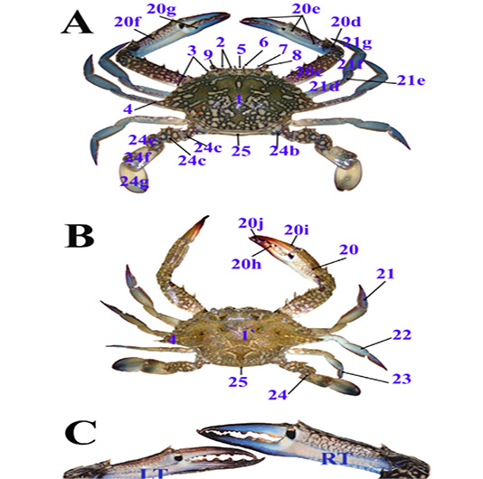
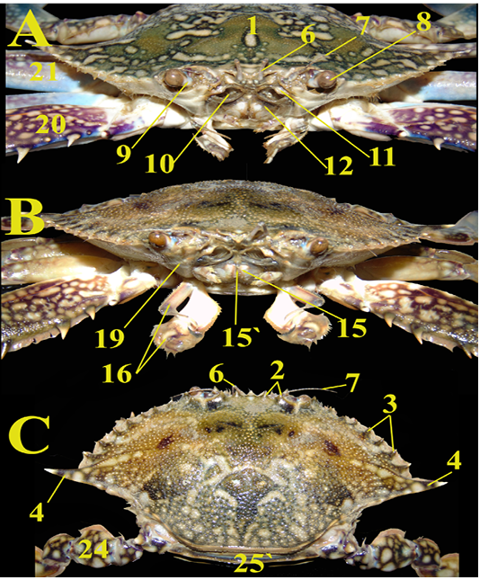
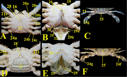
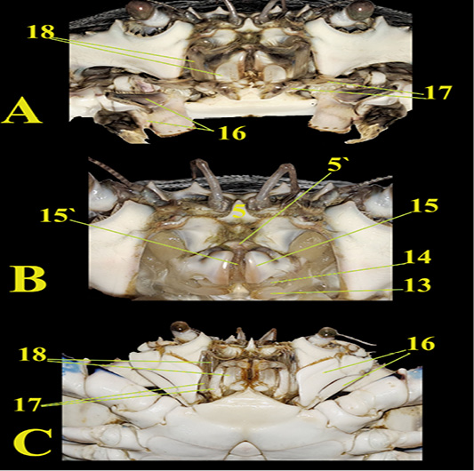
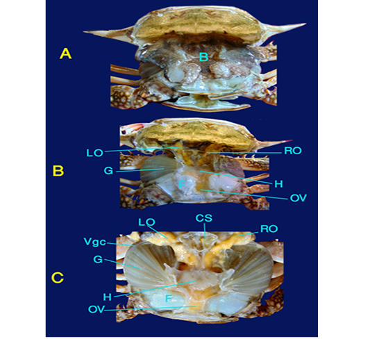
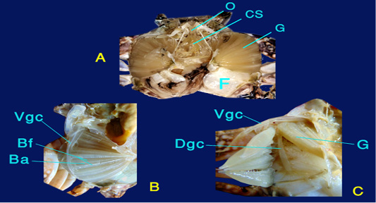
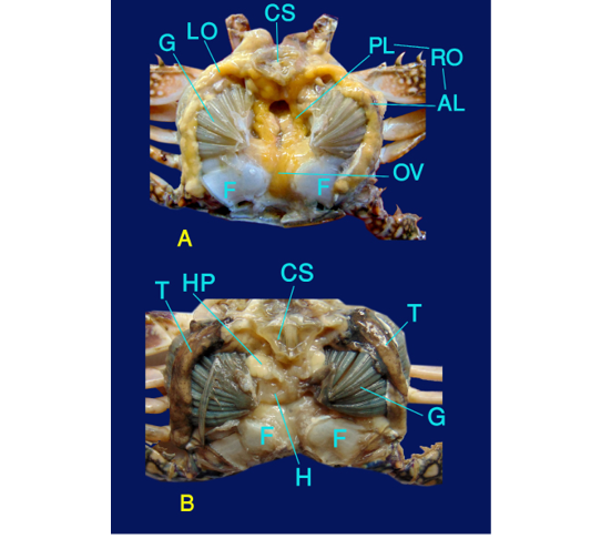
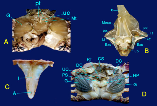
1, Male carapace; 1`, Female carapace; 2, Front teeth; 3, Anterolateral teeth 4; Lateral spine (ninth tooth); 5, Rostrum; 5`, Upper lip; 6, Antennules; 7, Antennae; 8, Compound eyes; 9, Eye stalk; 10, Inhalant aperture; 11, Operculum; 12, Exhalant aperture; 13, Second maxilla; 14, First maxilla; 15, Mandible; 15`, Mouth; 16, Third maxilliped; 17, Second maxilliped; 18, First maxilliped; 19, Cephalothorax; 20, Cheliped (first pereiopod); 20a, Coxa of first pereiopod; 20b, fused basis ischium; 20c, merus; 20d, Carpus; 20e, Claw; 20f, Palm; 20g, Pollex; 20h, Cutting conical teeth; 20i, Dactylus; 20j, Fingertip; 21, Second pereiopod (walking leg); 21a, Coxa, 21b, Basis; 21c, Ischium; 21d, merus; 21e, Carpus; 21f, Propodus; 21g, Dactylus; 22, Third pereiopod (walking leg); 23, Fourth pereiopod (walking leg); 24, Fifth pereiopod (natatory or swimming leg); 24a, Coxa; 24b, Basis; 24c, Ischium; 24d, merus; 24e, Carpus; 24f, Paddle like propodus; 24g, Paddle like dactylus; 25, Male abdomen (pleon); 25`, Female abdomen (pleon); 26, Thoracic sternum (pereion); 27, Abdominal segments; 28, Male telson; 28`, Female telson; 29a, First gonopod; 29b, Second gonopod; 30, Male gonopore; 30`, Female gonopore (vulva); 31, Penis; 32, Intestine; 33, Anus
Legs (pereiopods) of male blue swimmer crab are mixed blue and purple in coloration with off-white patches, in agreement with the description of Abbas et al. (2016). Conversely, Lai et al. (2010) described that male’s chelipeds had rust colored red tips. The female legs are dark brown with off-white patches, while Lai et al. (2010) described that the females had a uniformly brownish-green carapace with red tipped chelipeds.
The cheliped is the strongest and largest one, and functions as a movable finger and carries on its end a chela or claw (pincer), in agreement with Meyer (2014) that in many decapods the first peraeopods had enlarged pincers (chelae) called chelipeds (Brachyura). It is composed of six parts; the coxa (Fig. 2A/20a) proximally which articulates with the body, the fused basis ischium (Fig. 3A/20b), the merus (Fig. 3A/20c) with three spines on the inner margin while no spines are found on the outer margin, the carpus (Fig. 1A/20d) which has an inner carpal spine or tooth, the claw (Fig. 1A/20e) which has a palm, pollex, and cutting conical teeth (Fig. 1A,B/20f,20g,20h), and the dactylus (Fig. 1B/20i) as the fingertip (Fig. 1B/20j), in accordance with Abbas et al. (2016) and Lai et al. (2010). The right claw is slightly larger than the left claw (Fig. 1C).
Walking legs (second, third, and fourth pereiopods) (Fig. 1B/21,22,23) are flattened laterally and are composed of the coxa, basis, ischium, merus, carpus, propodus, and dactylus. The swimming leg (fifth pereiopod) (Fig. 1B/24) has a flat, paddle like dactyl and propodus, in the contrary, Lai et al. (2010) stated that in P. pelagicus, the dactylus of the fifth pereiopod was oval and relatively elongated. Moreover, Tavares (2003) mentioned that in swimming crabs, the last pair of legs was usually distinctly adapted for swimming, and was flattened and paddle-shaped, but in a few species it was flattened without being paddle-shaped.
The abdomen (Pleon) (Fig. 3) is present on the caudoventral surface of the crab body, and has six abdominal segments, the telson (terminal end of the abdomen), and pleopods, similarly to the observations of Tavares (2003), who observed that true crabs had a reduced, straight, and symmetrical abdomen bent under the cephalothorax, this abdomen lacked biramous uropods.
The abdomen of the male (Fig. 3A/27) is narrow rostrally with a small telson (Fig. 3A/28), and has a broad base, while the mature female abdomen is a broad triangle with convex sides. Additionally, the female telson (Fig. 3D/28`) has a small broad base. In males, the first and second segments of the abdomen are situated caudally after the end of the carapace, the third, fourth, and fifth segments are fused together, and the sixth segment is free and related to the terminal telson. In contrast, Lai et al. (2010) revealed that in P. pelagicus the sixth segment of the male abdomen was relatively short and broad. On the other hand, Meyer (2014) detected that the seven segmented pleon contained appendages; with each segment carrying a pair of biramous pleopods. The first pair of pleopods is the gonopods; while the last pleopods, together with the telson, form the tail fan forming uropods.
In mature females there are six separated abdominal segments (Fig. 3D/27) on the ventral surface of the abdomen; the intestine (Fig. 3B/32) is a small transparent tube that ends at the anus (Fig. 3E/33) located ventrally at the telson. The female vulva or gonopore (Fig. 3E/33`) is a small opening situated along the midline groove at the level of the sixth thoracic segment after reflection of the abdomen. In males, the penis (Fig. 3B/31) carries the male gonopore which is the male genital opening on the penis tip and is located near the fifth leg coxa cranial edge. The penis is a short, thin, transparent organ with a blunt end.
Abdominal appendages (pleopods) (Fig. 3B/29a, b) consist of two pleopods (gonopods) in the male, in accordance with Hidayani et al. (2018). The first and second appear on the ventral surface of the abdomen. The first gonopod (Fig. 3B/29a) is curved, tubular, and is the longer and more cranial one. It is the intromittent organ, While Lai et al. (2010) explored that P. pelagicus first gonopod was distinct and angled. Previously, Hidayani et al. (2018) illustrated that in Portunus armatus the basal part of first gonopod was rounded. The second gonopod (Fig. 3B/29b) is smaller and caudal to the first one. The female has several paired biramous pleopods (Fig. 3E/29`) situated on each of the abdominal segments except the last one.