Based on the results of the statistical analysis of the average spermatogonia cells, there were a significant difference between groups (C-) and a group (C+) (p <0.05). The administration of Kebar grass extract in groups T1, T2 and T3 had not had a significant effect (p>0.05). This research showed that the administration of Kebar grass extract could’n improve/increase the number of spermatogonia cells, although there was a tendency to increase the number of spermatogonia with the increase dose of Kebar grass extract administration (Table 1). The average primary spermatocyte cells were a significant difference between groups (C-) and a group (C+) (p <0.05). The administration of Kebar grass extract in the T1 and T2 groups had not had a significant effect on the average of primary spermatocyte cells. The Kebar grass extract could have a significant effect on the T3 group. This research showed that the administration of Kebar grass extract could increase the number of primary spermatocyte cells especially in administering Kebar grass extract at doses 0.135 mg/g BW (T3) (Table 1). The results of a similar statistical analysis were found in spermatid cell counts. The administration of Kebar grass extract had not had a significant effect on the T1 and T2 groups and only has a significant effect on the T3 group on average spermatid cells. This research showed that the administration of Kebar grass extract could increase the number of spermatid cells especially in administering Kebar grass extract at doses 0.135 mg/g BW (T3) (Table 1).
Table 1: Average of spermatogonia cells, primary spermatocytes and spermatids of male mice given TCDD exposed grass extracts.
Superscripts with different letters show significant differences (p<0.05). C (-): Group with aquades and CMCNa 0.5%, C (+): TCDD 7µg/Kg BB, T1: TCDD 7µg/Kg BW + Kebar Grass extract 0.045 mg/g BW/day in a 0.5% CMC-Na solution of 0.1 ml, T2: TCDD 7µg/Kg BW + Kebar Grass extract 0.08 mg/g BW / day in a 0.5% CMC-Na solution of 0.1 ml, T3: TCDD 7µg/Kg BW + Kebar Grass extract 0.135 mg/g BW/day in a 0.1% CMC-Na solution of 0.1 ml
Spermatogenic cells injury (spermatogonia, primary spermatocytes and spermatid cells) as a primary morphologic event is a common manifestation following administration of cytotoxic compounds. Spermatogenic cells not protected by the blood-testis barrier (BTB), are the most vulnerable to toxic effects (Meistrich, 1986). Effects of toxic compounds on the testis may be reversible following cessation of compound exposure through seminiferous epithelial reconstitution (Roeser et al., 1978).
Most of the toxicity of TCDD via the Aryl hydrocarbon Receptor (AhR) receptor, after binding to the AhR, the bonding complex changes and is translocated to the nucleus. Furthermore, it binds to the transcription factor, which is the Ribonucleic Acid Transferase (Arnt) in the nucleus and is involved in translating so that it encodes the cytochrome P450 gene and increases the production of the cytochrome P450 enzyme which results in cell damage (Dobrzyński et al., 2009). Damage to spermatogenic cells results in cells failing to divide, so the number of spermatogenic cells is reduced compared to normal (Gray et al., 1997). TCDD produces high free radicals, which can damage cells through lipid peroxidase. Testicular cell membranes are formed from lipids that are susceptible to free radicals (Alsharif et al., 1990). ROS reaction to unsaturated lipids in the cell membrane causes the formation of lipid peroxide, causing damage to body cells (Yin et al., 2012).
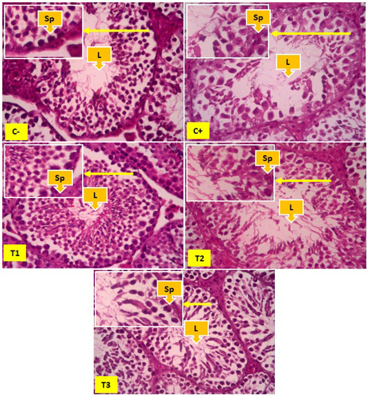
Figure 1: Overview of spermatogonia cells in seminiferous tubules, yellow arrows show spermatogonia cells (Sp), and Lumen (L). In Figure 1 it appears that the number of spermatonia was more common in the negative control group (C-) than the positive control group (C+). The number of spermatogonia appears to be increasing, although not significantly found in the T1, T2 and T3 groups.
The presence of lipid peroxidase in cell membranes due to TCDD free radicals resulted in a decrease in the number of spermatogonia, primary spermatocytes and spermatid cells in the control group exposed to TCDD (C+) compared to the control negative (C-). The administration of Kebar grass extract in groups T1, T2 and T3 had not had a significant effect of the average spermatogonia cells (p>0.05). The administration of Kebar grass extract in the T1 and T2 groups had not had a significant effect on the average of primary spermatocyte and spermatid cell counts. Only the Kebar grass extract could have a significant effect on the T3 group (primary spermatocyte and spermatid cell counts) (Table 1).
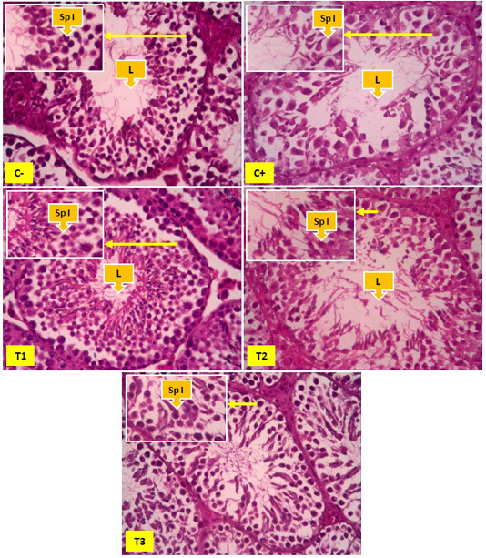
Figure 2: Overview of spermatocyte cells in the seminiferous tubules, yellow arrows indicate primary spermatocyte cells (Sp I) and Lumen (L). In Figure 2 it appears that the number of primary spermatocyte cells was more common in the negative control group (C-) than the positive control group (C+), T1 and T2. The number of primary spermatocyte cells appears to be increasing significantly found in the T3 groups.
Kebar grass extract as a therapy from TCDD exposure contains antioxidants that are used against the effects of TCDD. Kebar grass contains flavonoids and vitamin E which are high enough to be used to ward off ROS from TCDD. The content of Kebar Grass which is high in vitamin E and flavonoids is proven to be able to maintain the spermatogenic number of mice exposed to TCDD according to Leefan (2014) and Latchoumycandane and Marthur (2002). Kebar grass also contains high flavonoids that are useful as antioxidants in TCDD exposure because they have hydroxyl groups that can donate hydrogen atoms to free radical compounds and stabilize ROS (Rezaeizadeh et al., 2011).
Vitamin E were fat soluble so that it easily enters through cell membranes and protects Poly Unsaturated Fatty Acid (PUFA) so that it can protect cells against free radicals by breaking free radicals so that they cannot damage cells (Wati et al., 2014). Vitamin E is also able to convert peroxyl radicals resulting from lipid peroxidase into tocopherol radicals that are less reactive, so that cells are not damaged (Hariyatmi, 2004). Kebar grass contains Calcium which is sufficiently able to repair the influx of Ca and K ions in the testes whose permeability has been damaged by TCDD, thus maintaining spermatogenic cells to remain normal and avoid cell damage (Whitaker, 2006).
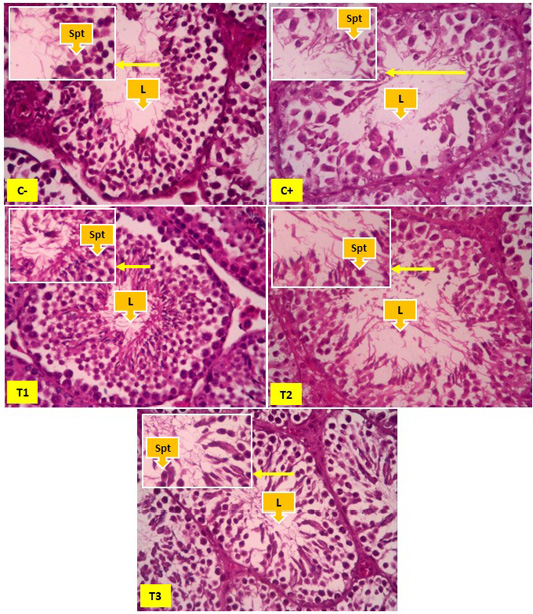
Figure 3: Spermatid cells in seminiferous tubules, yellow arrows showing spermatid cells (Spt) and Lumen (L). In Figure 2 it appears that the number of spermatid cells was more common in the negative control group (C-) than the positive control group (C+), T1 and T2. The number of spermatid cells appears to be increasing significantly found in the T3 groups.
The administration of Kebar grass extract had not had a significant effect on the average of spermatogonia (All treatment), cells primary spermatocyte and spermatid cell counts (T1 and T2), but could have a significant effect on the T3 group (Table 1). The treatment group with 0.135 mg/g BW/day (T1) Kebar Grass dose showed the best dose because it was not significantly different from the control group without TCDD exposure (C-). A similar study was carried out by Rusyawardani (2020) which stated that giving Kebar grass a dose of 0.135 mg/g BW/day gave the best effect in increasing the diameter of seminiferous tubules and the thickness of seminiferous epithelium exposed to TCDD. This might be because the dose of Kebar Grass extract can increase the hormone testosterone, according to Bearden and John (1980) that the number of spermatogenic cells is influenced by the number of cell division that depends on testosterone hormone as a trigger.

