
The isolations were due to the roots of the affected tomato plants that demonstrated the presence of F. oxysporum f. sp. lycopersici in all the isolates taken in different locations in Babil Governorate in culture in PDA medium. During the pathogenicity test of the fungal isolates on water agar (W.A) medium using radish seeds, the data depicted that all the fungal isolates that were pathogenic led to a high degree of reduction in the percentage of germination. The isolate of F. oxysporum f. sp. lycopersici in Babil Governorate (F1) of the seed, with the greatest seed lethality of 0.00 percent, was compared to the control treatment (seeds only, the absence of the pathogenic fungus), which had a germination percentage of 100 percent. The antagonistic activity test data show that Bacillus paramycoides, Lysinibacillus sphaericus and Pseudomonas fluorescens had a high efficacy level of 100 percent at the concentration of 5 x 10 8 CFU/mL, 8.5 x 10 8 CFU/mL, and 36 x 10 8 CFU/mL respectively since these bacterial isolates were able to fully inhibit the pathogenic growth of the fungus with a fungal This was followed by the fungi isolates Trichoderma harzianum and T. virens which showed the highest level of antagonistic activity as the percentages of inhibition were 96.6 and 96.5 respectively. The humic acid treatment showed the least antagonistic effect and an inhibition percentage of 18.1 and a fungal radial growth value of 7.3 cm with respect to other biocontrol agents. All of these findings suggest that the application of the biological control agents, especially bacteria secreting the bioactive compounds, and the Trichoderma spp. fungi is the effective and sustainable approach to disease management programs to decrease the severity of the disease, decrease the losses in production, and decrease the usage of the humic acid in the soils with the fungal pathogens.
Research Article · Volume 39, Issue 3, 7 September 2026 · Open Access
Testing the Antagonistic Ability of Some Biocontrol Elements and Humic Acid Against the Fungus Fusarium oxysporum f.sp. lycopersici, the Causal Agent of Tomato Wilt, In Vitro
·
Affiliations & Notes ⌄
- 1Biological Control Department, Al-Mussaib Technical College, Al-Furat Al-Awsat Technical University, 51006 Al-Mussaib, Babylon, Iraq
- 22Plant protection department, College of Agriculture ,University of basrah,61004 Basrah,Iraq. *Correspondence | Kadhim Z.K. Al-Karaawi, Biological Control Department, Al-Mussaib Technical College, Al-Furat Al-Awsat Technical University, 51006 Al-Mussaib, Babylon, Iraq
- 3Email: info.golden7farm@gmail.com
Article Info ⌄
- Journal:
- Pakistan Journal of Agricultural Research
- Published:
- 7 September 2026
From the article
- The pathogenic fungus isolates showed clear variation in pathogenicity, reflected in their differing effects on radish seed germination rates, indicating genetic or physiological differences among the isolates.
- The bacterial isolates of B. paramycoides, L. sphaericus, and P. fluorescens, along with the fungi T. harzianum and T. virens, demonstrated high antagonistic activity against the fungus F. oxysporum f.
- Humic acid, when used alone, showed a limited inhibitory effect on the growth of the pathogenic fungus compared to biological control agents, indicating that its role is secondary when used without combination with other agents.
Selected from the article's own text.
Abstract
Introduction
Tomato (Lycopersicon esculentum Mill.) is a plant of the Solanaceae family and it is ranked as one of the most significant vegetables because it is a high nutritive food. It is advisable to take it on a daily basis and in sufficient amounts due to the fact that it helps in sustaining a number of important functions. The Tomato is the one of the most effectively grown crops under greenhouse conditions, which is one of the main sources of food consumed by people every day because of its significance in nutrition. It has large quantities of carbohydrates, proteins and fats as well as mineral elements that include phosphorus, calcium and potassium. It is also an antioxidant pigment that includes lycopene, which is a protective factor against cancer . F. oxysporum Schl. f. sp. lycopersici is the causal agent of the Fusarium wilt that is among the most significant tomato diseases. This pathogen attacks the system of roots and results in discolouration of vascular tissue, yellowing of the leaves, wilting, and plant death which reduces the yield by about 30 to 40 per cent . Fusarium spp. have been regarded as significant facultative parasitic soil fungi, which exist saprophytically in the soil environment and live parasitically in the plants (when present), where they tend to prefer living tissues over plant residues . The Fusarium wilt is one of the determinants of tomato productions that has brought so much losses particularly when the soil and air temperatures are high in a warm month. It is a soil-borne fungus infecting roots and is difficult to control chemically, particularly when cultivating sensitive varieties. Factors that favor disease development include relatively high soil temperatures, low soil moisture, short day lengths, increased nitrogen and phosphorus fertilization, low potassium fertilization, and low soil pH; however, the optimal temperature for fungal growth is 28°C (Ojha and Chatterjee. 2012).In recent years, excessive and uncontrolled use of chemical pesticides has led to several problems, including the emergence of pest resistance, effects on non-target organisms, environmental pollution, and direct toxicity. This has motivated scientists in the field of managing plant diseases to minimize the application of chemicals and find safe alternatives . With the growing worldwide demand for environmentally accessible substitutes, the interest in utilizing a variety of strains of bacteria and fungi as biological control agents against numerous soil-borne pathogens, such as those causing root and seed rot and damping-off of seedlings, have been on the rise . Plant growth-promoting rhizobacteria are among the biological agents that are employed in the management of pathogenic fungi . Besides, T. virens when introduced into agricultural systems can be used to improve the productivity of crops, and therefore, is a sustainable substitute to the traditional use of chemical pesticides. Recent research has also suggested the application for humic acid, a non-toxic compound to soil organisms, as a way of promoting soil fertility as well as promoting the chemical properties of the soil.Humic acid is also important in biological control and plant growth promotion since it can attack pathogenic fungi and produce antifungal compounds, promote nutrient uptake in plants, and induced resistance in plants . The humic acid binds to ions, making nutrients become available to be absorbed by plants. Since Fusarium wilt is a serious disease affecting tomato and more detailed information is required to manage the disease and minimize its economic consequences, the research problem of this study was to examine the antagonistic effects of some biological control agents against F. oxysporum f. sp. lycopersici, which is the cause of tomato Fusarium wilt, on the bacteria Lysinibacillus sphaericus, Bacillus paramycodes, Pseudomonas fluorescens, the fungi T. virens and T. harzianum, and humic.
Materials and Methods
Isolation and identification of the pathogenic fungus fusarium spp.
The fungus Fusarium spp. was isolated and identified from wilted tomato plants collected from greenhouses in Babil Governorate. Seedlings were washed under running tap water for several hours, then rinsed multiple times with sterile distilled water and placed on sterile filter paper to remove excess moisture. Several pieces of roots from the infected plants and from the crown region, each measuring 1–2 cm in length, were taken using a sterile knife and forceps. These pieces were planted, with five pieces per Petri dish, and the inoculated dishes were incubated at 25 ± 2°C.After 2–3 days, the dishes were examined, and the fungal colonies growing from the infected tissues were purified. The fungus Fusarium spp. was identified based on standard taxonomic keys according to Booth (1977). The purified and identified isolates were stored in the refrigerator on slanted PDA medium in test tubes for subsequent studies.
Pathogenicity test of fusarium spp. isolates using radish seeds on water agar (W.A.) medium
The pathogenicity of Fusarium spp. isolates was tested according to the method of Bolkan and Butler (1974). Petri dishes (9 cm in diameter) containing 15–20 mL of water agar medium (W.A.) (20 g agar per 1 L distilled water) was prepared and sterilized using an autoclave for 20 minutes. Tetracycline antibiotic was added at a concentration of 250 mg/L. After solidification of the medium, the dishes were inoculated at the center with a 0.5 cm diameter disc taken from the edge of a 3-day-old fungal colony. The dishes were then incubated at 23 ± 2°C. After 3 days of fungal growth on the water agar medium, locally sourced radish seeds, surface-sterilized with 2% sodium hypochlorite solution (commercial concentration 6%), were rinsed several times with sterile distilled water to remove residual sterilant. The seeds were then placed in a circular pattern near the edge of the dish, at a rate of 25 seeds per dish. Four dishes were used for each isolate as replicates, in addition to a control treatment without the pathogenic fungus. The dishes were incubated at 25 ± 2°C for 7 days.The germination percentage was calculated using the following equation:

Test of the antagonistic ability of bacteria (B. paramycoides, L. sphaericus, and P. fluorescens) Against the fungus (Fusarium oxysporum f.sp. lycopersici)
This test was performed according to the following steps:
Preparation of bacterial suspensions for B. paramycoides, L. sphaericus, and P. fluorescens.
The bacterial isolates were obtained through prior isolation from soil and are maintained in the Plant Disease Biocontrol Laboratory of the Graduate Studies Department at Al-Mussaib Technical College, one of the formations of the Middle Technical University. The bacteria were cultured in Nutrient Broth medium (prepared at a concentration of 13 g/L in distilled water, according to the manufacturer’s instructions). This medium was used to prepare the bacterial suspension in 500 ml glass Erlenmeyer flasks. The flasks were sterilized in an autoclave at 121°C for 15 minutes. Subsequently, the medium was inoculated with each target bacterium by transferring four agar plugs (each 0.5 cm in diameter), using a sterile cork borer, from the bacterial growth on Nutrient Agar medium (prepared at a concentration of 28 g/L distilled water, according to the manufacturer’s instructions). The contents of the flasks were mixed thoroughly and then incubated at 35°C for 48 hours.
Determination of the effective concentration of the bacterial suspensions of b. paramycoides, l. sphaericus, and p. fluorescens in inhibiting the growth of the pathogenic fungus fusarium oxysporum f.sp. lycopersici
Ten test tubes were prepared, each containing 9 ml of sterile distilled water. One milliliter (1 ml) of the B. paramycoides bacterial suspension was added to the first tube using a sterile medical syringe, and the contents were mixed by hand agitation. Then, 1 ml was taken from the first tube and added to the second tube, and the contents were mixed thoroughly. This process was repeated for the remaining test tubes to obtain a dilution series from (10¯¹ to 10¯¹⁰) Subsequently, plates containing Potato Dextrose Agar (PDA) medium (without antibiotic addition) were inoculated. For each bacterial dilution, 1 ml per plate was taken using a sterile medical syringe and placed onto the PDA medium. The plates containing the bacterial suspension were swirled to ensure even distribution of the suspension over the agar surface. Then, a disk 0.5 cm in diameter, taken from the edge of a five-day-old colony of F. oxysporum f.sp. lycopersici (isolate 1, Al-Mahawil district) grown on PDA, was placed at the center of each plate. Four plates were used for each dilution, and another four plates were inoculated with the fungus alone without bacterial treatment (control treatment).All plates were incubated at a temperature of 25 ± 2°C until the fungus in the control treatment reached the edge of the plate. The percentage of fungal growth inhibition for the dilution that suppressed the growth of the pathogenic fungus was then calculated according to the formula by Montealegre et al. (2003).

And, following the same methodology, the effective concentration of L. sphaericus and P. fluorescens bacteria inhibiting the growth of the pathogenic fungus was determined.
Enumeration of bacterial cell density for B. paramycoides, L. sphaericus, and P. fluorescens
After identifying the minimum inhibitory dilution of the bacterial suspension against the fungus F. oxysporum f.sp. lycopersici for the three bacterial species, four sterile Petri dishes (9 cm diameter) containing Nutrient Agar medium were prepared for each bacterial type. The plates were then inoculated with the bacterial suspension from the fungistatic dilution, using 1 ml per plate. The plates were incubated at 25 ± 1°C for 48 hours. Subsequently, the number of colonies on each plate was counted, the average was calculated, and then multiplied by the reciprocal of the effective fungistatic dilution (Al-Anzi et al., 2024) Colony Forming Units (CFU) / 1 ml = Average number of developed colonies × (Reciprocal of the dilution) for the growth of the pathogenic fungus in the bacterial treatment
Testing the antagonistic ability of the biocontrol fungi T. virens and T. harzianum, and humic acid, against the Pathogenic Fungus F. oxysporum f.sp. lycopersici
An experiment was conducted to study the antagonistic relationship between the two biocontrol fungi (T. virens and T. harzianum) and the pathogenic fungus F. oxysporum f.sp. lycopersici using the dual-culture technique on Potato Dextrose Agar (PDA) medium in 9 cm diameter Petri dishes. One side of each plate was inoculated with a 0.5 cm diameter agar plug taken from the edge of a colony of the pathogenic fungus, while the opposite side was inoculated with a similar plug of the biocontrol fungus. The experiment was conducted with four replicates. The plates were placed in an incubator at 25 ± 2°C for 7 days, until the pathogenic fungus in the control treatment reached the edge of the plate. The percentage of inhibition was calculated according to the formula by Montealegre et al. (2003).Regarding testing the efficacy of humic acid against the pathogenic fungus, the food poisoning technique was employed. The solution was prepared by dissolving 2.5 grams of humic acid in one liter of PDA medium with continuous stirring until complete dissolution. The medium was sterilized in an autoclave, and the flasks were shaken well after cooling to approximately 45°C to ensure homogeneous distribution of the acid. The medium was then poured into sterile 9 cm diameter plastic Petri dishes. The center of each plate was inoculated with a 0.5 cm diameter agar plug taken from a colony of the pathogenic fungus. Each treatment was repeated four times. The control treatment involved inoculating the pathogenic fungus onto PDA medium only (without amendment). All plates were incubated at 25 ± 2°C for five days. Final results were recorded by measuring the average diameter of the fungal colony along two perpendicular axes, and the percentage of inhibition was calculated using the formula by Montealegre et al. (2003).
Design and analysis of experiments
Laboratory and greenhouse experiments were conducted according to a Completely Randomized Design (CRD). Significant differences between means were compared using the Least Significant Difference (LSD) test at the 0.05 probability level. The statistical analysis was performed using Genstat software (Al-Momin, 2024).
Results and Discussion
Isolation and identification
The outcomes of isolation and identification proved that F. oxysporum f. sp. lycopersici is the major causal organism that causes the occurrence of vascular wilt symptoms in tomato plants in all the surveyed regions in Babil Province. Symptoms of the disease were brown discoloration of roots with partial root rot, as well as infection of vascular tissue in the crown. Such symptoms are entirely aligned with classical and modern descriptions of Fusarium wilt of tomato in previous research ; .The numerous times this fungus was isolated in various geographical regions prove its high adaptation to the existing environment conditions in the agricultural soils. The main reason behind this prevalence has been marked by the common agricultural practices like continuous production of the same crop, and the resulting growth of the density of soil inoculums. The macroscopic and microscopic appearances of the fungal isolates also corroborated the outcomes of the identification in that the colonies had white mycelial growth with violet to purple pigmentation of the culture medium. In addition to that, the fungus generated three kinds of spores such as microconidia, macroconidia, and thick walled chlamydospores. These features confirm the known taxonomic characteristics of F. oxysporum f. sp. lycopersici in accepted taxonomic keys ; , and in line with the research that utilized morphological, microscopic, and molecular identifications of this pathogen .The observations support the idea that chlamydospores are significant in the survival and propagation of this pathogen in soil because they can survive in unfavorable environments and be viable over a long period on the debris This is the reason why the disease has been recurring in infested fields even without the presence of the host crop. The current results can be compared with those related to the same topic reported by Rahman et al. (2021) and with other recent molecular evidence showing that the pathogen causes vascular blockage by developing mycelium and its reproductive structures inside xylem tissues (ElGarhy et al., 2025).With regard to the mechanism of wilt development, the present results are consistent with recent findings that identified that the pathogen causes vascular blockage by growing mycelium and its reproductive structures on the inside of xylem tissues (ElGarhy et Moreover, the fungus releases novel phytotoxins and cell wall-degrading enzymes, including pectinolytic enzymes like tomatinase, to a large degree, damaging xylem cells and disrupting their important role in water and nutrient transport. This eventually results in wilting and The slow killing of the plant (Jirjis et al., 1992).The results of this research are in line with the findings of Wegulo et al. (2011) and current studies that have established that wilting and eventual death of the plant happens when the soil temperatures are at 25-28 0 C, conditions that usually prevail in the Iraqi farming setting. The aforementioned findings may also be summarized up to state that F. oxysporum f. sp. lycopersici is highly pathogenic and, therefore, it is among the most devastating causal agents of vascular wilt in tomato in the local environmental conditions, along with the presence of the pathogen in the form of irrigation water, agricultural equipment, and infected plant residues . This is largely explained by the fact that it has a long-term existence in agricultural soils over a matter of years, and this prompts the incorporation of integrated disease management programs. These programs should be based on crop rotation, use of tolerant or resistant cultivars, enhancement of agricultural practices as well as applying proper biological and chemical control measures (Figure 1).
Pathogenicity Test of the Pathogen F. oxysporum f. sp. lycopersici on Water Agar (W.A.)
The findings shown in Table 1 suggest that the percentage of radish seed germination on water agar (W.A.) was statistically reduced by all the isolates of the pathogen. The isolate of F. oxysporum f. sp. lycopersici of Al-Mahawil (F.1) had the greatest lethal effect on the radish seeds as the percentage germination was 0.00 as compared to the control treatment where it was 100 percent. The next isolate was the one of Kutha District / Al-Azawiya (F.3), which yielded a germination percentage of 4.00. Isolate of Al-Musayyib District / Abu Al-Jassim (F.2) exhibited the least lethal effect, whereby, the percentage germination was 6.00.
The obtained results are a clear indication that this fungus has a very high inhibitory and pathogenic potential. The difference in the pathogenicity of the isolates could either be due to differences in their genetics in respect to their virulence or due to the differences in environmental and biological factors that effect on the fungal activity. Additionally, the variations in the capacity of the isolates to secrete cell wall-degrading enzymes and phytotoxic metabolites, which are believed to be the major determinants of the pathogenesis of infection, could explain the differences in their impacts on the seed germination.
Table 1: Pathogenicity test of F. oxysporum f. sp. lycopersici isolates.
|
Treatment |
Isolate code |
Number of germinated seeds |
Percentage (%) |
|
Al-Mahawil |
F.1 |
0.00 |
0.00 |
|
Al-Musayyib district / Abu Al-Jassim |
F.2 |
1.50 |
6.00 |
|
Kutha district / Al-Azawiya |
F.3 |
1.00 |
4.00 |
|
Control |
— |
25.00 |
100 |
|
L.S.D. (0.05) |
— |
1.90 |
2.60 |
Each value in the table represents the mean of four replicates.
The Table 1 shows the difference in pathogenic potential of the tested isolates of F. oxysporum f. sp. lycopersici, which is expressed in the impact on the germination of radish seeds under the condition of controlled conditions.
Enumeration of bacterial density of B. paramycoides, L. sphaericus, and P. fluorescens
The findings showed that B. paramycoides and L. sphaericus had population densities of 5 ×10⁸ and 8.5 x10⁸ colony forming units per milliliter (CFU/mL), respectively. Moreover, the concentration of P. fluorescens was measured to be 36 × 10 8 CFU/mL.
Antagonistic Activity of B. paramycoides, L. sphaericus, P. fluorescens, the Biocontrol Fungi T. virens and T. harzianum, and Humic Acid against F. oxysporum f. sp. lycopersici
The findings indicated a great antagonistic potential of the bacterial isolate B. paramyoides and L. sphaericus towards the pathogenic fungus F. oxysporum f. sp. lycopersici. B. paramyoides with a concentration of 5 x 10 8 CFU/mL and L. sphaericus with a concentration of 8.5 x 10 8 CFU/ mL showed a 100 percent inhibitory potential, with the radial growth of the pathogen getting 0.0 cm PDA on medium in contrast to 9.0 cm in the control treatment. Based on this, the percentage of inhibition was 100% for both the isolates. The effect of this inhibition can be explained by the fact that the bacteria and the pathogen may be competing over nutrients because of the similarity in nutrient acquisition methods, or by the synthesis of antibiotics and cell wall-degrading proteins by the bacteria.
The latter is consistent with the research by Patwardhan et al. (2022), who found that B. paramycoides has a variety of defense mechanisms, such as the synthesis of lipopeptides that interfere with the cell membrane of fungi and the secretion of enzymes, including chitinase, glucanase, and cellulase. Likewise, Kathick Mohanraju (2020) proved that L. sphaericus could produce bioactive substances and hydrolytic enzymes, such as chitinase, glucanase, and protease, which give it a high antifungal effect. New research also confirms these findings; Dobrzyński and Jakubowska (2025) indicated that Pseudomonas protegens also generate secondary metabolites (DAPG and pyoluteorin and orfamides), hydrolytic enzymes (chitinases), that have role in fungi growth inhibition and cell wall degradation. Similarly, Vibha et al. (2025) demonstrated that B. velezensis isolate (CBMB205) had antifungal activity against F. oxysporum because it produced lipopeptide compounds that have an influence on fungal cell structure. Hussain (2025) indicated that lipopeptide fengycin produced by B. subtilis was a potent antifungal agent, as it disrupts the fungal cell membrane and degrades the cell wall, whereas He (2025) indicated that lipopeptides produced by B. velezensis dhm2 had an inhibitory effect on the growth of F. oxysporum, B. subtilis (36 × 10⁸ CFU/mL) with the radial growth of the pathogen being fully inhibited (0 This effect could be explained by the competition for nutrients or the production of antibiotics such as phenazine-1-carboxylate and hydrolytic enzymes like chitinolytic enzymes and catalase in addition to cyclic lipopeptides, amphisin, and endochitinase , recent studies of new lipopeptides and novel Pseudomonas isolates.The percentage of inhibition of the pathogen by T. harzianum was 96.6, the radial growth This antagonistic behavior is explained by direct mycoparasitism of the pathogen huphae or the secretion of enzymes 0, 3-glucanase, protease, and chitinase or a combination of these three factors with nutrient and space competition (Sivan et al., 1984; Limon et al., 1999; .
Similarly, T. virens was reported to have a high inhibitory efficiency (96.5%), whereas radial growth was 0.35 cm and not 9.0 cm like the control. Various mechanisms have been identified as responsible for this activity such as mycoparasitism, cell wall-degrading enzymes production, resource competition, and this process causes antibiotics production ; . This has been supported by other researchers who have emphasized the usefulness of T. virens as a biocontrol agent against various plant pathogens such as Pythium spp., Sclerotinia, Rhizoctonia and Fusarium .
Table 2: Antagonistic activity of B. paramycoides, L. sphaericus, P. fluorescens, the biocontrol fungi T. virens and T. harzianum, and humic acid against F. oxysporum f. sp. lycopersici (F.o1) on PDA medium.
|
Treatment |
Radial growth of the pathogen (cm) |
Inhibition of the pathogen (%) |
|
F.o + B. paramycoides |
0.00 |
100 |
|
F.o + L. sphaericus |
0.00 |
100 |
|
F.o + T. virens |
0.35 |
96.50 |
|
F.o + P. fluorescens |
0.00 |
100 |
|
F.o + T. harzianum |
0.70 |
96.60 |
|
F.o + Humic acid |
7.30 |
18.10 |
|
Control |
9.00 |
0.00 |
|
L.S.D. (0.05) |
0.03 |
0.72 |
Each value represents the mean of four replicates.
On the contrary, the humic acid treatment had the lowest percentage of inhibition (18.1) among the other biocontrol agents with a rather high radial growth of the pathogen (7.3 cm) on PDA medium Table 2. This lowered suppressive effect can be explained by the fact that the growth medium was acidified which might contribute to the growth of fungi because certain fungi that cause diseases in plants can also be cultivated in an acidic medium. Moreover, the organic substances found in humic acid can be used as a source of nutrients by pathogenic fungi. Humic acid might also be acting in an imbalanced way to microbial balance by generally increasing microbial activity in soil such as fungi; in unbalanced conditions or during high population of pathogenic fungi, humic acid may increase their proliferation. Moreover, humic acid enhances the water-holding capacity of soil which makes the surroundings damp giving an opportunity to some rotting fungi (Figure 2). The present findings are in line with those of Ren et al. (2022), who found that the humic acid application the rhizosphere soil of infected bayberry trees led to a significant increase in fungi belonging to the genera Fusarium and Coniosporium.
Conclusions and Recommendations
The fungus Fusarium oxysporum f. sp. lycopersici is the primary causative agent of vascular wilt disease in tomato plants across all areas included in the study within Babylon Governorate, due to its widespread distribution and high pathogenicity. The pathogenic fungus isolates showed clear variation in pathogenicity, reflected in their differing effects on radish seed germination rates, indicating genetic or physiological differences among the isolates. The bacterial isolates of B. paramycoides, L. sphaericus, and P. fluorescens, along with the fungi T. harzianum and T. virens, demonstrated high antagonistic activity against the fungus F. oxysporum f. sp. Lycopersici, achieving high inhibition rates, including complete inhibition in some isolates. Humic acid, when used alone, showed a limited inhibitory effect on the growth of the pathogenic fungus compared to biological control agents, indicating that its role is secondary when used without combination with other agents.
Acknowledgements
The researchers are thankful for the Department of Biological Control at MTC College. They provided labs and advice for the study.
Novelty Statement
It was shown in this study that some bacterial isolates, besides fungi, have antagonistic properties. showed high antagonistic activity against the fungus F. oxysporum f. sp. Lycopersici, achieving high inhibition rates, including complete inhibition in some isolates. For the extensive cultivation of tomato plants that are free from wilt, employing antagonistic substances.
Author’s Contribution
A.A.H Al-Moumen and R.A. Al-Janabi: The experiment, data collection, and manuscript preparation were conducted solely, Technical and laboratory support
K.Z. Al-Karaawi: Supervised the study and provided direction for the data analysis.
K.Z. Al-Karaawi, R.A. Al-Janabi, and A.A.H. Al-Moumen: Assisted with manuscript revision and data interpretation. The final version received endorsement from every author.
Generative AI and AI-assisted technology statement
No generative AI or AI-assisted technology was deployed by the authors for writing, data interpretation, or manuscript preparation, they announce.
Conflict of interest
The authors state that they have no conflicts of interest.
Figures
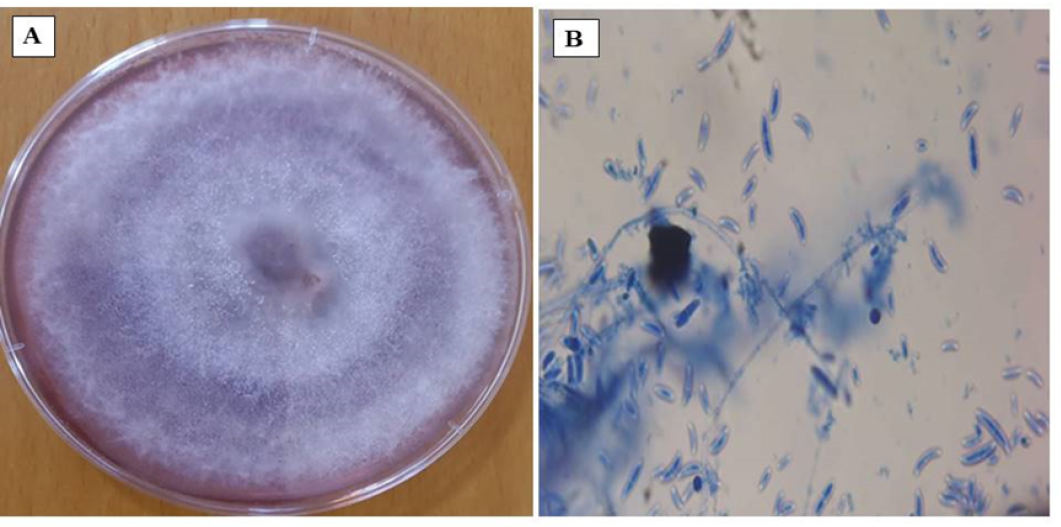
(A) shows the colony of F. oxysporum grown on potato dextrose agar (PDA), while (B) illustrates the microscopic structures of F. oxysporum.
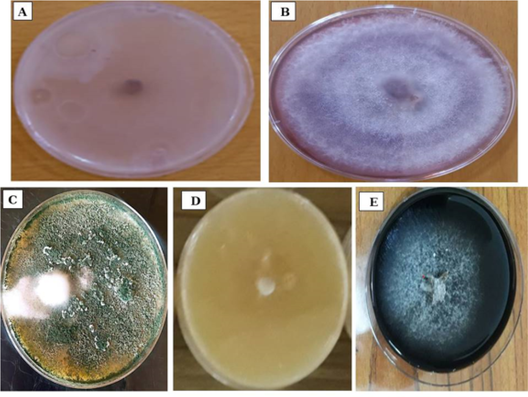
(A) Antagonistic activity assay between B. paramycoides and F. oxysporum. (B) Colony of Fusarium oxysporum grown alone (control treatment). (C) Antagonistic interaction between T. virens and F. oxysporum. (D) Antagonistic activity assay between P.fluorescens and F.oxysporum.(E) Evaluation of the Antagonistic Activity of Humic Acid against F. oxysporum.
References
- Abdo, A.M., Y.M. Heikal, S.A. Haroun, and H.M. Soliman. 2023. Isolation and identification of Fusarium oxysporum, the causal fungus of wilt disease of tomato (Solanum lycopersicum). Mansoura J. Biol., 62(1): 58-63. https://doi.org/10.21608/mjb.2023.447855[CrossRef]
- Agrios, G.N. 2005. Plant pathology (5th ed.). Academic Press.
- Al-Amari, G.M.G. 2025. Evaluation of the efficiency of some biological control agents in inducing systemic resistance to protect cucumber plants against infection with Pythium aphanidermatum causing root rot disease in Babil Province (Master’s thesis). Al-Musayyib Technical College, Al-Furat Al-Awsat Technical University, Iraq.
- Al-Mourrah, A., S. Mousa, and K. Al-Taweel. 2024. Evaluation of fifteen tomato varieties for resistance to Fusarium oxysporum f. sp. lycopersici. Afr. J. Biol. Sci., 6(8). https://doi.org/10.33472/AFJBS.6.8.2024.1536-1546[CrossRef]
- Al-Mu’min, A.A.H. 2024. Evaluation of the efficiency of effective microorganisms (EM-1), plant extracts, and chitosan in controlling some fungi causing root rot disease of faba bean. Iraqi J. Plant Sci., 12(1): 45–58.
- Al-Anzi, Y.A.S., K.Z. Al-Karaawi and I.J.Kadhim. 2025. Comparative Evaluation of Different Microbial Biocontrol Agents against Rhizoctonia solani Kuhn, the Causative Agent of Root Rot of Pepper Capsicum annuum L. Sarhad J. Agric.. 41(2): 674-683. https://dx.doi.org/10.17582/journal.sja/2025/41.2.674.683[Full text][CrossRef]
- Andersen, J.B., B. Koch, T.H. Nielsen, D. Sørensen, M. Hansen, O. Nybroe, C.Christophersen, J. Sørensen, S. Molin and M.Givskov. 2003. Surface motility in Pseudomonas sp. DSS73 is required for efficient biological containment of the root-pathogenic microfungi Rhizoctonia solani and Pythium ultimum. Microbiol., 149(1): 37–46. https://doi.org/10.1099/mic.0.25859-0[CrossRef]
- Babadoost, M. 2018. Fusarium: Historical and continued importance. Fusarium—Plant Diseases, Pathogen Diversity, Genetic Diversity, Resistance and Molecular Markers: Intech Open: 13-24. https://doi.org/10.5772/intechopen.74147[CrossRef]
- Batool, T., S. Ali, M.F. Seleiman, N.H. Naveed, A. Ali, K. Ahmed and M.Mubushar. 2020. Plant growth-promoting rhizobacteria alleviate drought stress in potato by suppressing oxidative stress and enhancing antioxidant enzyme activities. Sci Rep., 10(1): 16975. https://doi.org/10.1038/s41598-020-73489-z[CrossRef]
- Batson, A.M., S. Gyawali and L.J. du Toit. 2020. Evaluation of spinach and Beta vulgaris cultivars for differential susceptibility to two pathogenicity groups of Fusarium oxysporum f. sp. spinaciae. Plant Health 2020 Online.
- Bolkan, H.H. and E.E. Butler. 1974. Studies on heterokaryosis and virulence of Rhizoctonia solani. Phytopathol., 64: 513–522.
- Booth, C. 1977. Fusarium: Laboratory guide to the identification of the major species. Commonwealth Mycological Institute, Kew, Surrey, England.
- Chi, S.I., M. Akuma, R. Xu, V. Plante, M. Hadinezhad and J.T. Tambong. 2024. Phenazines are involved in the antagonism of a novel subspecies of Pseudomonas chlororaphis strain S1Bt23 against Pythium ultimum. Sci. Rep., 14(1): 20517. https://doi.org/10.1038/s41598-024-71418-y.[CrossRef]
- Clark, F.E. 1965. Agar-plate method for total microbial count. Methods of Soil Analysis: Part 2 Chemical and Microbiological Properties, 9, 1460-1466. https://doi.org/10.2134/agronmonogr9.2.c48.[CrossRef]
- Diniz, G.D.F.D., J.E.F .Figueiredo, K.M. Canuto, L.V. Cota, A.S.D.Q. Souza, M.L.F. Simeone and V.L. Dos Santos. 2024. Chemical and genetic characterization of lipopeptides from Bacillus velezensis and Paenibacillus ottowii with activity against Fusarium verticillioides. Front Microbiol.,15: 1443327. https://doi.org/10.3389/fmicb.2024.1443327.[CrossRef]
- Dobrzyński, J. and Z. Jakubowska. 2025. Pseudomonas protegens as a biocontrol agent against phytopathogenic fungi: A mini-review. World J. Microbiol. Biotechnol., 41(11): 1-10. https://doi.org/10.1007/s11274-025-04643-w[CrossRef]
- El-Garhy, H.A.S., T. Shoala, N.A.S. Messiha and S.E. El-Abeid. 2025 . The efficacy of nanoparticles on soil microbial biodiversity and the prevention of Fusarium wilt disease (Fusarium oxysporum f. sp. lycopersici). BMC Microbiol., 25(1): 305. https://doi.org/10.1186/s12866-025-04022-3.[CrossRef]
- Fayadh, M.A., J.T. AL-Kooranee, A.O. Manea and H.M. Abod. 2012. Effect of some Biological agent in control Fusarium wilt in tomato caused by Fusarium oxysporum f. sp. lycopersici. Basrah J. Agric. Sci., 25(2) : 4774
- Granada, D.L., R. Vibha, S. Skariyachan, P. Ujwal and K. Sandesh. 2025. In vitro and in silico investigation deciphering novel antifungal activity of endophyte Bacillus velezensis CBMB205 against Fusarium oxysporum. Sci Rep., 15(1):684. https://doi.org/10.1038/s41598-024-77926-1.[CrossRef]
- Guzmán-Guzmán, P., A. Kumar S. de Los Santos-Villalobos, F.I. Parra-Cota, M.D.C. Orozco-Mosqueda, A.E. Fadiji and G. Santoyo. 2023. Trichoderma species: Our best fungal allies in the biocontrol of plant diseases—A review. Plant, 12(3): 432. doi: https://doi.org/10.3390/plants12030432.[CrossRef]
- He, X., H. Duan, X. Liu, Z. Li, L. Yu, C. Zhou and H. Yu. 2025. Inhibitory Effect of Bacillus velezensis dhm2 on Fusarium oxysporum f. sp. cucumerinum and Synergistic Activity of Crude Lipopeptide Extract with Chemical Fungicides. Agric., 15(16): 1730. https://doi.org/10.3390/agriculture15161730[CrossRef]
- Hussain, S., M. Ali, A.H. Ghazy, A.A. Al-Doss, K.A. Attia, T.A. Shah and F. Li. 2025. Identification of antifungal lipopeptides from Bacillus subtilis Sh-17 targeting Fusarium oxysporum f. sp. lycopersici. Chem. Biol. Technol. Agric., 12(29). https://doi.org/10.1186/s40538-025-00744-8[CrossRef]
- Jackson, E. 2024. The ubiquitous wilt-inducing pathogen Fusarium oxysporum — a review of genes studied with mutant analysis. Pathogens, 13(10): 823. https://doi.org/10.3390/pathogens13100823[CrossRef]
- Jarjis, M. M., R.A. Al-Ani and A.A. Al-Hiti. 1992. Plant Diseases. Baghdad University, 569.
- Karthick, P. and R. Mohanraju. 2020. Antimicrobial compounds produced by Lysinibacillus odysseyi epiphytic bacteria associated with red algae. Braz. J. Microbiol., 51(4): 1683-1690. https://doi.org/10.1007/s42770-020-00341-x[CrossRef]
- Kumar, R., A. Khan, P. Singh, A. Singh, and A. Srivastava. 2024. Fusarium Infection of Eggplant: Disease Cycle and Management Strategies. In Molecular Dynamics of Plant Stress and its Management (pp. 281-306). Singapore: Springer Nature Singapore. https://doi.org/10.1007/978-981-97-1699-9_12[CrossRef]
- Limón, M.C., M.R. Chacón, R. Mejías, J. Delgado-Jarana, A.M. Rincón, and T. Benítez. 1999. Chitinase and related enzymes in Trichoderma antagonism: role in fungal cell wall degradation. Int. Microbiol., 2(4): 259–265.
- Montealegre, J.R., R. Reyes, L. M.Pérez, R. Herrera, P. Silva, and X. Besoain. 2003. Selection of bioantagonistic bacteria to be used in biological control of Rhizoctonia solani in tomato. Electron. J. Biotechnol., 6(2): 115–127. https://doi.org/10.4067/S0717-34582003000200006[CrossRef]
- Ojha, S. and N.C. Chatterjee. 2012. Induction of resistance in tomato plants against Fusarium oxysporum f. sp. lycopersici mediated through salicylic acid and Trichoderma harzianum. J. Plant Prot. Res., 52(2):220-225. https://doi.org/10.2478/v10045-012-0034-3[CrossRef]
- Patwardhan, R.B., P.S. Abhyankar, S.S. Gore, S.V. Kalekar and S.P. Umrani. 2022. Biofungicidal properties of Rhizobacteria for plant growth promotion and plant disease resistance. In Antifungal Metabolites of Rhizobacteria for Sustainable Agriculture (pp. 103-133). Cham: Springer International Publishing. Fungal Biol., https://doi.org/10.1007/978-3-031-04805-0_6[CrossRef]
- Podgórska-Kryszczuk, I., E. Solarska and M.Kordowska-Wiater. 2022. Reduction of Fusarium mycotoxins by selected non-conventional yeast strains in wheat grains and bread. Molecul., 27(5): 1578. https://doi.org/10.3390/molecules27051578[CrossRef]
- Ptaszek, M., L. Canfora, M. Pugliese, F. Pinzari, G. Gilardi, P. Trzciński and E.Malusà. 2023. Microbial-based products to control soil-borne pathogens: Methods to improve efficacy and assess impacts on microbiome. Microorgan., 11(1): 224. https://doi.org/10.3390/microorganisms11010224[CrossRef]
- Rahman, M.Z., A. Ahmad, Y.B. Kutawa, N. Siddiqui, T. Saad, E.M. Geok Hun, K. Hata and M.I. Hossain. 2021. Biology, diversity, detection, and management of Fusarium oxysporum f. sp. niveum: A review. Agron., 11: 1310–1331. https://doi.org/10.3390/agronomy11071310[CrossRef]
- Ren, H., M.S. Islam, H. Wang, H. Guo, H., Z. Wang, X. Qi, S. Zhang, J. Guo, Q. Wang and B. Li. 2022. Effect of humic acid on soil properties and microbial community structure in decline-diseased bayberry. Int. J. Mol. Sci., 23(23): 14707. https://doi.org/10.3390/ijms232314707[CrossRef]
- Rodrigues, M.H.P. and E.B. Furlong. 2022. Fungal diseases and natural defense mechanisms of tomatoes (Solanum lycopersicum): A review. Physiological and Molecular Plant Pathology, Physiol. Mol. Plant Pathol., 122: 101906. https://doi.org/10.1016/j.pmpp.2022.101906[CrossRef]
- Saad, M.T., A.M. El-Saadony, M.A. El-Tahan, S. Sayed, M.A. Moustafa, A.E. Taha and M.M. Ramadan. 2021. Polyphenolic extracts from pomegranate and watermelon wastes as substrate to fabricate sustainable silver nanoparticles with larvicidal effect against Spodoptera littoralis. Saudi J Biol Sci., 28(10): 5674–5683. https://doi.org/10.1016/j.sjbs.2021.06.011[CrossRef]
- Sambrook, J., E.F. Fritsch, and Tom Maniatis. 1989. Molecular Cloning : A Laboratory Manual. 2nd ed. Cold Spring Harbor: Cold Sprng Harbor Laboratory Press.
- Shoresh, M., G.F. Harman and F. Mastouri . 2010. Induced systemic resistance and plant responses to fungal biocontrol agents. Annu Rev Phytopathol., 48(1): 21-43. https://doi.org/10.1146/annurev-phyto-073009-114450[CrossRef]
- Sivan, A. and I. Chet. 1989. Degradation of fungal cell walls by lytic enzymes of Trichoderma harzianum. Microbiol., 135(3): 675-682. https://doi.org/10.1099/00221287-135-3-675[CrossRef]
- Sivan, A., Y. Elad and I. Chet. 1984. Biological control effects of a new isolate of Trichoderma harzianum on Pythium aphanidermatum. Phytopathol., 74(4):498-501. https://doi.org/10.1094/Phyto-74-498[CrossRef]
- Torbati, M., M. Arzanlou and A.C. da Silva Santos. 2021. Fungicolous Fusarium species: ecology, diversity, isolation, and identification. Curr Microbiol., 78(8): 2850-2859. https://doi.org/10.1007/s00284-021-02584-9[CrossRef]
- Vibha, R., D.L. Granada, S. Skariyachan, P. Ujwal and K. Sandesh. 2025. In vitro and in silico investigation deciphering novel antifungal activity of endophyte Bacillus velezensis CBMB205 against Fusarium oxysporum. Sci. Rep., 15: 684. https://doi.org/10.1038/s41598-024-77926-1[CrossRef]
- Vinale, F., K. Sivasithamparam, E.L. Ghisalberti, R. Marra, S.L. Woo and M.Lorito. 2008. Trichoderma–plant–pathogen interactions. Soil Biol. Biochem., 40(1): 1-10. https://doi.org/10.1016/j.soilbio.2007.07.002[CrossRef]
- Wegulo, S.N., W.W.Bockus, J.H. Nopsa, E.D. De Wolf, K.M. Eskridge, K.H. Peiris and F.E.Dowell. 2011. Effects of integrating cultivar resistance and fungicide application on Fusarium head blight and deoxynivalenol in winter wheat. Plant Dis., 95(5): 554-560. https://doi.org/10.1094/PDIS-07-10-0495[CrossRef]
Services
Article usage statistics
Loading…