
Tannic acid, as a polyphenol, is widely present in dicotyledonous plants and angiosperms. The study found that tannins could affect the ovarian development of Brandt’s voles. However, the specific effects of tannic acid on reproduction and its underlying mechanisms are still poorly understood. Therefore, this study explored the effects of tannic acid on the reproduction of Brandt’s voles. We treated female Brandt’s voles from four weeks postpartum with varying tannic acid doses (0 (control), 0.3% (low dose) or 0.6% (high dose) tannic acids) until 8 weeks (puberty) and 13 weeks (sexual maturity)). Ovarian specimens were collected for immunohistochemical analysis and RT-PCR. The treated Brandt’s voles were mated with male Brandt’s voles of the same age and we recorded the number of embryos, litters and the male to female ratio of the offspring. We found that the female Brandt’s voles in the low-dose tannic acid group had enhanced reproductive abilities, characterized by reduced follicular atresia and increased numbers of embryos and litters of offspring. The different doses of tannic acid altered the female-male ratio of the Brandt’s voles’ offspring. While the low-dose of tannic acid enhanced the level of ovarian autophagy by significantly increasing the expression of Beclin1 and LC3 proteins in the ovaries of the Brandt’s voles during sexual maturity. In short, tannic acid can promote the reproduction of Brandt’s voles by affecting the level of ovarian autophagy and improving the development of follicles. Tannins can boost the reproduction of Brandt’s voles.
Research Article · Volume 57, Issue 1, 20 December 2024 · Open Access
Tannic Acid Regulates Autophagy in the Ovary of Female Brandt’s Voles Affecting their Reproduction
· · · ·
Affiliations & Notes ⌄
- 1Department of College of Biological Science and Technology, Yangzhou University, Yangzhou 225009, China * Corresponding author: smyang@yzu.edu.cn
Article Info ⌄
- Journal:
- Pakistan Journal of Zoology
- Published:
- 20 December 2024

From the article
- Therefore, it is very important to explore the effect of TA on the reproduction of Brandt’s voles.
- The results of this study showed that low-dose TA could increase the number of embryos and litters of adult female Brandt’s voles.
- The results of the female-male ratio of the offspring showed that adolescent female Brandt’s voles have more male offspring after TA treatment.
- The low-dose TA content is similar to the TA content contained in Brandt’s voles plants food sources.
Selected from the article's own text.
Abstract
Introduction
Polyphenols are the most abundant dietary antioxidants and are common components of many plant-based food sources . Polyphenols are known to prevent diabetes and neurodegenerative diseases . Additionally, they appear to also influence ovarian function and lifespan as polyphenols have been shown to increase the follicle reserve and extend the lifespan of the ovaries in rats . Tea polyphenols inhibit follicular atresia during ovarian development from birth into the early years . Thus, the positive effect exhibited by polyphenols may not only be due to intervention in the transformation of primordial follicles to primary follicles, but also due to an inhibitory effect on follicular atresia . Tannic acid (TA) is a naturally occurring water-soluble polyphenol compound , which is synthesized and accumulated by higher plants and is widely present in various plant tissues, seeds and peels . Many studies have reported the beneficial effects of TA and its extracts on mammals . These studies have demonstrated that this molecule has anti-oxidant, anti-aging and anti-inflammatory properties . However, the effect of TA and its extracts on reproduction is rarely report.
It has been shown that polyphenols can affect autophagy through diverse mechanisms . Autophagy is a highly regulated self-degradation process that, with the help of lysosomes, eliminates damaged, unwanted or redundant subcellular proteins and organelles . Autophagy is an important regulator of cell homeostasis, and it encompasses three main types: chaperone-mediated autophagy, microgametophyte and macroscopical. Macroscopical autophagy has the highest turnover rate of the three different types of autophagy . According to reports, autophagy exists in the ovaries of mice, rats, pigs, geese and quail. Autophagy plays an important role in maintaining and regulating the reserve of ovarian primitive follicles ; as studies have shown that autophagy actively participates in the depletion of rat ovarian oocytes . Autophagy contributes to the renewal of organelles, energy management, and immune regulation. From the development of primordial follicles to embryogenesis and placental development , autophagy is a double-edged sword. When subjected to oxidized low density lipoprotein, granulose cells induce autophagy causing cell death, which is the underlying mechanism of follicular atresia . It has now been established that the regulation of autophagy is very important for all stages of oocyte maturation, implantation, pregnancy, placental physiology and postpartum remodeling. All this indicates that changes in the level of autophagy plays a very important role in follicular development and has a profound impact on reproduction . However, there are no reports of long-term polyphenol treatment on mammalian ovarian autophagy and reproduction.
Brandt’s voles are a social species, mainly found in the grasslands of inner, central and eastern Mongolia, and southern Russia . The population of Brandt’s voles has been irregular over the years . Brandt’s voles are generally considered a harmful animal during a population outbreak as they are detrimental to grasslands . In recent decades, this species has received considerable attention due to its important role in the ecosystem of the grasslands (the main food source for most carnivores) and its potential conflict with humans . Sex ratio is an important statistical parameter of animal population dynamics. It is a fundamental standard to explain the fluctuations of small rodent populations . The local resource competition (LRC) hypothesis holds that if local food resources are abundant, females will selectively produce more offspring and make better use of resources; on the contrary, if local food sources are limited, females will give birth to more males to avoid potential resource competition with the females they give birth .
To the best of our knowledge, the specific effects of TA on the reproduction and its underlying mechanisms are still poorly understood. Here, we determined the underlying mechanisms of TA on the ovaries of female Brandt’s voles and the resulting reproductive effects in addition, we also explored whether TA could affect reproduction through ovarian autophagy and follicular development. Importantly, low-dose tannins can enhance ovarian autophagy and increase the reproductive ability of female Brandt’s voles during sexual maturity, and different doses of tannins can change the ratio of male to female offspring of Brandt’s voles.
Materials and Methods
Experimental animals
In this study, Brandt’s voles were captured from an Inner Mongolia grassland and paired for breeding in captivity while being fed ad libitum. The photoperiod was 14L:10D and the temperature was maintained at 22°C ±1. When the female Brandt’s voles offspring reach 21 days-old, they were separated from their parents and housed in a cage alone. The female Brandt’s voles were allowed to adapt to the experimental conditions for one week, and at 28 days-old were weighed. The female Brandt’s voles with a body weight of 25-30 g were selected as test subjects.
Experimental design
All experiments were conducted between 09:00 and 16:00 h. 96 female Brandt’s voles (48 for perfusion fixation, 48 for blood samples) were divided into 6 groups with 16 in each group, as follows: 8 weeks-groups (control group, low-dose TA treatment group, high-dose TA treatment group), and 13 weeks-groups (control group, low-dose TA treatment group, high-dose TA treatment group). Simultaneously, another 78 female Brandt’s voles were mated with male Brandt’s voles of the same age after being treated with the same TA as described above. We found that wild Brandt’s voles consumed on average 0.03-0.069 g of TA every day, alongside the consumption of 10 ml of drinking water, so we choose 0.3% and 0.6% as the low and high dose treatments of TA, respectively.
Histological analysis of ovaries
The ovaries (n= 8 per group) were fixed overnight with 4% paraformaldehyde, and then histologically processed according to a standard protocol. The tissue was serially sliced (5 µm thick) throughout the ovary, and every fifth section was placed on a glass slide and stained with hematoxylin and eosin (H and E) stain. Then observe the entire field of view of each part under a microscope to count the number of follicles.
Real-time PCR
Real-time PCR was performed on a ABI 7500 real time PCR system (Applied Biosystems, USA) of SYBR Premix Ex TaqTM II (TaKaRa, China). After initial denaturation (95°C 60 s), 40 cycles of amplification were performed in a total volume of 20 μL, with the following parameters: 95°C 15, 58°C 30 s and 72°C 30 s. The primer sequences are listed in Table I, and GAPDH was used as the internal control. To quantify the relative mRNA expression by comparing the cycle threshold (Ct) values, we used the 2-ΔΔCt method to process the experimental data: ΔΔCt = (Ct target-Ct internal control) experimental group-(Ct target-Ct internal control) control group. Each experiment was repeated three times.
Table I. The primer sequences used for real-time quantitative PCR.
| Gene | Primer | Sequence (5’–3’) |
| LC3A | F | GCTTCGCCGACCGCTGTAA |
| R | ATCCGTCTTCATCCTTCTCCTG | |
| Beclin1 | F | GGTCGCTTGCCCAGTGTT |
| R | ACGGCAACTCCTTAGATT | |
| P62 | F | GGAAGAGCTTCGGAAGGG |
| R | GGCATGGGCCATAAGAGC | |
| GAPDH | F | TGGCAAAGTGGAGATTGTTGCC |
| R | AAGATGGTGATGGGCTTCCCG |
Immunochemistry
The ovaries were left at room temperature for 24 h, and then sectioned and frozen. Serial sections (5-μm) were prepared on a modified Leica CM 1950 rotary microtome (Leica Microsystems Nussloch Gmbh, Germany) and treated with 0.3% H2O2 to eliminate endogenous peroxidase activity. The sections were then incubated in BSA for 1 h at room temperature to reduce non-specific binding, followed by incubation) at 4°C overnight with the primary antibodies: anti-Beclinl, LC3B, and P62 polyclonal antibodies (bs-1353R 1:400, bs-4843R 1:600, bs-2951R, Bioss, Beijing, China, 1:400). Following this, the sections were incubated with rabbit serum as a negative control. Then, sections were incubated with SABC and developed with DAB. Images were acquired with an Image Pro Plus 6.0 photomicrograph analysis systems (Olimpus, Japan). In order to specify the staining intensity of Beclinl, LC3B, and P62, three independent observers outside the study group evaluated the pictures. Staining was quantified by measuring the immunoreactive area (IA) in µm² and the integrated optical density (IOD). The staining intensity (SI) for each image was calculated as SI = IOD/IA.
Transmission electron microscope
The paraformaldehyde samples were cut into 1 cm3 sections with a scalpel, and the ovarian tissue was fixed with glutaraldehyde for 2 h at room temperature. The tissue was then washed 3 times with PBS (pH 7.4) for 15 min, and then fixed with 1% osmic acid (6 drops of 0.2MPBS + 6 drops of 2% osmic acid) for 2 h. The samples were then again washed 3 times with PBS. After dehydration and concentration in ethanol and acetone, respectively, the ovarian tissue was inserted into pure Epon 812. Then the tissue block was cut into thin (0.07μm) slices on an ultramicrotome and mounted onto a copper plate. The sections were then stained with leading citrate and uranyl acetate. The slices were observed and photographed with a Philips Tecnai 12 Electron Microscope.
Statistical analysis
All variables were tested for normality and homogeneity by the Shapiro–Wilk and Levene tests. Data are presented as the mean ± SEM. One-way variance analysis (ANOVA) and Bonferroni tests were used to determine the effects of TA on qPCR, immunohistochemistry and all reproduction parameters. When P <0.05, the difference is considered statistically significant. All analyses were performed using SPSS 22.0.
Results
Effect of TA on ovarian follicles
We evaluated the effect of TA on follicular development. It was observed that the ovaries of the Brandt’s voles were normal after ingesting TA, the follicles were in different stages, and the CL formation was normal (Fig. 1). Studies have found that low-dose TA can reduce follicular atresia in adult female Brandt’s voles.
Expression of ovarian autophagy-related genes after TA treatment
Beclin1, LC3 and P62 are key biomarkers of autophagy. There was significant differences in LC3 mRNA levels between the three treatment groups. Compared with the control group, both low-dose and high-dose TA significantly increased the expression of LC3 mRNA in the ovaries during puberty and sexual maturity. With increased tannin concentration, the degree of LC3 mRNA expression becomes more obvious. TA has no effect on the expression of Beclinl1 mRNA in the ovaries of adolescent Brandt’s voles, but low-dose TA can increase the expression of Beclinl1 mRNA in the ovaries at sexual maturity. Treatment with any dose of TA had no effect on the expression level of P62 mRNA (Fig. 2).
TA enhances ovarian autophagy in the Brandt’s voles
To evaluate the effect of TA on autophagy in the Brandt’s voles, autophagy was observed with transmission electron microscopy (TEM), which is the gold standard method. The control group contained relatively few autophagosomes, while in the low-dose treatment group, autophagic vesicles containing degraded organelles
(such as mitochondria and endoparasitic reticulum) increased (Fig. 3A). These results indicate that low-dose TA can increase autophagy levels in the ovaries of Brandt’s voles.
In addition, in this study, immunohistochemistry was used to measure the protein levels of Beclinl, LC3B, and p62 in the ovarian tissue. As shown in Figure 3B, the positive cells are brown. Immunohistochemical statistics showed that compared with the control group, low-dose TA treatment increased the levels of Beclinl and LC3 proteins, but there was no significant change in the levels of autophagy substrate SQSTM1/P62 (Fig. 3C). In conclusion, low-dose TA promotes autophagic flux in the ovary.
The effect of TA on the reproduction of Brandt’s Voles
The number of embryos and litters as well as the sex ratio of the offspring are important indicators for measuring reproduction and population dynamics. We found that after treatment with low-dose TA, the number of embryos and litters of adult Brandt’s voles increased significantly. Low-dose TA increased the proportion of female offspring from adult female Brandt’s voles, while high-dose TA increased the proportion of male offspring from adult female Brandt’s voles (Fig. 4). This may be due to the breeding strategy under different environmental conditions.
Discussion
The potential for females to conceive depends on the development and growth of ovarian follicles, and animal species with higher fertility usually show more mature follicles in the ovaries and have a higher ovulation rate than animals with lower fertility . However, because most follicles go through a process called atresia before ovulation from the ovary, the utilization of follicles is extremely low. The ovary is the main regulator of female mammals’ reproduction, which regulates follicle development and secretion of reproductive hormones to produce mature oocytes (Zhou et al., 2019b). A large number of studies have shown that in the ovaries of female mammals, more than 99% of the developing follicles are atresia (Zhou et al., 2019a).
The results of this study showed that the ovaries of Brandt’s voles were normal after ingestion of TA, the follicles were in different stages and low doses of TA could reduce the atresia of the follicles in adult female Brandt’s voles. The reduction of follicular atresia led to an increase in the development of mature follicles, which further affected the reproduction of these mammals. More and more evidence show that autophagy plays a very important role in follicular atresia and GC proliferation . But there are few studies on how the regulation of autophagy affects follicular atresia and further affects reproduction. The level of autophagy in the ovary plays a very important role in the development of follicles We determined three key genes and proteins related to ovarian autophagy. The autophagy gene Beclin 1 (BECN1) plays an important role in the nucleation of autophagosomes through its association with phosphatidylinositol . The ubiquitin-like conjugation pathway results in the cleavage of the microtubule-associated protein light chain 3 (LC3) α/β, exposing a glycine residue at the C-terminus. This process leads to the conjugation of LC3 with phosphatidylethanolamine, which eventually forms LC3-II. The production of LC3-II is a well-known marker of mammalian autophagy . P62 is also known as SQSTM1 protein. As a regulatory factor, it participates in the formation of autophagosomes. It has substrate specificity and is a bridge connecting LC3-II with the ubiquitinated substrate to be degraded. P62 binds to the ubiquitinated protein and enters the autophagosome and finally fuses with the lysosome to form an autophagolysosome to be eliminated. When autophagy flux is inhibited, P62/SQSTM1 will accumulate in cells with impaired autophagy flux, and the overall expression of P62 in the cell negatively correlates with autophagy activity . The results of this study showed that low-dose TA could increase the expression levels of Beclinl mRNA and LC3 mRNA in the ovaries of adult female Brandt’s voles. Compared with the control group, the expression levels of P62 mRNA did not significantly change after TA treatment. The levels of the three autophagy-related proteins detected by immunohistochemistry were consistent with the gene levels. We found that TA has no effect on the P62 protein. At the same time, transmission electron microscopy showed that the number of autophagosomes increases but the volume does not increase, which proves that the degradation process of autophagosomes is proceeding normally. At the same time, TA can increase the protein levels of LC3 and Beclin1. In summary, we showed that TA can increase the autophagy level in the ovaries of adult female Brandt’s voles. As mentioned earlier, the level of ovarian autophagy plays an important role in follicular development and follicular atresia. Increase levels of ovarian autophagy may be responsible for a reduction of atretic follicles.
Our results show that TA has an effect on the number of embryos, litter size and the ratio of female to male offspring in Brandt’s voles. The number of embryos and litter size are the most intuitive indicators of species reproduction. We found that low-dose TA can increase the number of embryos and litters of Brandt’s voles during sexual maturity, but has no significant effect on adolescent Brandt’s voles. This may be due to the stronger ability of Brandt’s voles in sexual maturity to cope with changes in the external environment than that of adolescent Brandt’s voles.
Sex ratio is an important demographic parameter of animal population dynamics. It is an important criterion that explain the fluctuations of small rodent populations . The first group dominance hypothesis (FCA) predicts that there will be fluctuations in food resources during the breeding season, and the sex ratio is affected by the breeding season; therefore, females tend to give birth to more male offspring early on in the breeding season as too few females would reduce the birth rate. Similarly, because there are more males and fewer females, there would be a struggle for female mating rights, which would increase the mortality rate . As a chemical defense substance, TA is widely present in dicotyledonous plants and angiosperms. Therefore, it is very important to explore the effect of TA on the reproduction of Brandt’s voles. The results of this study showed that low-dose TA could increase the number of embryos and litters of adult female Brandt’s voles. The results of the female-male ratio of the offspring showed that adolescent female Brandt’s voles have more male offspring after TA treatment. For adult female Brandt’s voles, female Brandt’s voles had more female offspring after treatment with low-dose TA and more male offspring after treatment with high-dose TA. This is consistent with resource competition. The low-dose TA content is similar to the TA content contained in Brandt’s voles plants food sources. Therefore, the population of Brandt’s voles will tend to produce more female offspring when the environment is suitable, and the poor environment will produce more male offspring.
In short, low-dose TA can promote the reproduction of female Brandt’s voles in a variety of ways, including inhibiting follicular atresia and increasing the level of autophagy in the ovaries. In addition, different concentrations of TA represent a kind of environmental pressure for Brandt’s voles. At the appropriate concentration, the Brandt’s voles population adapts to environmental changes by adjusting the number of embryos and the ratio of male to female offspring. Finally, some measures could be considered to prevent Brandt’s vole from flooding their environements.
Funding
This work was supported by the National Natural Science Foundation of China (no. 31971418).
Ethical approval
All procedures were approved by the Animal Care and Use Committee of the Faculty of Veterinary Medicine of Yangzhou University (No: SJXY-2).
Data availability
The data that support this study will be shared upon reasonable request to the corresponding author.
Statement of conflict of interest
The authors have declared no conflict of interest.
Figures
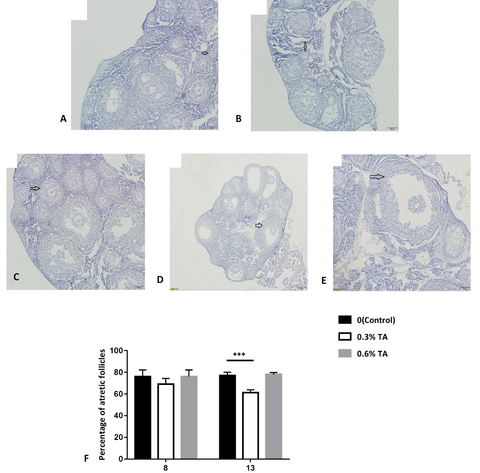
Histology of ovaries treated with different doses of tannic acid. Morphological classification of follicles. (A) primordial follicle (oocyte surrounded by a single layer of flattened granulosa cells), (B) primary follicle (oocyte surrounded by a single layer of cuboidal granulosa cells), (C) secondary follicle (oocyte surrounded by two or more layers of cuboid granulosa cells without a visible antrum) and (D) tertiary follicle (follicles with a clearly defined antral space and multiple layers of granulosa cells around the oocyte). (E) atretic follicle (identified as those follicles that appear to enter the degenerative process without ovulation. In atretic follicles, the oocytes shrink or are missing, and the granulosa cells are replaced by fibrous material). (F) Proportion of ovarian atresia follicles in Brandt’s voles treated with different doses of tannic acids (n=8). Data are expressed as the mean ± sem. ***P < 0.001.

The relative abundance of mRNA of ovarian autophagy-related genes after tannic acid treatment. (A) the effect of different doses of tannic acid on the relative abundance of ovarian Beclinl gene mRNA(n=8), (B) the effect of different doses of tannic acid on the relative abundance of ovarian LC3 gene mRNA (n=8), (C) the effects of different doses of tannic acids on the relative abundance of ovarian P62 gene mRNA (n=8). Data are expressed as the mean ± sem. * P < 0.05, **P < 0.01.
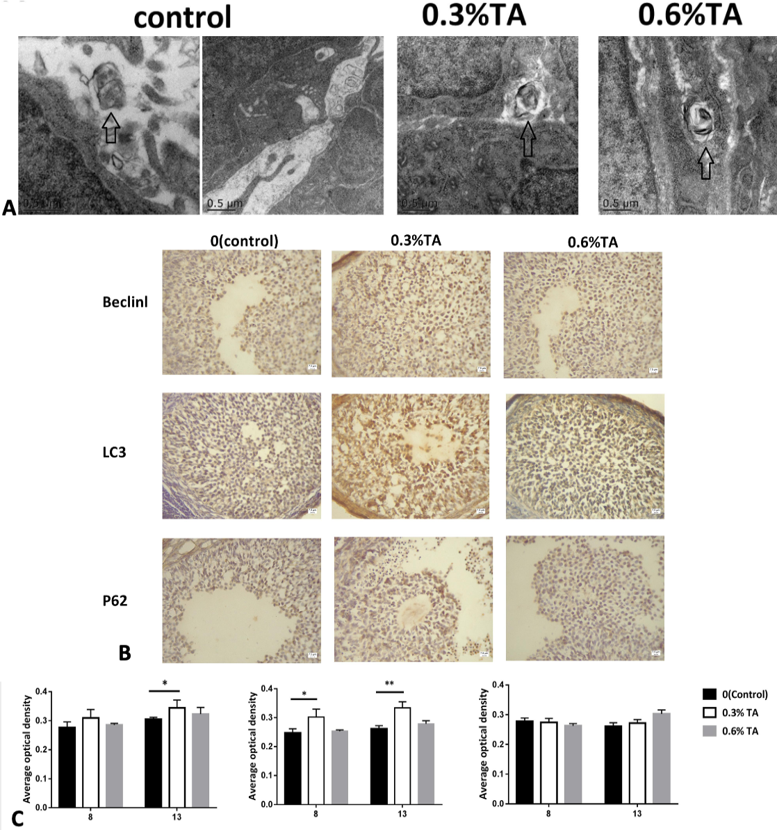
(A) Electron microscopy of the ovaries treated with different doses of tannic acids. The arrows represent autophagosomes. (B) Immunohistochemical staining of Beclin1 protein in the ovary. The immunostaining of the ovaries of the control animals was weaker. The ovaries in the adult low-dose group had stronger immunostaining. Immunohistochemical staining of LC3 protein in the ovary. The immunostaining of the ovaries of the control animals was weaker. Strong immunostaining of the ovaries in the low-dose and high-dose groups. In the ovaries, granulosa cells are strongly stained. Immunohistochemical staining of P62 protein in the ovary. There was no significant difference in immunostaining in each treatment group. (C) The effect of different doses of tannic acid on Beclinl, LC3 and P62 proteins during adolescence and sexual maturity (n=8). Data are expressed as the mean ± sem. * P < 0.05., **P < 0.01
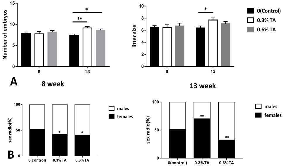
Effects of different doses of tannins on the reproduction of Brandt’s vole (A) The effect of embryo number and litter size of Brandt’s vole treated with different doses of tannin. (B) Female-male ratio of offspring of Brandt’s voles treated with different doses of tannic acid (n=13). Data are expressed as the mean ± sem. * P < 0.05, **P < 0.01.
References
- Barra, J.M., Kozlovskaya, V., Kharlampieva, E. and Tse, H.M., 2020. Localized immunosuppression with tannic acid encapsulation delays islet allograft and autoimmune-mediated rejection. Diabetes, 69: 1948-1960. https://doi.org/10.2337/db20-0248[CrossRef]
- Chen, Z.G., Luo, L.L., Xu, J.J., Zhuang, X.L., Kong, X.X. and Fu, Y.C., 2010. Effects of plant polyphenols on ovarian follicular reserve in aging rats. Biochem. Cell Biol., 88: 737-745. https://doi.org/10.1139/O10-012[CrossRef]
- Hewison, A.J.M. and Gaillard, J.M., 1996. Birth-sex ratios and local resource competition in roe deer, Capreolus capreolus. Behav. Ecol., 7: 461-464. https://doi.org/10.1093/beheco/7.4.461[CrossRef]
- Huang, R. and Liu, W., 2015. Identifying an essential role of nuclear LC3 for autophagy. Autophagy, 11: 852-853. https://doi.org/10.1080/15548627.2015.1038016[CrossRef]
- Kihara, A., Kabeya, Y., Ohsumi, Y. and Yoshimori, T., 2001. Beclin-phosphatidylinositol 3-kinase complex functions at the trans-Golgi network. EMBO Rep., 2: 330-335. https://doi.org/10.1093/embo-reports/kve061[CrossRef]
- Kumsta, C., Chang, J.T., Lee, R., Tan, E.P., Yang, Y., Loureiro, R., Choy, E.H., Lim, S.H.Y., Saez, I., Springhorn, A., Hoppe, T., Vilchez, D. and Hansen, M., 2019. The autophagy receptor p62/SQST-1 promotes proteostasis and longevity in C. elegans by inducing autophagy. Nat. Commun., 10: 5648. https://doi.org/10.1038/s41467-019-13540-4[CrossRef]
- Li, G., Yin, B., Wan, X., Wei, W., Wang, G., Krebs, C.J. and Zhang, Z., 2016. Successive sheep grazing reduces population density of Brandt’s voles in steppe grassland by altering food resources: A large manipulative experiment. Oecologia, 180: 149-159. https://doi.org/10.1007/s00442-015-3455-7[CrossRef]
- Linklater, W.L., Law, P.R., Gedir, J.V. and du Preez, P., 2017. Experimental evidence for homeostatic sex allocation after sex-biased reintroductions. Nat. Ecol. Evol., 1: 88. https://doi.org/10.1038/s41559-017-0088[CrossRef]
- Liu, H., Wang, D.H. and Wang, Z.W., 2003. Energy requirements during reproduction in female brandt’s voles (Microtus brandtii). J. Mammal., 84: 1410-1416. https://doi.org/10.1644/BRG-030[CrossRef]
- Luo, L.L., Huang, J., Fu, Y.C., Xu, J.J. and Qian, Y.S., 2008. Effects of tea polyphenols on ovarian development in rats. J. Endocrinol. Invest., 31: 1110-1118. https://doi.org/10.1007/BF03345661[CrossRef]
- Moilanen, J., Karonen, M., Tahtinen, P., Jacquet, R., Quideau, S. and Salminen, J.P., 2016. Biological activity of ellagitannins: Effects as anti-oxidants, pro-oxidants and metal chelators. Phytochemistry, 125: 65-72. https://doi.org/10.1016/j.phytochem.2016.02.008[CrossRef]
- Nakashima, A., Tsuda, S., Kusabiraki, T., Aoki, A., Ushijima, A., Shima, T., Cheng, S.B., Sharma, S. and Saito, S., 2019. Current understanding of autophagy in pregnancy. Int. J. mol. Sci., 20: 13. https://doi.org/10.3390/ijms20092342[CrossRef]
- Pasquariello, R., Verdile, N., Brevini, T.A.L., Gandolfi, F., Boiti, C., Zerani, M. and Maranesi, M., 2020. The role of resveratrol in mammalian reproduction. Molecules, 25: 16. https://doi.org/10.20944/preprints202009.0003.v1[CrossRef]
- Provenza, F.D., Burritt, E.A., Perevolotsky, A. and Silanikove, N., 2000. Self-regulation of intake of polyethylene glycol by sheep fed diets varying in tannin concentrations. J. Anim. Sci., 78: 1206-1212. https://doi.org/10.2527/2000.7851206x.[CrossRef]
- Shen, M., Jiang, Y., Guan, Z., Cao, Y., Li, L., Liu, H. and Sun, S.C., 2017. Protective mechanism of FSH against oxidative damage in mouse ovarian granulosa cells by repressing autophagy. Autophagy, 13: 1364-1385. https://doi.org/10.1080/15548627.2017.1327941[CrossRef]
- Shi, Y.N., Zhou, S.W., Fan, S.F., Ma, Y.H., Li, D.D., Tao, Y. and Han, Y.B., 2021. Encapsulation of bioactive polyphenols by starch and their impacts on gut microbiota. Curr. Opin. Fd. Sci., 38: 102-111. https://doi.org/10.1016/j.cofs.2020.11.001[CrossRef]
- Sobhani, M., Farzaei, M.H., Kiani, S. and Khodarahmi, R., 2020. Immunomodulatory; anti-inflammatory/antioxidant effects of polyphenols: A comparative review on the parental compounds and their metabolites. Fd. Rev. Int., pp. 1-53. https://doi.org/10.1080/87559129.2020.1717523[CrossRef]
- Sun, Y.C., Wang, Y.Y., Sun, X.F., Cheng, S.F., Li, L., Zhao, Y., Shen, W. and Chen, H., 2018. The role of autophagy during murine primordial follicle assembly. Aging (Albany NY), 10: 197-211. https://doi.org/10.18632/aging.101376[CrossRef]
- Tang, Z., Xu, R., Zhang, Z., Shi, C., Zhang, Y., Yang, H., Lin, Q., Liu, Y., Lin, F., Geng, B. and Wang, Z., 2021. HIF-1alpha protects granulosa cells from hypoxia-induced apoptosis during follicular development by inducing autophagy. Front. Cell Dev. Biol., 9: 631016. https://doi.org/10.3389/fcell.2021.631016[CrossRef]
- Verheyden-Tixier, H. and Duncan, P., 2000. Selection for small amounts of hydrolysable tannins by a concentrate-selecting mammalian herbivore. J. chem. Ecol., 26: 351-358. https://doi.org/10.1023/A:1005401203954[CrossRef]
- Watanabe, R. and Kimura, N., 2018. Non-suckling starvation of neonatal mice promotes primordial follicle formation with activation of ovarian autophagy. J. Reprod. Dev., 64: 89-94. https://doi.org/10.1262/jrd.2017-126[CrossRef]
- Weldy, M.J., Epps, C.W., Lesmeister, D.B., Manning, T., Linnell, M.A. and Forsman, E.D., 2019. Abundance and ecological associations of small mammals. J. Wildl. Manage., 83: 902-915. https://doi.org/10.1002/jwmg.21641[CrossRef]
- Weng, X.C., Ma, X.S., Wang, Q.Z., Xu, K.L., Hu, X.S., Liu, W.B. and Zhang, C., 2019. Effect of hypothyroidism on CYP51 and FSHR expression in rat ovary. Theriogenology, 138: 145-151. https://doi.org/10.1016/j.theriogenology.2019.07.012[CrossRef]
- Wishart, A.E., Williams, C.T., McAdam, A.G., Boutin, S., Dantzer, B., Humphries, M.M., Coltman, D.W. and Lane, J.E., 2018. Is biasing offspring sex ratio adaptive? A test of Fisher’s principle across multiple generations of a wild mammal in a fluctuating environment. Proc. R. Soc. B Biol. Sci., 285. https://doi.org/10.1098/rspb.2018.1251[CrossRef]
- Xie, X., Yi, W., Zhang, P., Wu, N., Yan, Q., Yang, H., Tian, C., Xiang, S., Du, M., Getachew, A.E., Zuo, X. and Ying, C., 2017. Green tea polyphenols, mimicking the effects of dietary restriction, ameliorate high-fat diet-induced kidney injury via regulating autophagy flux. Nutrients, 9: 14. https://doi.org/10.3390/nu9050497[CrossRef]
- Yadav, P.K., Tiwari, M., Gupta, A., Sharma, A., Prasad, S., Pandey, A.N. and Chaube, S.K., 2018. Germ cell depletion from mammalian ovary: Possible involvement of apoptosis and autophagy. J. biomed. Sci., 25: 36. https://doi.org/10.1186/s12929-018-0438-0[CrossRef]
- Yang, S., Shao, S., Huang, B., Yang, D., Zeng, L., Gan, Y., Long, D., Chen, J. and Wang, J., 2020. Tea polyphenols alleviate tri-ortho-cresyl phosphate-induced autophagy of mouse ovarian granulosa cells. Environ. Toxicol., 35: 478-486. https://doi.org/10.1002/tox.22883[CrossRef]
- Yim, W.W. and Mizushima, N., 2020. Lysosome biology in autophagy. Cell Discov., 6: 6. https://doi.org/10.1038/s41421-020-0141-7[CrossRef]
- Yin, B., Li, G., Wan, X., Shang, G., Wei, W. and Zhang, Z., 2017. Large manipulative experiments reveal complex effects of food supplementation on population dynamics of Brandt’s voles. Sci. China Life Sci., 60: 911-920. https://doi.org/10.1007/s11427-017-9114-9[CrossRef]
- Zhihan, T., Xinyi, M., Qingying, L., Rufei, G., Yan, Z., Xuemei, C., Yanqing, G., Yingxiong, W. and Junlin, H., 2019. Autophagy participates in cyst breakdown and primordial folliculogenesis by reducing reactive oxygen species levels in perinatal mouse ovaries. J. Cell Physiol., 234: 6125-6135. https://doi.org/10.1002/jcp.27367[CrossRef]
- Zhong, W., Wang, G., Zhou, Q. and Wang, G., 2007. Communal food caches and social groups of Brandt’s voles in the typical steppes of Inner Mongolia, China. J. Arid Environ., 68: 398-407. https://doi.org/10.1016/j.jaridenv.2006.06.008[CrossRef]
- Zhou, J., Peng, X. and Mei, S., 2019a. Autophagy in ovarian follicular development and atresia. Int. J. Biol. Sci., 15: 726-737. https://doi.org/10.7150/ijbs.30369[CrossRef]
- Zhou, Z., Lin, Q., Xu, X., Illahi, G.S., Dong, C. and Wu, X., 2019b. Maternal high-fat diet impairs follicular development of offspring through intraovarian kisspeptin/GPR54 system. Reprod. Biol. Endocrinol., 17: 13. https://doi.org/10.1186/s12958-019-0457-z[CrossRef]
Services
Article usage statistics
Loading…