
Cabbage cyst nematode, Heterodera cruciferae Franklin, 1945 causing serious damage on different cabbage cultivars and other host plants. High incidence of this nematode revealed by soil and root samples in the vegetable growing farmlands in Tabriz, Iran. Different populations of H. cruciferae were collected from the region and characterized based on morphological, morphometric and molecular features, four out of those populations isolated from the rhizosphere and roots of four different cabbage cultivars were subjected to further studies. All the populations showed same range of morphometric and morphological characters fitted thoroughly with the identity of H. cruciferae. Different primers for amplification of ITS1-5.8S-ITS2, D2-D3 rDNA and hsp 90 genes, showed similarity between the nematode populations. ITS-RFLP profiles generated by 16 different restriction enzymes did not differentiate the populations, too. In phylogenetic analyses of partial sequences of 28S rDNA D2-D3, a clade clustering the four populations of cabbage cyst nematode was formed along with the sequences of H. cruciferae populations deposited in NCBI. These results showed morphometrical, morphological and molecular characters of under study populations are same and the host is the only differences between pathogenicity populations.
Research Article · Volume 42, Issue 1, 21 March 2024 · Open Access
Study on Populations of Cabbage Cyst Nematode Heterodera cruciferae Franklin, 1945 from Iran
Affiliations & Notes ⌄
- 1Department of plant protection, Faculty of Agriculture, University of Maragheh, Maragheh, East Azarbaijan , Iran. Citation | Jabbari, H., 2024. Study on populations of cabbage cyst nematode Heterodera cruciferae Franklin, 1945 from Iran. Pakistan Journal of Nematology, 42(1): 14-24.
Article Info ⌄
- Journal:
- Pakistan Journal of Nematology
- Published:
- 21 March 2024
Abstract
Introduction
Cyst forming nematodes, heteroderids, are sedentary endoparasites of plants having ability to establish specific feeding sites in their host cells to achieve necessary nutrition for their development and growth. Some species such as Heterodera glycines Ichinohe, 1952 and H. schachtii (Schmidt, 1871) Örley, 1880 cause high damages and yield losses on different host plants worldwide . Cabbage cyst nematode, H. cruciferae Franklin, 1945 has been reported from different continents ; causing economically significant damages on cabbage and some other crops such as radish, cauliflower, etc. ; . In Iran, occurrence of the nematode has been reported from rhizosphere and roots of two different varieties of cabbages (Brassica oleracea L. var. capitata and Brassica oleracea L. var. gongylodes) , and rhizosphere of sugar beet and peanut (Mirgasemi et al., 2014). Chenopodium album, Raphanus sativus, Coriandrum sativum, Sonchus oleraceus, Sisymbrium loeselii and Lepidium sativum are also naturally infected by the nematode in the region .
Study on occurrence and presence of H. cruciferae based on morphometric and morphological data in different regions of the world have been documented ; ; ; Oostenbrink and den Ouden, 1953; ; ; ; ; ; ; ; ; but reports on molecular diagnosis of the nematode, are very scare (Chizhov et al., 2010; ; Escobar et al., 2018; ; . Although different cyst nematodes have been subjected of molecular identification studies in Iran , 2007), H. cruciferae was not included in the studies. Molecular studies based on ribosomal and mitochondrial genes are frequently used in different groups of nematode species identification , 2007; Chizhov et al., 2010; ; Escobar et al., 2018; ; . Ribosomal ITS genes having high variability in the region, so can be use for nematode identification even at subspecies level . On the other hand, only using some sequences for nematode identification is not sufficient for identification at species level and morphological and morphometrical characters are needed for accurate and reliable identification. In current research, the main aims were (i) characterization of main populations of the nematode recovered from roots of four different cabbage cultivars in the region by using ITS1-5.8S-ITS2 of rRNA, D2-D3 rDNA and hsp 90 sequences, (ii) ITS-RFLP, (iii) morphologic and morphometric study on populations.
Materials and Methods
Nematode samples
Cysts were isolated from roots and rhizosphere of four different varieties of cabbage (Table 1) in vegetable farmlands in Tabriz, East Azarbaijan province, Iran (GPS coordinates 38˚ 06΄ N, 46˚15΄E, by mean temperature of 6.6-20˚C and sandy-loam soil type) during 2020-2022. Cysts were extracted by Fenwick (1940) method and were picked up from extracted material with a fine brush or needle under steriomicroscope.
Table 1: List of cabbage cultivars grown in Tabriz vegetable farmlands.
| S. | Cultivars | Growing period | Cultivars code |
| 1 | Brassica oleracea L. var. gongylodes | Six months | K6 |
| 2 | Brassica oleracea L. var. gongylodes | Three months | K3 |
| 3 | Brassica oleracea L. var. capitata | Six months | C6 |
| 4 | Brassica oleracea L. var. capitata | Three months | C3 |
Nematode identification
Morphological and morphometric characters: Species identification was carried out based on morphological and morphometrical characters of cysts, J2 and male according to Subbotin et al. (2010).
Molecular diagnosis
DNA Extraction and PCR: The genomic DNA was extracted from J2 by lysing method . Extracted DNA from the four populations were kept in -20ºC until use.
The PCR reaction carried out in 25µl per reaction containing 1μl DNA, 16.35μl ddwater, 5μl PCR buffer (Promega, USA), 0.5 dNTPs (Promega, USA), 0.15 Taq DNA polymerase (5U μl/1- Promega, USA) and 2μl of each primer (10 pmol/μl). In all PCR reactions, positive and negative controls were included. PCR products were run on 1% agarose gel, stained with ethidium bromide and the results visualized using Bio Rad Molecular image®-Gel DocTMXR+ Gel documentation. The expected bands were extracted from gel using cutting tools and purified using illustrate GFX® PCR DNA and Gel Band Purification Kit, GE Healthcare-UK. Quality and quantity of purified DNA were evaluated by nanodrop (Nanodrop 2000c- USA) and the process followed by cloning. Out of nine different primer combinations applied for molecular diagnosis and phylogenetic studies of cyst forming nematodes , three primers (Table 2) were used for amplifying ITS1-5.8S-ITS2 of rRNA, D2-D3 28S rRNA and hsp 90 genes. The PCR was programmed for all the reactions as shown in Table 3.
PCR-RFLP
For each enzyme, 2µl of each PCR product (0.2-1µg),
Table 2: List of primers used for amplification of rRNA-ITS, D2-D3 and hsp 90 of Heterodera cruciferae populations in this study.
| Target region | Primers | Reference |
| ITS1-5.8S-ITS2 | F TW81: GTTTCCGTAGGTGAACCTGC | Curran et al. 1994 |
| R AB28: ATATGCTTAAGTTCAGCGGGT | ||
| D2-D3 28S rRNA | F D2A: ACAAGTACCGTGAGGGAAAGTTG | Nuun, 1992 |
| R D3B:TCGGAAGGA ACCAGCTACTA | ||
| hsp 90 | F U831: GAYACVGGVATYGGNATGACYAA | Skantar and Carta, 2005 |
| R L1110:TCRCARTTVTCCATGATRAAVAC |
Table 3: PCR program for amplification of the ITS1-5.8S-ITS2 of rRNA, D2-D3 28S rRNA and hsp 90 genes (A: Calculated based on primer sequences; B: Depends on expected size of amplification in PCR, for each 1Kb equal to 1 min.).
| Initial denaturation | Denaturation | Annealing | Extension | Final extension |
| 3 min - 94˚C | 1 min - 94˚C | 1.5 min - (A) ˚C | (B) min - 72˚C | 10 min - 72˚C |
| 30- <40X |
Table 4: The restriction enzymes and related cutting sites used in PCR-RFLP.
| S. | Enzyme | Cutting site | S. | Enzyme | Cutting site | S. | Enzyme | Enzyme |
| 1 | Hinf I | 5' -G ANTC- 3' | 7 | Alu I | 5' -AG CT- 3' | 12 | Bsp143I | 5' - GATC- 3' |
| 2 | Mva I | 5' -CC WGG- 3' | 8 | Msp I | 5' -C CGG- 3' | 13 | Hind III | 5' -A AGCTT- 3' |
| 3 | Xba I | 5' -T CTAGA- 3' | 9 | Sa II | 5' -G TCGAC- 3' | 14 | Pvu I | 5' -CGAT CG- 3' |
| 4 | Hin6 I | 5' -G CGC- 3' | 10 | Kpn I | 5' -GGTAC C- 3' | 15 | BamH I | 5' -G GATCC- 3' |
| 5 | EcoR I | 5' -G AATTC- 3' | 11 | Rsa I | 5' -GT AC- 3' | 16 | Pst I | 5' -CTGCA G- 3' |
| 6 | BusR I | 5' -GG CC- 3' |
1µl restriction enzyme (2-10 unit), 1µl buffer 10X and 6µl ddwater were added to a sterilized PCR tube and all tubes incubated at least eight hours in 37°C for. None of the used enzymes (Table 4) had star activity. Digested DNA fragments present in PCR tubes (10 µl) were loaded on a 1.5% agarose gel and the results were visualized under Bio Rad Molecular image®-Gel DocTMXR+ Gel documentation.
Cloning and sequencing
PCR products were cloned by pGEM-T® easy vector system (pGEM®-Tand pGEM®-TEasy vector System, Promega-USA). Ligation process was carried out based on the kit instruction. Transformation was achieved using heat shock by 3µl of ligation mix and 50µl of Escherichia coli competent cell (Homemade, DH5α). The transformed bacteria were cultured in liquid LB media contained in special falcon tubes and were shaken at the speed of 120 rpm for 90 minutes. The bacteria were cultured on plates containing solid LB with agar. White-blue screening was implemented after keeping the plates in 37ºC at least for 12 hours. Colony PCR for blue colonies as DNA template along with specific primers of inserted sequences (Tables 2 and 3) was done. The insertion of the sequence into the bacteria was visualized on PCR products. The residue of the colonies cultured in liquid LB media under the same situation that cited above were kept overnight (at least 8 hours). The plasmid extraction of the cultured bacteria carried out using Nucleospin®, Plasmid, DNA, RNA and protein purification kit, Mchery-Nagel, Germany according to the kit manual. In order to more confirmation of sequences insertion into plasmids, one more PCR reaction was conducted on the extracted plasmid as DNA template in the PCR and its specific primers (Tables 2 and 3). All transformed plasmids were sent to GATC Biotech-Germany for sequencing by Sanger method
. The sequences were aligned and blasted by selected sequences from NCBI and phylogenetic trees created using Mega-6® and CLC®Genomic Workbench, v7 (CLC Inc, Aaraus, Denmark) softwares considering the default parameters.
Results and Discussion
Nematode identification
Morphology and morphometric characters: The morphometric and morphological features of the populations collected from the region were shown in Table 5. In comparison with previous records and descriptions (Chizhov et al., 2010; ; it was found that all the populations collected from the region was H. cruciferae and their morphometrical and morphological data were fully corresponded with those of the other reports documented for this species. No significant variation was found among the populations as the morphological and morphometrical characteristics.
Molecular diagnosis
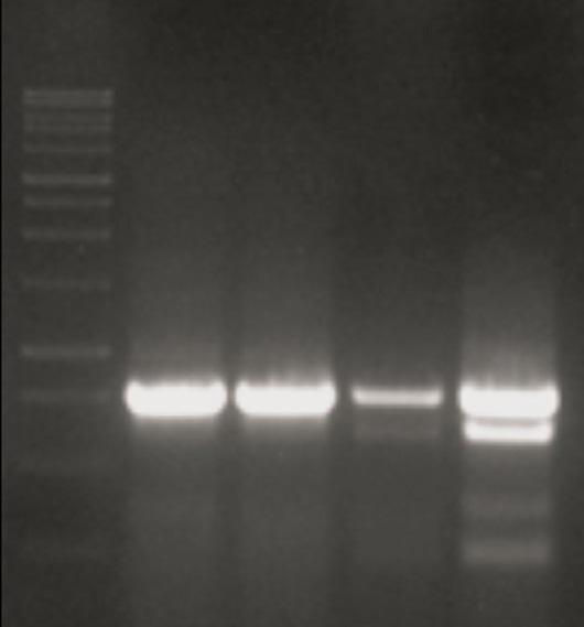
ITS1-5.8S –ITS2 region: Amplification of the ITS1-5.8S-ITS2 regions of rRNA gene yielded a single fragment (1250 bp) for all studied populations (Fig. 1). Sequencing, blasting and alignment results approved it. Alignment of the sequence with similar sequences, 1041 bp , 1040 bp (Chizhov et al., 2009), ~ 1000 bp and 816-825 bp showed more than 95 percent similarity between the sequences. The results of sequencing and blasting of ITS1-5.8S-ITS2 of rRNA obtained from the four populations of the cabbage cyst nematode revealed high (98 and more than 95 percent) similarity between the populations and the sequences of ITS1-5.8S-ITS2 of rRNA gene of H. cruceferae deposited to NCBI. Since the ITS sequences of the Iranian populations of H. cruciferae showed high similarity to each other, only one sequence (yielded from K6 population) submitted to NCBI by Kp114545 accession number.
ITS-RFLP: The ITS-RFLP profile of all four populations digested with the enzymes (Table 4), displayed similar restriction sites. The resulted bands for four (Mva I, BusR I, Alu I, Rsa I) out of eight enzymes used by Subbotin et al. (2010) were identical with their results. Concerning the enzymes that were used for the first time in our study (Tables 4) only three enzymes namely Pst I (173, 340 and 512 bp), Hin 61 (69, 174, 287 and 495 bp) and Bsp 1431(42, 355 and 628 bp) yielded RFLP patterns in the gene region (Figure 2).
No differences were observed among the RFLP-ITS-rDNA profiles of the four populations of H. cruciferae (Figure 2). ITS-RFLP is one of the reliable methods for analyzing the differences and similarities among populations and used frequently for identification of nematode populations ; Baklava et al., 2015).
Table 6: The restriction enzymes (in bold) used for the first time in present study for the ITS-RFLP of four populations of H. cruciferae.
| S. | Enzyme | S. | Enzyme | S. | Enzyme | S. | Enzyme |
| 1 | Hinf I (No cutting site) | 5 | Hin6 I (69, 174, 287, 495 bp) | 10 | Hind III (No cutting site | 14 | Rsa I (21, 130,321, 569 bp) |
| 2 | Pst I (173, 340, 512 bp) | 6 | Pvu I (No cutting site) | 11 | Kpn I (No cutting site) | 15 | Mva I (771, 270 bp) |
| 3 | EcoR I (No cutting site) | 7 | Sa II (No cutting site) | 12 | BamH I (No cutting site) | 16 | BsuR I (24,1070,325,522 bp) |
| 4 | Xba I (No cutting site) | 8 | Msp I (No cutting site) | 13 | Alu I | - |
Phylogeny based on ITS1-5.8S-ITS2 of rRNA
The phylogenetic tree (Figure 3) was generated based on the ITS1-5.8S-ITS2 of rRNA sequence for the under studied populations and seven other sequences belonging to H. cruciferae, H. carotae and H. urticae used by Sasanelli et al. (2013). Similar to ITS-RFLP profiles, there are very low differences between different populations of H. cruciferae and even H. carotae and H. urticae populations.
Heterodera carotae and H. urticae are morphologically distinguished from each other but their different populations could not be precisely differentiated using ITS1-5.8S-ITS2 of rRNA gene sequence (Figure 3).
D2-D3 of 28S rRNA
The amplified fragment of D2-D3 of 28S rRNA region showed identical size in all the four populations (Figure 4). The similarity between the sequences was more than 95 percent, the length of sequence of D2-D3 of 28S rRNA obtained from the populations was 750 bp that was deposited to GenBank under Kp114546 accession number.
Phylogenetic tree of the D2-D3 of 28S rRNA sequence was created and it was found that all the four Iranian populations of the cabbage cyst nematode were clustered at same clade and there was no intraspecific polymorphism among H. cruciferae populations but other populations of the cabbage cyst nematode reported by Sasanelli et al. (2013) and the outgroup were clustered in different groups (Figure 5).
hsp 90 gene
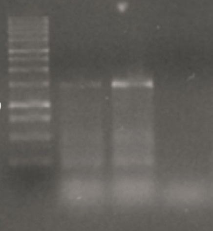
The hsp 90 gene was amplified and it was revealed that the length of hsp 90 gene in H. cruciferae was about 1250 bp. Since the primers used in the PCR was degenerated, amplification of the region was only completed in two (K6 and K3) populations. The PCR product profile of both populations was found to be similar and sequences of the two populations had 96 percent similarity in alignment (Fig. 6). The partial sequence lenght of the hsp 90 gene of Iranian populations of H. cruciferae was 826 bp and was submitted to NCBI under Kp114552 accession number.
As there was no previous record of hsp 90 gene for the H. cruciferae in NCBI, creating of phylogenetic tree by using the hsp 90 gene sequences was not possiable.
Cabbage cyst nematodes host plants are mostly in Brassicaceae and Lamiaceae families. Brassica oleracea, Chenopodium album, Raphanus sativus, Coriandrum sativum and Sisymbrium loeselii as main cultivated cruciferous species in the vegetable farmlands of Tabriz naturally and annually infected by H. cruciferae . Tabriz vegetable farmland are under cultivation of different vegetables for many years. For this long time, the nematode has enough time to establish itself on the crops as a pathogen. Because of the availability of different host plants and weeds belonging to the two above mentioned families in this region as well as cultivation of a so long time, it was thought that especial and certain differentiations of the nematode might have been occurred among various host plants. But, the results of this study showed similarity of morphological, morphometrical and molecular characters between the main and dominant populations of the nematode on cabbages. The populations of H. cruciferae were not clustered in a single clade based on ITS1-5.8S-ITS2 rRNA sequences. PCR-ITS-RFLP results showed that all four studied populations are similar to each other and to PCR-ITS-RFLP profiles generated by effect of 16 restriction enzymes on H. cruciferae populations, too. Because these results may relate to the selected gene region for studies which is not appropriate for diagnosis of differences among the populations or may be related to high similarity of the genome region in H. crucifeare, H. carotae and H. urtica species as well. Mehaline et al. (2020) showed that while H. cruciferae and H. carotae are very close species to each other. On the other hand, the results show different of ITS1, ITS2 regions sequences capacity to making distinguish between different populations of the nematodes, are different ; Mehaline et al., 2020). On the other hand, researchers like Escobar et al. (2018) mention that ITS rRNA and COI partial sequences do not have potential to make differentiation between H. cruciferae and H. carotae and host range is very crucial for the species separation.
D2-D3 of 28S rRNA sequences also is frequently used, mostly at species level, by some researchers for molecular studies in different groups of nematodes (Van Den Berg et al., 2016; ; ; . However, it used at a species level in different Globodera pallida and G. rostochiensis populations by Douda et al. (2010). The results of this study revealed that the ability of the gene for differentiation between populations is far better than ITS1-5.8S-ITS2 region. It could be due to shorter length and less variability in D2-D3 28SrRNA of the gene region, in current study all the four populations made a separate clade compared to the populations reported from other localities based on the region sequence and it confirmed that all populations are same.
Skantar and Karta (2004) used hsp 90 gene sequence for molecular characterization and phylogenetic evaluation of some nematodes including H. glycines. Skantar et al. (2012) also used the gene for molecular identification of different populations of Hetrodera. zeae. In current study he results of hsp 90 sequences were similar to those of D2-D3 of 28S rRNA. Since there is not any similar study on cabbage cyst nematode using this part of nematode genome, it was not possible to do any more comparison.
Sequence-based methods may involve analyses of nucleotide sequence either the nuclear DNA, mitochondrial DNA (mtDNA) or the whole genome of nematodes. The rDNA and mitochondrial cytochrome c oxidase subunit I (COI) genes are preferred by most studies on nematode ; . Mitochondrial genes have great ability to diagnose different populations and races of cyst nematodes . Subbotin et al. (2015) noticed the different populations of Longidorus discordance in phylogenetic relationships inferred from the ITS1 rRNA and COX- I gene sequence datasets. Toktay et al. (2022) recently used COX -I and rDNA-ITS region sequences for identification of cabbage cyst nematode in Nigde province and Turkish populations were separated from each other using ITS and COX -I genes sequences however, except one of the studied populations which showed rRNA-ITS sequences similarity with populations from Italy, Algeria, the Netherlands and Russia, the other studied populations were similar to Iranian H. cruciferae populations. Escobar et al. (2018) by working on H. carotae and H. cruciferae populations also confirmed the ability of COX -I gene for the population’s differentiation. While the results of current study confirm similarity in all four populations of H. cruciferae in Tabriz farmlands, it sounds that the ability of different sequences to making differentiation between populations is not a fix and it can show different capability upon to species. Morphometrical and morphological data in all cases are crucial and should be the main and important part of any identification.
Acknowledgement
The author expresses her gratitude to University of Bonn (Germany), University of Tabriz and Agricultural Research Center (Iran) who provided financial assistance in parts of this research.
Novelty Statement
Novelty of the research are:
- First study about identification of different population of the nematode based on traditional and molecular methods.
- Identification of some restriction enzymes cutting sites on the nematode.
- Identification of partially sequence of hsp 90 gene in H. cruciferae
Conflict of interest
The author has declared no conflict of interest.
Figures
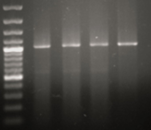
The amplified ITS1-5.8S-ITS2 of rRNA region of four different populations of H. cruciferae using universal primers (TW81- AB28). (1: Ladder, 2: K6, 3: K3, 4: C6, 5: C3).
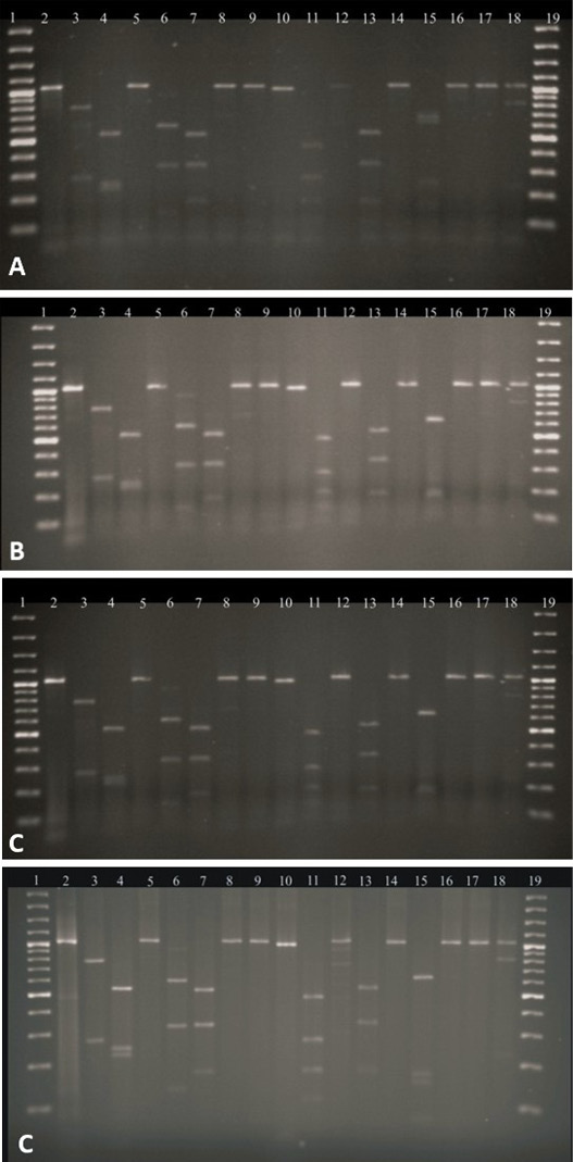
RFLP-ITS-rDNA profiles of four populations of Heterodera cruciferae collected from Tabriz vegetables growing area against restriction enzymes. A: K6, B: K3, C: C6 and D: C3 (1: Ladder, 2: Intact PCR product, 3: Mva I, 4: Alu I, 5: Xba I, 6: Pst I, 7: Bsu R I, 8: Pvu I, 9: Sa II, 10: Hinf I, 11: Hin 6 I, 12: Eco RI, 13: Rsa I, 14: Bam H I, 15: Bsp 143 I, 16: Hind III, 17: Kpn I, 18: Msp I, 19: Ladder).
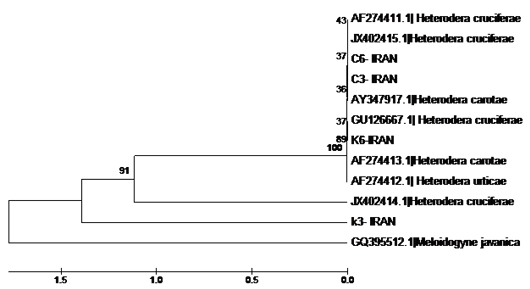
Phylogenetic tree of Heterodera cruciferae as inferred from the ITS1-5.8S-ITS2 rRNA sequences of populations collected from Tabriz-Iran and other populations using the Maximum Likelihood. The bootstrap consensus tree inferred from 1000 replicates.
The amplified D2-D3 of 28S rRNA region obtained from the studied populations of H. cruciferae using specific primers). (1: Ladder, 2: K6, 3: K3). (4: C6, 5: C3) after 3: K3.
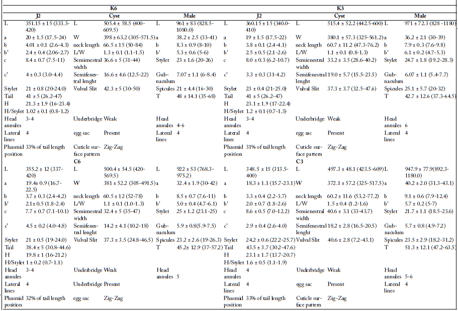
Morphological and morphometric features of Heterodera cruciferae populations collected from Tabriz vegetable growing farmlands (ll measurements are in µm).
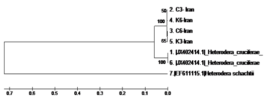
Phylogenetic tree as inferred from D2-D3 of 28S rRNA sequences of H.cruciferae populations collected from Tabriz-Iran and other populations, using the Maximum Likelihood method. The bootstrap consensus tree inferred from 1000 replicates.
References
- Baklawa, M., Niere, B., Heuer, H. and Massoud, S., 2015. Characterization of cereal cyst nematodes in Egypt based on morphometrics. RFLP rDNA-ITS Sequence. Analyses Nematol., 17: 103–115. https://doi.org/10.1163/15685411-00002855[CrossRef]
- Bello, A., Escuer, M. and Sanz, R., 1999. El género Heterodera en hortalizas en España. Boletín de Sanidad Vegetal. Plagas, 25: 321–334.
- Brzeski, M.W., 1998. Nematodes of Tylenchida in Poland and temperate Europe. Muzeum i Instytut Zoologii, Polska Akademia Nauk. Warszawa.
- Chizov, V.N., Pridannikov, M.V., Nasonova, L.V. and Subbotin, S.A., 2010. Heterodera cruciferae Franklin, 1945 of Brassica oleraceae L. from flood land fields in the Moscow region Russia. Rus. J. Nematol., 17(2): 107-113.
- Curran, J., Driver, F Ballard, J.W.O. and Milner R.J. 1994. Phylogeny of metarhizium-analysisi of ribosoma DNA-sequence data. Myco. Res., 98: 547-552.
- Derycke, S., Vanaverbeke, J., Rigaux, A., Backeljau, T. and Moens, T. 2010. Exploring the use of cytochrome oxidase c subunit 1 (COI) for DNA barcoding of free-living marine nematodes. PLoS One, 5(10): e13716. https://doi.org/10.1371/journal.pone.0013716[CrossRef]
- Douda, O., Zouhar, M., Nováková, E., Mazáková, J. and Ryšánek, P., 2010. Variability of D2/D3 segment sequences of several populations and pathotypes of potato cyst nematodes (Globodera rostochiensis, Globodera pallida). Plant Prot. Sci., 46(4): 171–180. https://doi.org/10.17221/1/2010-PPS[CrossRef]
- Escobar-Avila, I.M., López-Villegas, E.Ó., Subbotin, S.A. and Tovar-Soto, A. 2018. First report of Carrot cyst nematode Heterodera carotae in Mexico: Morphological, molecular characterization, and host range study. J. Nematol., 50(2): 229-242. https://doi.org/10.21307/jofnem-2018-021[CrossRef]
- Fenwick, D.W., 1940. Methods for the recovery and counting of cysts of Heterodera schachtii from soil. 1940. J. Helminthol., 18: 155-172. https://doi.org/10.1017/S0022149X00031485[CrossRef]
- Fenwick, D.W. and Franklin, M.T., 1951. Futher studies on the identification of Heterodera species by larval length. Estimation of the length parameters for eight species and varieties. J. Helminthol., 25: 57-76. https://doi.org/10.1017/S0022149X00018988[CrossRef]
- Franklin, M.T., 1945. On Heterodera cruciferae n. sp. of brassicas, and on a Heterodera strain infecting clover and dock. J. Helminthol., 21: 71-84. https://doi.org/10.1017/S0022149X00031965[CrossRef]
- Franklin, M.T., 1951. The cyst-forming species of Heterodera. England, Commonwealth Agricultural Bureau (CAB).
- Grujicic, G. and Krnjaic, D., 1966. Heterodera cruciferae Franklin, 1945, štetna nematoda na korenu kupusa u Jugoslaviji. Zašt. Bilja, 91: 309–313.
- Jabbari, H. and Niknam, G., 2008. SEM observations and morphometrics of the cabbage cyst nematode, Heterodera cruciferae Franklin, 1945, collected where Brassica spp. are grown in Tabriz, Iran. Turk. J. Zool., 32: 253-262.
- Jones, F.G.W., 1950. Observations on the beet eelworm and other cyst forming species of Heterodera. Ann. App. Biol., 37: 407-440. https://doi.org/10.1111/j.1744-7348.1950.tb00966.x[CrossRef]
- Khanzad-Bonab and Jabbari, H., 2019. Infection of some medicinal plants with Cabbage Cyst Nematode, Heterodera cruciferae, Franklin, 1945 under greenhouse conditions. Iran. J. Plant Pathol., 54(3): 255-258.
- Macdougal, M.G., 1960. The morphology of the brassica root eelworm Heterodera cruciferae Franklin, 1945. Nematologica, 5: 158-165. https://doi.org/10.1163/187529260X00307[CrossRef]
- Mahdikhani, E., 1998. Reports of H. cruciferae and H. carotae and distribution of other cyst forming nematodes in Mashhad sugerbeet fields. Procciding of the 13th Iranian Congress of Plant Protection. Karaj, Iran. pp. 139
- Mehalaine, K., Imren, M., Özer, G., Hammache, M. and Dababat, A., 2020. A molecular identifcation and phylogenetic diversity of cereal cyst nematodes (Heterodera spp.) populations from Algeria. Nematropica, 50(2): 134–143.
- Mennan, S. and Handoo, Z., 2012. Heterodera cruciferae Franklin, 1945 (Tylenchida: Heteroderidae) ile bulaşık Brassica oleracea var. capitata subvar. alba nın histopatojisi. Turk. J. Entomol., 36(3): 301–310.
- Mirghasemi, S.N., Jmali, S. and Sharifi, K., 2014. Identification of plant parasitic nematodes of peanut (Arachis hypogaea) in Guilan province. Procciding of the 21th Iranian Congress of Plant Protection. Urmia, Iran. p. 315.
- Nunn G.B., 1992. Nematode molecular evolution. PhD dissertation, University of Nottingham, Nottingham, UK.
- Oostenbrink, M., and den Ouden, H., 1953. Het koolcystenaaltje, Heterodera cruciferae Franklin, 1945, in Nederland. Tijdschrift Over Plantenziekten, 59: 95–100. https://doi.org/10.1007/BF02106326[CrossRef]
- Ravichandra, N.G., 2014. Horticultural nematology. New Dehli, Springer India. https://doi.org/10.1007/978-81-322-1841-8[CrossRef]
- Riggs, R.D., Rakes, L. and Hamblen, M.L., 1982. Morphometric and serologic comparisons of a number of populations of cyst nematodes. J. Nematol., 14: 188-199.
- Sanger, F. and Coulson, A.R. 1975. A rapid method for determining sequences in DNA by primed synthesis with DNA polymerase. J. Mol. Biol., 94(3): 441-448. https://doi/org/ 10.1016/0022-2836(75)90213-2
- Sasanelli, N., Vovlas, N., Trisciuzzi, N., Cantalapiedra Navarrete, C., Palomares-Rius, J.E. and Castillo, P., 2013. Pathogenicity and host-parasite relationships of Heterodera cruciferae in cabbage. Plant Dis., 97(3): 333-338. https://doi.org/10.1094/PDIS-07-12-0699-RE[CrossRef]
- Shahina, F. and Maqbool, M.A., 1995. Cyst nematodes of Pakistan (Heteroderidae). National Nematological Research Centre, Karachi, University of Karachi.
- Shurtleff, M.C. and Averre, C., 2000. Diagnosing plant disease caused by nematodes. USA, The American Phytopathological Society.
- Skantar, A. and Carta, L., 2004. Molecular characterization and phylogenetic evaluation of the Hsp90 gene from selected nematodes. J. Nematol., 36: 466-80.
- Skantar, A.M., Handoo, Z.A.L., Zanakis, G.N. and Tzortzakakis, E.A., 2012. Molecular and morphological characterization of the corn cyst nematode, Heterodera zeae, from Greece. J. Nematol., 44(1): 58–66.
- Stone, A.R. and Rowe, J.A., 1976. Heterodera cruciferae. C.I.H. Descriptions of plant parasitic nematodes. Set 6, No. 90. Farnham Royal, UK: Commonwealth Agriculture Bureaux.
- Sturhan, D. and Das K., 1960. (Heterodera cruciferae Franklin, 1945) in Bayern. Pflanzenschutz.; 12: 142-144.
- Subbotin, S.A., Mundo-Ocampo, M. and Baldwin, J.G., 2010. Systematic of cyst nematodes (Nematoda: Heteroderinae). 8 A. Netherland, Brill Leiden-Boston. https://doi.org/10.1163/ej.9789004164345.i-512[CrossRef]
- Subbotin, S.A., Vierstraete, A., De ley, P., Rowe, J., Waeyen berge, L., Moens, M. and Vanfleteren, J.R., 2001. Phylogenetic relationships within the cyst-forming nematodes (Nematoda, Heteroderidae) based on analysis of sequences from the ITS regions of ribosomal DNA. Mol. Phylogenet. Evol., 21: 1-16. https://doi.org/10.1006/mpev.2001.0998[CrossRef]
- Subbotin, S.A., Vovlas, N., Yeates, G.W., Hallmann, J., Kiewnick, S., Chizhov, V.N., Manzanilla-Lopez, R.H., Inserra, R.N. and Castillo, P., 2015. Morphological and molecular characterization of Helicotylenchus pseudorobustus (Steiner, 1914) Golden, 1956 and related species (Tylenchida: Hoplolaimidae) with a phylogeny of the genus. Nematology, 17(1): 27–52. https://doi.org/10.1163/15685411-00002850[CrossRef]
- Tamura, K., Peterson, D., Stecher, G., Nei, M., Kumar and S. and Peterson, N., 2011. MEGA5: Molecular evolutionary genetics analysis using maximum likelihood, evolutionary distance, and maximum parsimony methods. Mol. Biol. Evol., 28(10): 2731-2739. https://doi.org/10.1093/molbev/msr121[CrossRef]
- Tanha-Maafi, Z., Moens, M. and Subbotin, S.A., 2003. Molecular identification of cyst-forming nematodes (Heteroderidae) from Iran and a phylogeny based on ITS-rDNA sequences. Nematology, 5(1): 99-111. https://doi.org/10.1163/156854102765216731[CrossRef]
- Tanha-Maafi, Z., Sturhan, D., Handoo, Z., Mordehi, M., Moens and M., Subbotin, S.A., 2007. Morphological and molecular studies of Heterodera sacchari, H. goldeni and H. leuceilyma (Nematoda: Heteroderidae). Nematology, 9: 483-497. https://doi.org/10.1163/156854107781487242[CrossRef]
- Toktay, H., Akyol, B.G., Evlice, E. and İmren, M. 2022. Molecular characterization of Heterodera cruciferae Franklin, 1945 from cabbage fields in Niğde province, Turkey. Mol. Biol. Rep., PMID: 36097115. https://doi.org/10.1007/s11033-022-07860-w[CrossRef]
- Van den Berg, E., Tiedt, L.R., Palomares-Rius, J.E., Vovlas, N. and Castillo, P., 2016. Morphological and molecular characterisation of one new and several known species of the reniform nematode, Rotylenchulus Linford and Oliveira, 1940 (Hoplolaimidae: Rotylenchulinae), and a phylogeny of the genus. Nematology, 18(1): 67-107. https://doi.org/10.1163/15685411-00002945[CrossRef]
- Van Megen, H., Van Den Elsen, S., Holterman, M., Karssen, G., Mooyman, P., Bongers, T., Holovachov, O., Bakker J. and Helder, J., 2009. A phylogenetic tree of nematodes based on about 1200 full-length small subunit ribosomal DNA sequences. Nematology, 11: 927–950. https://doi.org/10.1163/156854109X456862[CrossRef]
- Wang, K., Xiei, H., Li, Y., Xu, Ch., Yu, L. and Wang, D., 2013. Paratylenchus shenzhenensis n. sp. (Nematoda: Paratylenchinae) from the rhizosphere soil of Anthurium andraeanum in China. Zootaxa, 3750(2): 167–175. https://doi.org/10.11646/zootaxa.3750.2.4[CrossRef]
Services
Article usage statistics
Loading…