
A comparative study of stem epidermis of fourteen sugarcane varieties (i.e., L-l18, CP72-2086, CP43-33, HSF-240, CPF-237, SPF-213, COjv-84, CO-1148, COL-54, COL-29, CO-975, BL-4, Triton and BF-162) was carried out using Schultz’s maceration method followed by staining with chloloiodide of Zinc. The results indicated substantial differences among the sugarcane varieties for various stem epidermal cell combinations and structures like number of stomata, number and shape of cork, silica, long, and pointed elongated cork cells. It is therefore contended that the structure of the epidermis in sugar cane can be used as a characteristic of variety differentiation.
Research Article · Volume 33, Issue 2, 14 May 2017 · Open Access
Stem Epidermal Anatomy of Fourteen Sugarcane Varieties of the Punjab
·
Affiliations & Notes ⌄
- 1Institute of Pure and Applied Biology, Bahauddin Zakariya University, Multan, Pakistan. , Email: nosheenilahi@yahoo.com
Article Info ⌄
- Journal:
- Sarhad Journal of Agriculture
- Published:
- 14 May 2017
From the article
- Among the stem epidermal cells of CP43-33, higher number of pointed elongated cork cells and presence of hook like cork cell (Figure 1D) are the distinguishing characters.
- When comparing the number of stomata in the stem epidermal cells of all the sugarcane varieties, few stomata were observed in COL-29 and CO-975, while in varieties COjv-84, CO-1148, COL-54 and Triton stomata were absent (Table 2).
- In the stem epidermal cells of all the 14 sugarcane varieties width of long cell at its maximum and minimum point ranged from 23.5µm-13.25µm and 7.25µm -5µm, respectively, while length of long cells ranged from a minimum of 65.15 µm in the variety HSF-240, to the maximum of 187.75 µm in CP43-33 (Table 1).
- So it is suggested that characters of the sugarcane stem epidermal cells are diagnostic and can provide additional reliable characters for identification of varieties when combined with morphological characters.
Selected from the article's own text.
Abstract
Materials and Methods
The study was conducted at the Institute of Pure and Applied Biology, Bahauddin Zakariya University, Multan. Mature stalks of the fourteen sugarcane field varieties of Punjab i.e., L-118, CP72-2086, CP43-33, HSF-240, CPF-213, Cojv-84, CO-1148, COL-54, COL-29, CO-975, BL-54, Triton and BF-162 were obtained from the Ayyub Agricultural Research Center, Faisalabad, Pakistan.
Cotological Study of Stem Epidermis
Twelve months old mature stalks of the sugarcane varieties were collected from experimental sites. Ten individual samples of every variety were tested. Care was taken to select the material at similar phase of growth. Central portion of the mature inter node which had reached their absolute length and were no longer enclosed by the protective leaf cover were used for this study.
Maceration
Schultz’s method of maceration was used (Subramanayam, 1996). A portion of about 5 mm2 of the stem epidermis with some of the fibrous and cortical tissues was cut from the selected part of the internodes and placed it in a test tube then 4 ml concentrated nitric acid, 2 gm potassium chlorate and 1 ml of distilled water was added. Then mixture was boiled in a test tube and when epidermis detached, the contents were transferred into a Petridish having some water.
Staining and Slide Preparation
The stem epidermis was mounted on a slide and stained with chloloiodide of Zinc (Van Dillewijin, 1952). Different cytological characters were observed under NIKON Leitzoptiphot research microscope and photographs were taken on Konica Monochrome VX 400.
Statistical Analysis
The data was statistically analyzed by using Analysis of Variance. While for the significance of means, Duncan’s multiple range (DMRT) test was used.
Results and Discussion
Stem epidermis of fourteen sugar cane varieties (i.e., L-118, CP72-2086, CP43-33, HSF-240, CPF-237, SPF-213, COjv-84, CO-l148, COL-54, COL-29, CO-975, BL-4, Triton and BF-162) were studied with the help of light microscope (Figure 1, 2, 3, 4, 5, 6 and Tables 1, 2). The stem epidermis seemed to be made up of several types of cells inclined in a uniform design. The long cells, which occupy the larger portion of the epidermal cells, look like a four-sided prism. They differ largely in length while differences in width were less apparent.
Table 1: Various stem epidermis characteristics of fourteen sugar cane varieties. Letters indicate statistically significant differences by Duncan’s multiple range tests at P < 0.05
| Name of variety | Width of long Cells at its Max point(µm) | Width of long cells at its Min point (µm) | Length of long cells (µm) | Extra cork cells per square (mm) |
| L-118 | 17de | 6.25bc | 90.25b | Scarce |
| CP72-2086 | 21b | 6.25bc | 67b | Abundant |
| CP43-33 | 18.45c | 7.5a | 187.75a | Scarce |
| HSF-240 | 13.25f | 5.5cd | 65.15b | Scarce |
| CPF-237 | 18.2cd | 5d | 103.75b | Absent |
| SPF-213 | 14f | 6.25bc | 77.75b | Absent |
| COjv-84 | 18.55c | 6cd | 81.76b | Abundant |
| CO-1148 | 18.75c | 6.25bc | 131.7ab | Scarce |
| COL-54 | 18.8c | 6.255bc | 112.5b | Absent |
| COL-29 | 23.5a | 6.5abc | 108.75b | Absent |
| CO-975 | 18.5c | 7.25ab | 97.25b | Absent |
| BL-4 | 18.2cd | 6.25bc | 103b | Absent |
| Triton | 14.5f | 5.5cd | 136.25ab | Absent |
| BF-162 | 16.25e | 5d | 88.75b | Absent |
Table 2: Various stem epidermis characteristics of fourteen sugar cane varieties
| Name of variety | Solitary Cork cells/square (mm) | Pointed elongated cork cells/ square (mm) | Stomata/square (mm) |
| L-118 | Absent | Absent | Scarce |
| CP72-2086 | Scarce | Absent | Scarce |
| CP43-33 | Scarce | Abundant | Scarce |
| HSF-240 | Absent | Scarce | Scarce |
| CPF-237 | Scarce | Abundant | Scarce |
| SPF-213 | Scarce | Scarce | Scarce |
| COjv-84 | Scarce | Scarce | Absent |
| CO-1148 | Scarce | Absent | Absent |
| COL-54 | Absent | Scarce | Absent |
| COL-29 | Scarce | Scarce | Scarce |
| CO-975 | Scarce | Scarce | Scarce |
| BL-4 | Scarce | Abundant | Scarce |
| Triton | Absent | Absent | Absent |
| BF-162 | Abundant | Scarce | Scarce |
The differences in epidermal cell structure is divided into two groups; i.e., quantitative and qualitative. Qualitative changes consist of the absence of any type of cell as in the varieties L-118, HSF-240, COL-54, Triton, solitary cork cells and in varieties CPF-237, SPF-213, COL-54, COL-29, CO-975, BL-4, Triton, BF-162 extra cork cells were absent (Table 1, 2) in the stem epidermal cell structure which differentiate them from all other varieties. Structural differences of stem epidermal cells among different sugar cane varieties are sometimes so much striking that many varieties can be recognized by only one character. For example, in the epidermal cell structure of sugarcane varieties Triton (Figure 5C, D) and BF-162 (Figure 6) two cork and two silica cells mostly form short cell group, while in HSF-240, the short cell group mostly formed with two cork and one silica cell (Figure 2A, B). Dahlgren and Clifford (1982) stated that the size, shape and distribution scheme of silica bodies on various plant epidermal structures varies from one species to another, and this difference has always been thought of high taxonomic significance.
There are also quantitative differences as there are partial absences or abundance of any type of cells in different varieties as shown in Table 1 and 2. Among the stem epidermal cells of CP43-33, higher number of pointed elongated cork cells and presence of hook like cork cell (Figure 1D) are the distinguishing characters. This increase may be brought about by the omission of long or silica cells, so that two short cell groups join each other directly . The inconsistencies of the number of stomata among the different varieties play an important role in classification . The distribution of stomata, though erratic, offers a valued diagnostic feature. When comparing the number of stomata in the stem epidermal cells of all the sugarcane varieties, few stomata were observed in COL-29 and CO-975, while in varieties COjv-84, CO-1148, COL-54 and Triton stomata were absent (Table 2). Various kinds of epidermal cells and their supplementary design may help in creating a more natural grouping of the genera and tribes of the Gramineae , 1963; . Among quantitative variations of epidermal cells, the width and length of long cells, the number and distribution of stomata are very important ; . In the stem epidermal cells of all the 14 sugarcane varieties width of long cell at its maximum and minimum point ranged from 23.5µm-13.25µm and 7.25µm -5µm, respectively, while length of long cells ranged from a minimum of 65.15 µm in the variety HSF-240, to the maximum of 187.75 µm in CP43-33 (Table 1). According to Metcalfe (1960), the long cells exist in different shapes, and can be used for the solution of various taxonomic problems. Van Dillewijn (1952) stated that the sugar cane stem epidermis display a fascinating patterns, which differ significantly in various varieties. Rao and Balasubramanian (1953) suggested that quantitative dissimilarities in size of many organs and cell - types are sometimes linked to chromosome number of the clone. According to Artschwager (1930), it would be necessary to study the epidermis of a large number of canes, in order to trace group relationships and investigates the parental influence in the different crosses.
Conclusion
The diagnostic importance of sugarcane stem epidermal cell structure has been examined on 14 varieties of commercial value. It is clear from the present study that the design of the epidermis of sugarcane varies greatly among varieties and remains nearly constant within variety. So it is suggested that characters of the sugarcane stem epidermal cells are diagnostic and can provide additional reliable characters for identification of varieties when combined with morphological characters. Further studies should be designed to verify that this epidermal cell configuration remains constant under diverse environmental situations.
Acknowledgement
We gratefully acknowledge the Ayyub Agricultural Research Center, Faisalabad for providing the mature stalks of the varieties used for this study.
Authors’ Contributions
Nosheen Noor Elahi designed the experiments, helped with interpretation of data and contributed in conducting experiments. Misbah Mughal conducted the experiments and wrote the manuscript.
Conflict of Interest
Authors have declared no conflict of interests.
Figures
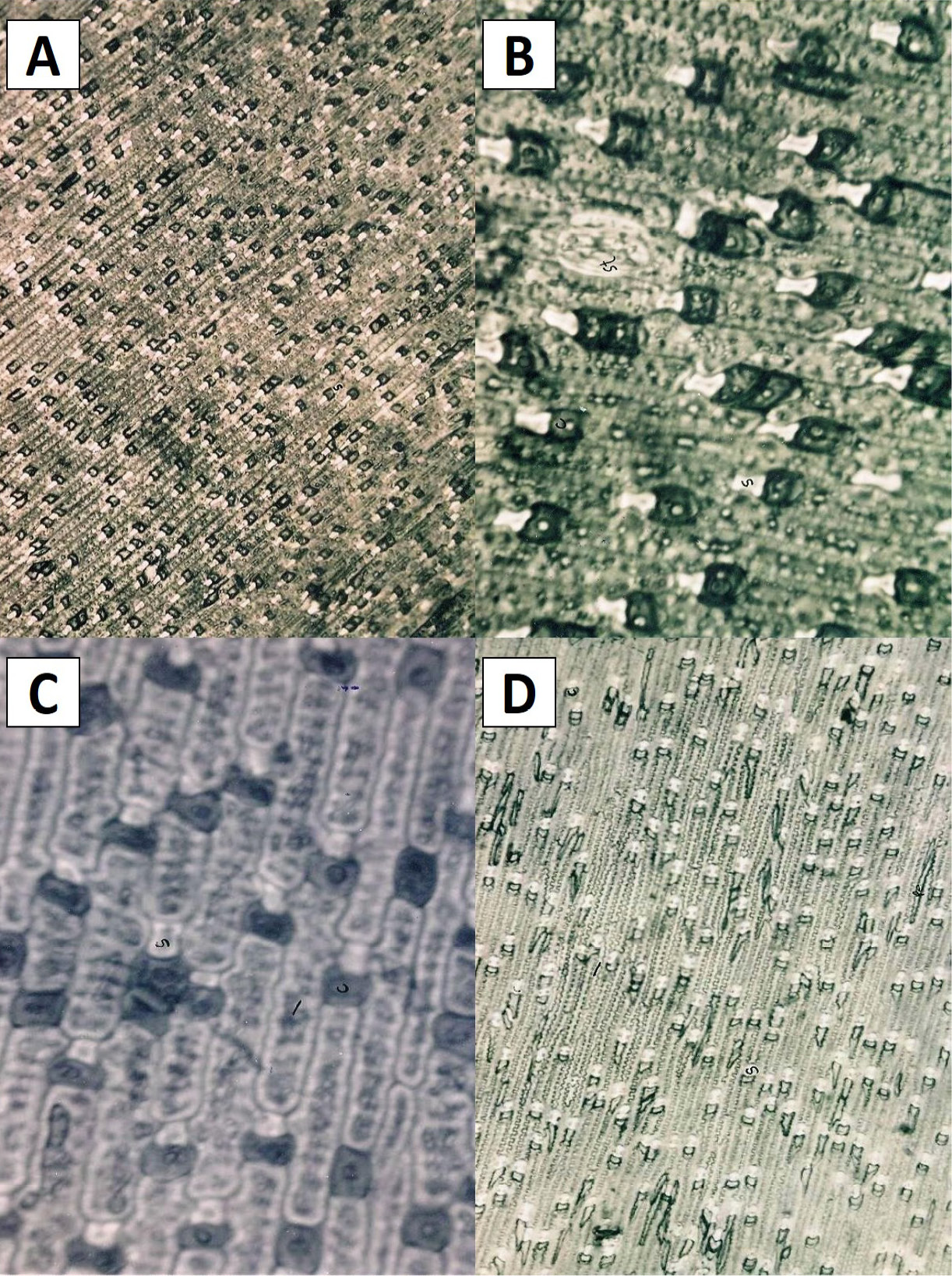
Photomicrographs of the stem epidermis of the sugarcane variety: A: (X 208); B: L-118 (X 416); C: CP72-2086 (X 416); D: CP43-33 (X 208). c, cork cell; s, silica cell; st, stomata; l, long cell; pe, pointed elongated
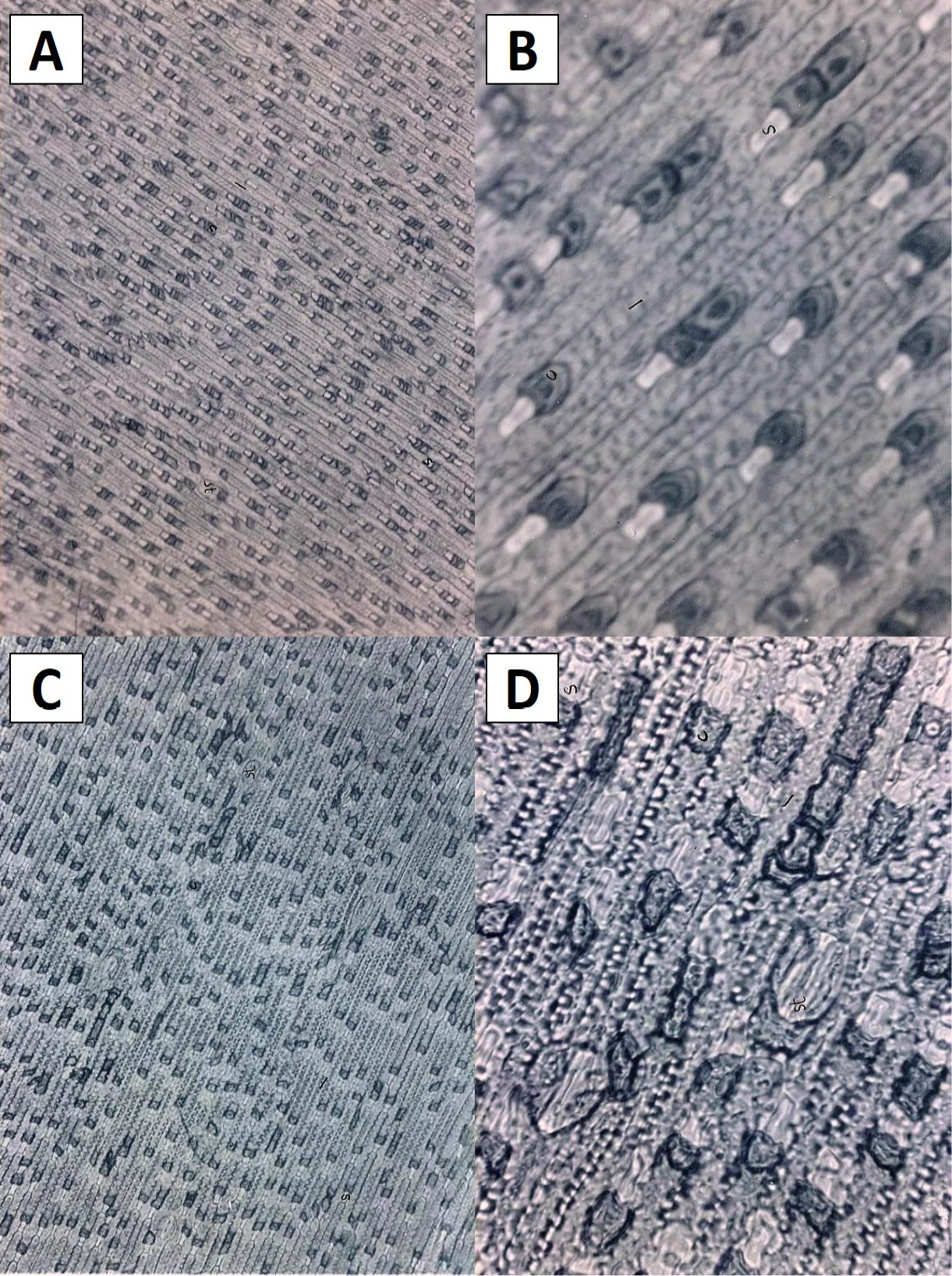
Photomicrographs of the stem epidermis of the sugarcane variety: A: HSF-240 (X 208); B: HSF-240 (X 416); C: SPF-213 (X 208); D: SPF-213 (X 416). c, cork cell; s, silica cell; l, long cell; st, stomata
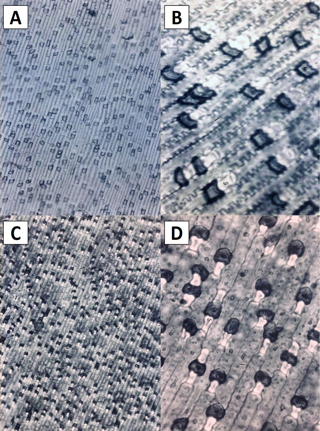
Photomicrographs of the stem epidermis of the sugarcane variety: A: CO-1148 (X 208); B: CO-1148 (X 416); C: CoL-54 (X 208); D: CoL-54 (X 416). c, cork cell; s, silica cell; l, long cell
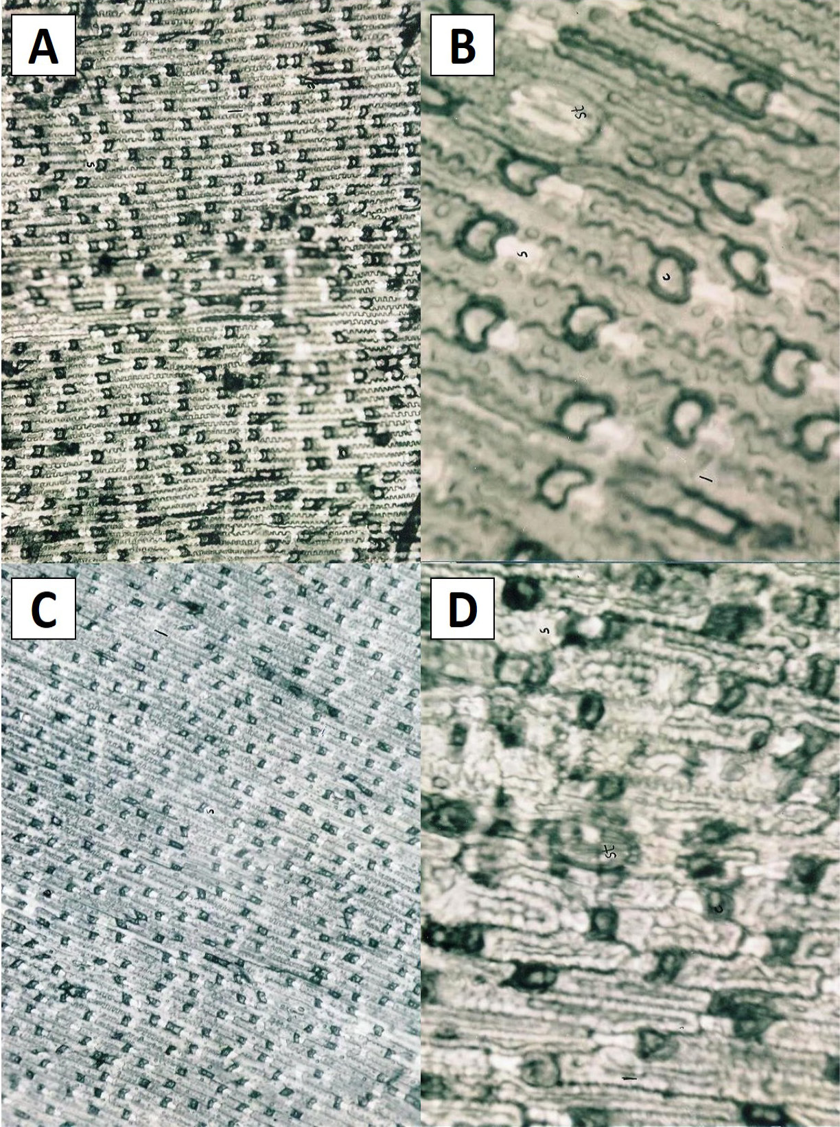
Photomicrographs of the stem epidermis of the sugarcane variety: A: CoL-29 (X 208); B: CoL-29 (X 416); C: CO-975 (X 208); D: CO-975 (X 416). c, cork cell; s, silica cell; I, long cell; st, stomata
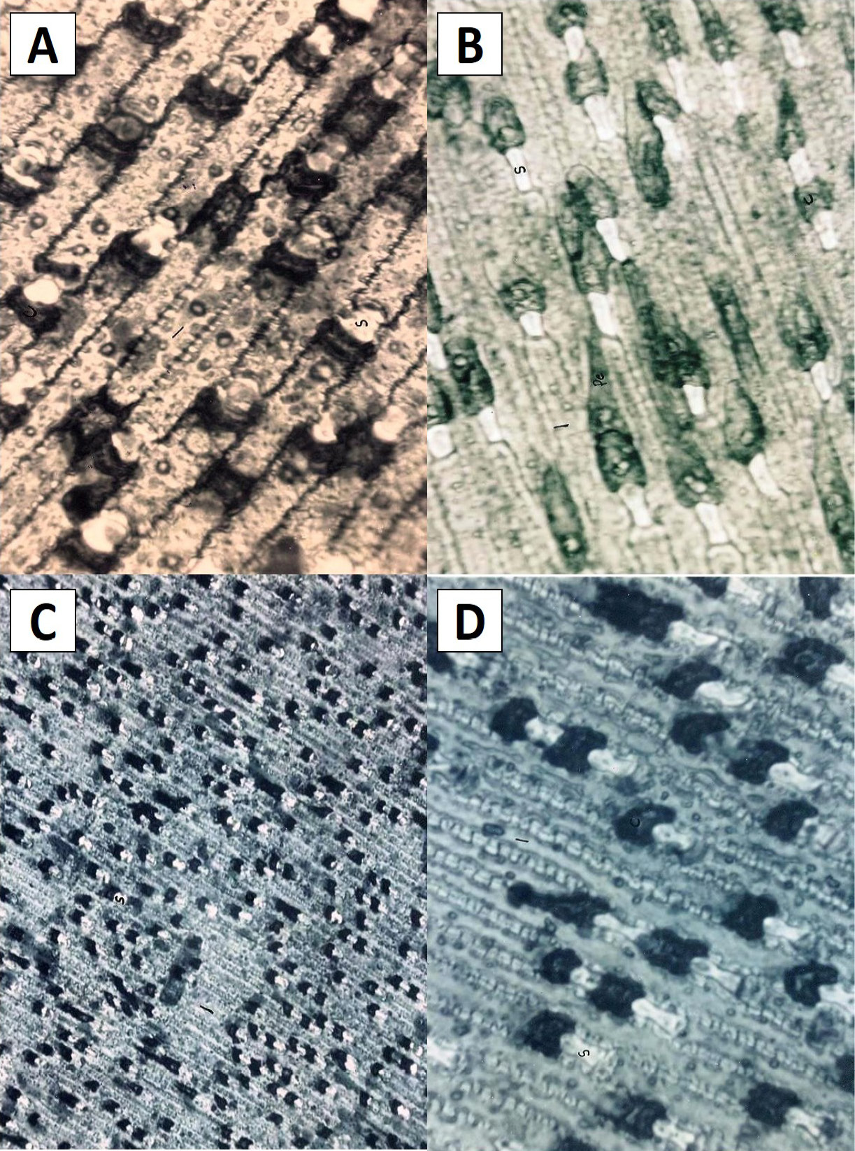
Photomicrograph of the stem epidermis of the sugarcane variety: A: Cojv-84 (X 416); B: BL-4 (X 416); C: Triton (X 208) D: Triton (X 416). c, cork cell; s, silica cell; l, long cell; pe, pointed elongated
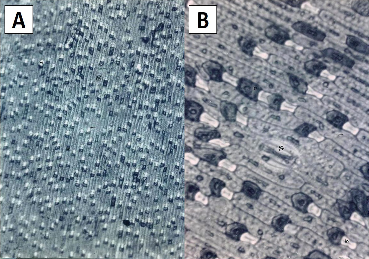
Photomicrographs of the stem epidermis of the sugarcane variety BF-162: A: (X 208); B: (X 416). c, cork cell; s, silica cell; I, long cell; st, stomata
References
- Artschwager, E. 1939. Illustrated outline for use in taxonomic description of sugarcane verities. Proc. Int. Soc. Sugarcane Technol., 6: 116-128.
- Artschwager, E. 1930. A comparative study of the stem epidermis of certain sugarcane varieties. J. Agric. Res. 41: 853-865.
- Barber, C.A. 1915. Studies in Indian sugarcanes. I. Punjab canes. Indian Dept. Agric. Mem. Bot. Ser. 7: 1–112
- Chaudhari, K.S., M. Arshad, G. Mustafa, S. Fatima, M.S. Amjad and F. Yasmeen. 2014. Foliar epidermal anatomy of grasses from Thal desert, district Khushab, Pakistan. Int. J. Biosci. 4(8): 62-70.
- Cowgill, H.B. 1917. A method of identification and description of sugarcane Varieties and its application to types grown in Portu Rico. J. Dept. Agric. Porto Rico. Pp. 119-140, ISSN 2308-1759. Available at: http://revistas.upr.edu/index.php/jaupr/article/view/3352 (Accessed on: 10 may 2017).
- Dahlgren, R.M.T., and H.T. Clifford. 1982. The monocotyedons: A comparative study. Academic Press, London, Pp. 378.
- Elahi, N.N., and M. Ashraf. 2002. A comparative study of stem epidermis of six sugarcane varieties. Biol. Pak. 48(1-2): 175–184.
- Frohnmeyer, M. 1914. Die entstehung und ausbildung der kieselzellen bei den gramineen. Biblthca. Bot. 21(86): 1-41.
- Hubbard, C.E. 1948. In: British Flowering Plants (ed. J. Hutchison), Vol. VIII, P.R. Gawthorn Ltd. Pp. 374.
- Khan, I., K.B. Marwat, I.A. Khan, I.J. Khan, H. Ali, K. Dawar and H. Khan. 2011. Invasive weeds of southern districts of Khyber Pakhtunkhwa-Pakistan. Pak. J. Weed Sci. Res. 17(2): 161-174.
- Metcalfe, C.R. 1960. Anatomy of the monocotyledons, Volume 1. Clarendon Press, Oxford.
- Metcalfe, C.R. 1963. Comparative anatomy as a modern botanical discipline. In: Advance botanical research (ed. R.D. Preston), Volume 1. Academic Press, London. Pp. 101. https://doi.org/10.1016/s0065-2296(08)60180-9[CrossRef]
- Prat, H. 1936. La Systématique des Graminées. Ann. Sci. Nat. Bot. 18: 165–258.
- Rao, J.T., and A. Balasubramanian. 1953. Relation between anatomical characters and chromosome numbers in Saccharum Spontaneum. Curr. Sci. 22: 247.
- Steel, R.G.D. and J.H. Torrie. 1980. Principles and procedures of statistics, 2nd edn. McGraw-Hill Book Co., New York.
- Subrahmanyam, N.S. 1996. Laboratory manual of plant taxonomy. Vikas Publishing House (Pvt.) Ltd., New Dehli.
- Ullah, Z., M.A. Khan, A. Mushtaq, Z. Muhammad and U. Kifayat. 2011. Systematic implications of foliar epidermis in andropogoneae (poaceae) from Hindukush-himalayas Pakistan. J. Med. Plants Res. 5(6): 949-957.
- Van Dillewijn, C. 1952. Botany of the sugarcane. Waltham, MASS., USA.
Services
Article usage statistics
Loading…