Research Article · Volume 10, Issue 2, 19 June 2022 · Open Access
Spreading Phenomena of Mobile Colistin Sulfate Resistant (Mcr-1) in Broiler Chickens and its Residue In Chicken Meat
· ·
Affiliations & Notes ⌄
1Reference Laboratory for Veterinary Quality Control on Poultry Production, Animal Health Research Institute (AHRI), Agriculture Research Centre (ARC), Dokki, Giza, Egypt. *Correspondence | Azhar G Shalaby, Reference Laboratory for Veterinary Quality Control on Poultry Production, Animal Health Research Institute (AHRI), Agriculture Research Centre (ARC), Dokki, Giza, Egypt
The results presented in Table (2) showed that the incidence of colistin sulfates residues in chicken muscles is 33.7% (28 out of 83) for the total samples examined.
The incidence of mcr-1 gene amongst the studied E. coli strains was 53%.
Our study recorded a percentage of 25.5% E. coli in diseased examined broiler chickens, and these isolated E. coli strains were described to have a high frequency of colistin resistance.
The obtained results in the current study regarding the detection of mcr-1-positive E. coli in chicken and chicken meat in Egypt, indicating the likelihood of spreading of this gene to humans through consumption of chicken meat that may lead to resistance against the colistin sulfate.
Selected from the article's own text.
Abstract
Colistin is considered the lastly used antimicrobial drug for the treatment of many infections. As the extensive use of colistin, resistance has been increasing rapidly, thus, 200 different chicken samples were examined for isolation of E. coli and for colistin resistance gene. Eighty-three selected samples (chicken breast) were subjected to detection of colistin residues by HPLC (High-performance liquid chromatography). In this research, we used polymerase chain reaction (PCR) for the detection of the mcr-1 gene, and phylogeny was performed on three isolates to detect mcr-1 gene mutations and relationships. The percentage of isolated E. coli was 25.5%. All isolates showed resistance to colistin in disc-diffusion assay, while in MIC (minimum inhibitory concentration) method 68.8% exhibited resistance. Colistin was recorded as 33.7% in chicken breast by HPLC. Furthermore, mcr-1gene was detected 54% using PCR. Sanger sequencing revealed the same identity (100%) between the three examined strains despite coming from different sources. It could be concluded that the practices inside poultry farms might be a possible source of spread of antimicrobial resistance to the food chain in Egypt.
Antimicrobial resistance is reported as a large problem in animal and human health worldwide. The extensive use of antibiotics in poultry has led to the development of resistance against many types of antibiotics, even to the last alternative of drugs like colistin . Colibacillosis is a disease caused by different strains of pathogenic E. coli strains named Avian Pathogenic E. coli (APEC) which causes two forms of the disease: acute form, which is characterized by high mortality, and septicemic picture. While, subacute or localized being caused inflammation in the internal organs in addition to, egg yolk peritonitis in layers , . Also, the extended use of cephalosporin is leading to E. coli resistant strains that are transferred from food animals to humans causing a public health threat . As the global population is increasing, so there is significant pressure on poultry production. Furthermore, China is considered a major producer of poultry. The animals could also be a source of transmission of colistin-resistant bacteria to humans. This privilege is very important to be known the likelihood of resistance to colistin in some diseased persons . Mcr-1 genes are distributed widely between animals and transferred rapidly between Enterobacteriaceae . The transformed therapeutic role is now theoretically susceptible by a number of plasmid genes mcr-1, to mcr-5 that facilitate the resistance to colistin , . In this work, we aimed to screen the incidence of E. coli APEC and try to seek the problem of colistin-resistant and its residual in chicken meat with a phylogenetic analysis of the mcr-1 gene.
Materials and Methods
Bacteriological investigation for detection of avian colibacillosis
Two hundred samples of (liver, heart and lungs) were collected from colibacillosis-infected broiler chickens suffering from omphalitis, coli-septicemic form, and pericarditis. The chicken organs were pooled altogether and then examined.
Isolation of the main causative agent of avian colibacillosis from infected chickens were carried out using the Ethylene Methylene Blue (EMB), and MacConkey agar media. The microbiological screening for 200 samples was done according to CLSI (2017) and Pasteran, et al. (2020).
Antimicrobial sensitivity testing
Isolated APEC strains were tested against colistin by using two tests i.e., disc diffusion test and MIC (minimum inhibitory concentration) test. Disc diffusion was performed according to the Clinical and Laboratory standards institute (CLSI)(2017).MIC was performed according to Clinical and Laboratory standards institute (CLSI) andEMA/AMEG (EU cast) 2016.
MIC interpretation according to (CLSI) is: resistant (>2 μg/ml) and sensitive (≤ 2 μg/ml),
while in EU cast interpretation: (>4 μg/ml) is resistant and sensitive (≤ 4 μg/ml).
Analytical methodology
Chemical reagents: Analytical standard of colistin sulfate was obtained from HEBEI Co. (China), while other reagents viz., 9-fluorenylmethyl chloroformate (FMOC-Cl), trichloroacetic acid, sodium hydroxide, acetone, sodium hydrogen carbonate, boric acid, HPLC grade methanol, acetonitrile, and tetrahydrofuran was also purchased from same source. Ultrapure water was produced using a Milli-Q system (Millipore, Bedford, MO, USA). The blank muscle employed for quality control (QC) was purchased from Animal Production Institute.
Device and chromatographic parameters: The drug was analyzed by HPLC (Agilent 1200) by reversed-phase Kinetex® XB C18 column (4.6 mm i.d., 250 mm, 5 µm). All measurements were carried out at excitation and emission wavelengths of 260 nm and 315 nm, respectively, and the column temperature was 40 °C. The mobile phase components (acetonitrile/ tetrahydrofuran/ water (50:14:20, v/v/v)) at 1.6 ml/min flow rate.
Standard and quality control samples preparation: Stock standard solution (1 mg/mL) was prepared in HPLC- water. It was diluted to obtain the fortification solution at a concentration of 10 ppm that should be prepared freshly. The calibration curve was created by fortifying blank chicken muscle with various volumes of fortification solution to yield a concentration range of 50- 1000 ppb (part per billion) (calibration samples) and spike blank muscle to prepare quality control (QC) samples at a low level of 75 ppb, moderate level 150 ppb and at a high level 300 ppb, then analyzed as mentioned below.
Sample preparation
The extraction of colistin from muscle samples and calibration samples was done as described by Hanai et al. (2018) using solid-phase extraction (SPE) followed by derivatization using FMOC-Cl solution.
Intra-lab validation of the HPLC- assay
This method was validated according to (USP 41-NF 36, 2018). The validation parameters were determined using quality control samples (QC).
Molecular examination
DNA extraction: DNA extraction from samples was performed using the QIAamp DNA Mini kit (Qiagen, Germany, GmbH) according to manufacture instructions. The Primers used were obtained from Metabion (Germany) phoA gene forward (CGATTCTGGAAATGGCAAAAG), reverse primer (CGTGATCAGCGGTGACTATGAC) the product was observed at 720 bp according to Hu et al. (2011). The mcr-1 gene forward (CGGTCAGTCCGTTTGTTC), reverse primer (CTTGGTCGGTCTGTAGGG), the reaction and cycles for PCR was described by Newton-Foot et al. (2017). The reaction was performed in an Applied biosystem 2720 thermal cycler.
Analysis of the amplified Products: The products of PCR were separated by electrophoresis on 1.5% agarose gel according to Sambrook et al. (1989) (Applichem, Germany, GmbH). A gene ruler 100 bp ladder (Fermentas, Germany) was used to determine the fragment sizes. The gel was photographed by a gel documentation system (Alpha Innotech, Biometra) and the data was analyzed through computer software (Automatic Image Capture Software (Protein Simple, formerly Cell Biosciences, San Jose, CA, USA).
Genetic and phylogenetic analyses: The PCR products were purified by using a QIA-quick PCR Product extraction kit (Qiagen, Gmbh, Germany). The reactions were achieved using a Bigdye Terminator V3.1 cycle sequencing kit (Perkin-Elmer), and purification was performed using Centri-Sep spin columns (Thermo Fisher, Germany). The Basic Local Alignment Search Tool (BLAST®) was used for determining the phylogenetic distances between the tested strains. The MegAlign module of Lasergene DNA-Star version 12.1 was used to determine phylogenetic distances among the analyzed strains , and MEGA6 was used to create a phylogenetic tree .
Results
Two hundred samples of diseased broiler chickens were examined for avian pathogenic E. coli APEC. Among these 25.5% (51/200) birds were diagnosed as avian colibacillosis as shown in Table (1). Total of 83 samples out of 200 (33.7%) were positively selected by microbiological screening for HPLC examination. An antimicrobial sensitivity test was done with colistin sulfate. 100% of isolates were resistant to colistin sulfate, with disc diffusion test and it was 68.8% with MIC method.
Table 1: Incidence of Avian Pathogenic E. coli (APEC) in chickens.
No. of positive samples
% of positive samples
Lesion
Age of chicken (day)
19
19/51
(37.2 %)
Omphilits
1
18
18/51
(35.2%)
Colisepticemia
1
14
14/51 (27.4%)
Periheptitis – Pericarditis
20-33
Total: 51 positive samples out of 200
Intra lab validation of HPLC assay
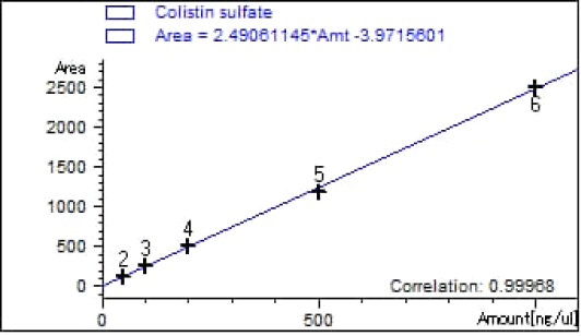
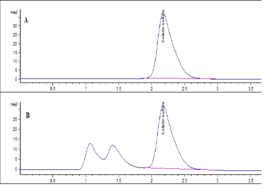
Colistin sulfate standards at a range of 50-1000 ppb were prepared in blank chicken muscles with a correlation coefficient= 0.99968 as shown in Figure (1). The obtained chromatograms showed the specificity of the analytical method as there is no peak of the matrix that interferes with the intended peak as shown in Figure (2).
Colistin sulfate recovery from chicken muscle was ranged from 85% to 93%. The coefficient of variance (CV) of the intra- and inter-day precisions were 0.12 and 1.3%, respectively. The pooled CV for robustness was not exceeding 4.3%. The detection and quantification limits were 3.6 and 10.8 ppb, respectively for 100 µL injection volume.
Results of colistin residues
The results presented in Table (2) showed that the incidence of colistin sulfates residues in chicken muscles is 33.7% (28 out of 83) for the total samples examined. The 28 samples that contain colistin sulfate residues exceed the maximum residue limits (150 ppb) recommended by EMEA.
MRL: maximum residual limit; PPB: part per billion)
Results for PCR
All the isolated strains were positive and confirmed by PCR test by examination of the phoA gene. The incidence of mcr-1 gene amongst the studied E. coli strains was 53%.
Nucleotide sequencing results
Three randomly selected strains carrying mcr-1 gene were subjected to sequencing using Bigdye Terminator V3.1. The phylogenetic tree was created and analyzed for the examined strains by neighbor-joining, and maximum parsimony. The examined strains were deposited under three accession numbers (OM362242 AZHM 8, OM362243 AZHM 9, and OM362244 AZHM 17).
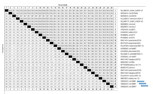
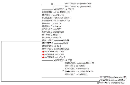
It was obvious that there was maximum identity (100%) between the three examined strains as shown in the identity percentage Figure (3) and also in the phylogenetic tree as shown in Figure (4).
Discussion
Colistin has been evaluated as a vital antimicrobial that is used extensively in human treatment due to its effectiveness against multi-drug resistant bacteria of negative Gram (Apostolako and Piccirillo, 2018). Also, the distribution of colistin-resistant bacteria carrying colistin resistance genes is considered a warning for infection treatments . The high occurrence of the mcr-1 gene in Escherichia coli from meat (14.9%) and birds reached 28%, so it can be easily transmitted to man . Our study recorded a percentage of 25.5% E. coli in diseased examined broiler chickens, and these isolated E. coli strains were described to have a high frequency of colistin resistance. It reaches 100% in the case of those tested with the disc diffusion test and 68.8% with the minimum inhibitory concentration method. The current results are well-matched to Ferreira et al. (2012), who studied data from animal products and isolated colibacillosis (19.8%) as the leading cause of condemnations of bird meat in 2010 in South Brazil. Wang et al. (2021) reported about 563 isolates of E. coli that were recovered from tissues and raw chicken meat samples in eastern China.
Numerous antibiotic classes are widely used in animals as growth promoters, including tetracyclines, fluoroquinolones, sulfonamides cephalosporins and beta-lactams, . About 90% of antibiotics used as the sub-therapeutic doses in poultry and animals production. However, our findings are nearly as high as those of Dawadi et al. (2021), who reported a high colistin resistance percentage of up to 30%, compared to another finding in Europe that reach (12.4%) as described by Kempf et al. (2013). In developing countries, the risk of residue is very high as a result of a lack of detection methods and monitoring that control the residue of the drug level in foods in the form of extreme residue limits . Multiple researchers have proven that the existence of residual antibiotics in farm animal foods is the main human health impact .
In the present study, 83 samples were positively selected out of 200 to be analyzed by HPLC. The percentage of colistin was 33.7% (28 out of 83). There were different studies that mentioned the colistin residue. Thus, one of them shown by Thuat et al. (2002) who discussed the residues of antibiotics in animals, especially in chicken meat and pork. In addition, they reported that 26 different antibiotics were extremely used in pig and chicken production, from which colistin appeared as a residue by 13%. However, a closely matched study was conducted by Bristy et al. (2019), who found that colistin residue was 50% (18 out of 36) in broiler meat using Thin Layer Chromatography assay.
The mcr-1 gene was mostly recovered from enterobacteria isolated from different sources like food animals, humans, and the environment in different countries all over the world , . Subsequently, more variants of the genes (mcr-1 to mcr-9) related to colistin resistance have been identified in family Enterobacteriaceae . On examination of mcr-1 gene, we reported a percentage of 53% presence of genes. This finding was consistent with that of Moawad et al. (2018), who confirmed and identified the presence of mcr-1 gene from E. coli isolates recovered from healthy broilers. Also, the presence of the mcr-1 gene was found to be very high amongst E. coli that isolated from diseased broiler which died from colibacillosis in Tunisia . Hence, we are in contrast to that recorded by Yang et al. (2017) who found the mcr-1 gene was detected with a percentage of 5.11% (58/1136) of Escherichia coli isolates of chicken origin from different places in China. The occurrence of the colistin resistance mcr-1 gene with a percentage of 60% (6/10) in E. coli strains from the chicken was mentioned by Uddin et al. (2022); also they added that the high percentage indicated the predominant in food animals in Bangladeshi. Another study supports our results where the percentage of E. coli isolation was 61.7% from poultry guts and the incidence of mcr-1 was 36.4% .
The current study sequenced three strains of positive mcr-1 and found that they were Geno-typically identical with a percentage of 100%. Also, as shown in Figures 3 and 4 The detailed sequence analysis of that was almost of high identity to the mcr-1-positive plasmid pCFSAN061769_01, which reached 99.9% (with GeneBank accession no. CP042970.1) in an E. coli isolated from cheese in Egypt. And that indicates that this plasmid type is circulating between different sources . The examined strains were showed different identity percentages with different strains randomly selected from the GeneBank. As a result, it provided a maximum identity percentage of 100% with (NG052893.1, MK620991, and KU934209), as well as a percentage of 99.3% with a Chinese strain (KU934208), and the same percentage with MW811403 Klebsilla pnemonaie and MW811404 Pseudomonas aurignosa. Also, it gave a percentage of 95.8 with the Salmonella enterica strain (NG.067235). This variation of identity percentage may be due to the different geographical distribution as discussed by Yang et al. (2017) during the comparison of the multilocus sequence types (MLST) of mcr-1 from different areas. However, there was a deficiency of data concerning the epidemiology of the colistin-resistant mcr genes in E. coli of food origin, especially in Egypt . The results of the current study are compatible with those obtained by Ahmed et al. (2019) who studied the correlation between humans, wild birds and water sources using phylogenetic analysis of mcr-1. The current results agreed with different studies in Africa that mcr-1-positive E. coli has been found in food matters, including chicken meat and carcasses ; Hassan et al., 2020).
Conclusion
The obtained results in the current study regarding the detection of mcr-1-positive E. coli in chicken and chicken meat in Egypt, indicating the likelihood of spreading of this gene to humans through consumption of chicken meat that may lead to resistance against the colistin sulfate.
Conflict of interest
The authors manifested that they have no conflicts of interest (not applicable).
Authors’ contributions
HK Sorour, MAM Saleh and AG Shalaby established the plan and ideas for work, H K Sorour isolated E. coli isolates, AG. Shalaby executed the molecular detection of the mcr-1 gene by PCR technique and also sequence and phylogenies for the selected isolates, MAM Saleh was responsible for the screening by HPLC. All the authors wrote and revised the manuscript. All the authors read and approved the manuscript.
Figures
Figure 1. Figure 1.
Standard curve of Colistin sulfate in blank chicken muscle
Figure 2.
Chromatograms showing 200 ppb of Colistin sulfate in water (A) and in blank muscle (B) at a retention time 2.16 min.
Figure 3.
The Genetic distance between the three E.coli strains carrying mcr-1 and randomly selected strains from Gene BANK
Figure 4.
The phylogenetic tree between isolated E. coli strains carrying mcr-1 and other randomly selected strains from Gene BANK.
References
Abiola OO, Serge M, Jean-Marc R (2015). Emergence of Colistin-Resistant Bacteria in Humans Without Colistin Usage: A New Worry and Cause for Vigilance, Inter. J. Antimicrob. Age.· https://doi.org/10.1016/j.ijantimicag.2015.11.009[CrossRef]
Ahmed ZS, Elshafiee EA, Khalefa HS, Kadry M, Hamza DA (2019) Evidence of colistin resistance genes (mcr-1and mcr-2) in wild birds and its public health implication in Egypt. Antimicrob. Resis Infect. con. 8:197. https://doi.org/10.1186/s13756-019-0657-5.[CrossRef]
Altschul SF, Gish W, Miller W, Myers EW, Lipmanl DJ (1990). Basic local alignment search tool. J. Mol. Biol., 215(3): 403-410. http//doi.org/10.1016/S0022-2836(05)80360-2[CrossRef]
Apostolakos I, Piccirillo A (2018). A review on the current situation and challenges of colistin resistance in poultry production. Avian Path. 47(6):546-558. https://doi: 10.1080/03079457.2018.1524573[CrossRef]
Borowiak M, Fischer J, Hammer JA, et al. (2017). Identification of a novel transposon-associated phosphoethanolamine transferase gene, mcr-5, conferring colistin resistance in d-tartrate fermenting Salmonella enterica subsp. enterica serovar Paratyphi B. J. Antimicrob. Chemother. 72(12):3317–3324. https//doi.org/10.1093/jac/dkx327.[CrossRef]
Bristy NI, Das S, Al Noman Z, Ferdous J, Sachi S, Kabir SL, Sikder MH (2019). Colistin residue in broiler: Detection in different growth stages. Asian-Aust. J. Food Safe. Secu. 3(1): 43-47. https://doi.org/10.3329/aajfss.v3i1.55926.[CrossRef]
Carattoli A, Villa L, Feudi C, Curcio L, Orsini S, Luppi A, et al. (2017) Novel plasmid-mediated colistin resistance mcr-4 gene in Salmonella and Escherichia coli, Italy 2013, Spain and Belgium, 2015 to 2016. Euro. Surveill. 22 (31). https://doi.org/10.2807/1560-7917.ES.2017.22.31.30589[CrossRef]
Clinical and Laboratory Standards Institute (2017). Performance standards for antimicrobial susceptibility testing. 27th Informational Supplement Document M100- S27, CLSI, Wayne. Vol. 37(1).
Clinical and Laboratory Standards Institute CLSI (2017). Performance standards for antimicrobial susceptibility testing. 27th Informational Supplement Document M100- S27, CLSI, Wayne. Vol. 37(1).
Dawadi P, Bista S, Bista S (2021). Prevalence of Colistin-Resistant Escherichia coli from Poultry in South Asian Developing Countries. Vet. Med. Inter. Article ID 6398838, 5. https://doi.org/10.1155/2021/6398838.[CrossRef]
Dhaouadi S, Soufi L, Hamza A, Fedida D, Zied C, Awadhi E, Mtibaa M, Hassen B, Cherif A, Torres C (2020). Co-occurrence of MCR-1 mediated colistin resistance and _-lactamase-encoding genes in multidrug-resistant Escherichia coli from broiler chickens with colibacillosis in Tunisia. J. Glob. Antimicrob. Resist. 22: 538–545. https://doi.org/10.1016/j.jgar.2020.03.017.[CrossRef]
EMA/AMEG (European Medicines Agency - Antimicrobial Advice Ad Hoc Expert Group), 2016. Updated advice on the use of colistin products in animals within the European Union: development of resistance and possible impact on human and animal health (EMA/ EU CAST (2016). European Committee on Antimicrobial Susceptibility Testing Breakpoint tables for interpretation of MICs and zone diameters Version 6.0, valid from 2016-01-0.
Ferreira TZ, Sesterhenn R, Kindlein L (2012). Economic losses of main causes of condemnation of the broiler carcass in a slaughterhouse under federal inspection on rio grande do sul, Brazil. Acta Sci. Vet. 40 (1): 01-06.
Gharaibeh MH, Shatnawi SQ (2019). An overview of colistin resistance, mobilized colistin resistance genes dissemination, global responses, and the alternatives to colistin: A review. Vet. World. 12(11): 1735–1746. https://doi.org/10.14202/vetworld.2019.1735-1746.
Hanai Y, Matsuo K, Kosugi T, Kusano A, Ohashi H, Kimura I, Yoshio T (2018). Rapid, simple, and clinically applicable high-performance liquid chromatography method for clinical determination of plasma colistin concentrations. J. Pharma. Heal. Care Sci. 4(1): 1-9. https://doi.org/10.1186/s40780-018-0119[CrossRef]
Hassen B, Abbassi MS, Ruiz-Ripa, L, Mama OM, Hassen A, Torres C, Hammami S (2020). High prevalence of mcr-1 encoding colistin resistance and first identification of blaCTX-M-55 in ESBL/CMY-2-producing Escherichia coli isolated from chicken faeces and retail meat in Tunisia. Int. J. Food Microbiol. 318: 108478. https://doi.org/10.1016/j.ijfoodmicro.2019.108478.[CrossRef]
Hu Q, Tu J, Han X, Zhu Y, Ding C, Yu S (2011). Development of multiplex PCR assay for rapid detection of Riemerella anatipestifer, Escherichia coli, and Salmonella enterica simultaneously from ducks. J. Microbiol. Meth. 87 (1): 64–69. https://doi.org/10.1016/j.mimet.2011.07.007[CrossRef]
Islam S, Urmi, UL, Rana, M, Sultana, F, Jahan, N, Hossain B, Iqbal S, Hossain MM, Mosaddek MB, Nahar S (2020). High abundance of the colistin resistance gene mcr-1 in chicken gut-bacteria in Bangladesh. Sci. Reports. 10:17292 https://doi.org/10.1038/s41598-020-74402-4[CrossRef]
Jank L, Martins MT, Arsand JB, Motta TMC, Feij, TC, Castilhos, TDS (2017). Liquid chromatography-tandem mass spectrometry multiclass method for 46 antibiotics residues in milk and meat: Development and validation. Food Anal. Meth. 10:2152–2164. https://doi.org/10.1007/s12161-016-0755-4[CrossRef]
José MS, la Rosa M.Q, Maky M.A, Dandrawy M.K, Nordmann P, Poirel L (2021) Genomic Features of MCR-1 and Extended-Spectrum Lactamase-Producing Enterobacterales from Retail Raw Chicken in Egypt Microorganisms. 9: 195.
Kebede G, Zenebe T, Disassa H, Tolosa T (2014). Review on detection of antimicrobial residues in raw bulk milk in dairy farms. African J. Bas. Appl. Sci. 6(4): 87-97. http//doi.org/10.5829/idosi.ajbas.2014.6.4.8642[CrossRef]
Kempf I, Fleury, MA, Drider D (2013). “What do we know about resistance to colistin in Enterobacteriaceae in avian and pig production in Europe?” Inter. J. Antimicrob. Agents. 42 (5): 379–383. http//doi.org/10.1016/j.ijantimicag.2013.06.012.[CrossRef]
Moawad AA, Hotzel H, Neubauer H, Ehricht R, Monecke S, Tomaso H, Hafez HM, Roesler U El-Adawy H (2018). Antimicrobial resistance in Enterobacteriaceae from healthy broilers in Egypt: Emergence of colistin-resistant and extended-spectrum _-lactamase-producing Escherichia coli. Gut Pathog. 10: 39. http//doi.org/10.1186/s13099-018-0266-5[CrossRef]
Newton-Foot M, Snyman Y, Maloba MRB and Whitelaw AC (2017). Plasmid-mediated mcr-1 colistin resistance in Escherichia coli and Klebsiella spp. Clinical isolates from the Western Cape region of South Africa. Antimicrob. Resist. Infec. Con. 6 (78):1-7. http//doi.org/10.1186/s13756-017-0234-8
Nolan LK, Vaillancourt JP, Barbieri NL, Logue CM (2020). Colibacillosis,” in Diseases of Poultry, 14 Edn, eds D. E. Swayne, M. Boulianne,C. M. Logue, L. R.McDougald, N. Venugopal, and D. Suarez (Ames, IW: Wiley and Sons). https://doi.org/10.1002/9781119371199.ch18[CrossRef]
Pasteran F, Danze D, Menocal A, Cabrera C, Castillo I, Albornoz E, Lucero C., Rapoport M, Paola Ceriana P, Corsoa A. (2020). Simple Phenotypic Tests to Improve Accuracy in Screening Chromosomal and Plasmid-Mediated Colistin Resistance in Gram-Negative Bacilli. J. Clin. Microbiol. 59 (1) e01701-20. http//doi.org/ 10.1128/JCM.01701-20.
Poirel L, Jayol A, Nordmann P (2017). Polymyxins: antibacterial activity, susceptibility testing, and resistance mechanisms encoded by plasmids or chromosomes. Clin. Microbiol. Rev. 30(2):557–596. http//doi.org/10.1128/CMR.00064-16[CrossRef]
Sadek, M, la Rosa MQ, Maky MA, Dandrawy MK, Nordmann P, Poirel L (2021). Genomic Features of MCR-1 and Extended-Spectrum Lactamase-Producing Enterobacterales from Retail Raw Chicken in Egypt. Microorganisms. 9(1):195. http//doi.org/10.3390/9010195.[CrossRef]
Sambrook J, Fritscgh EF, Mentiates T (1989). Molecular Cloning. A Laboratory Manual. Vol. 3. Laboratory Press, Cold Spring Harbor, New York. p55-57.
Tamura K, Stecher G, Peterson D, Filipski A, Kumar S (2013). MEGA6: Molecular evolutionary genetics analysis version 6.0. Mol. Biol. Evol., 30(12): 2725-272. http//doi.org/10.1093/molbev/mst197.[CrossRef]
Thuat DT, Tuan NN, N, VTT, Hien LT, Lam VB, Ninh KT (2002). Study on antibiotic use and antibiotic residues in pork and broiler meat of Binh Duong province. J. Vet. Sci., 50–57.
Thompson JD, Higgins DG, Gibson TJ (1994). CLUSTAL W: Improving the sensitivity of progressive multiple sequence alignment through sequence weighting, position-specific gap penalties and weight matrix choice. Nucl. Acids Res., 22(22): 4673-4680. http//doi.org/10.1093/nar/22.22.4673[CrossRef]
Touati A, Mairi A (2021). Plasmid-determined colistin resistance in the North African countries: A systematic review. Microb. Drug Resist. 27(1):121-133. http//doi.org/10.1089/mdr.2019.0471.[CrossRef]
United States Pharmacopoeial Convention. (2018). USP 41-NF 36, chapter (1225) Validation of compendial procedures; United States Pharmacopoeia: North Bethesda.
Wang Y, Jiang M, Wang Z, Chen R, Zhuge X, Dai J (2021). Characterization of antimicrobial resistance in chicken-source phylogroup F Escherichia coli: similar populations and resistance spectrums between E. coli recovered from chicken colibacillosis tissues and retail raw meats in Eastern China. Poul. Sci. 100:101370 https://doi.org/10.1016/j.psj.2021.101370[CrossRef]
Wang Y, Tian GB, Zhang R et al (2017). Prevalence, risk factors, outcomes, and molecular epidemiology of mcr-1-positive Enterobacteriaceae in patients and healthy adults from China: an epidemiological and clinical study. Lancet Infect Dis. 17(4):390–399. http//doi.org/10.1016/S1473-3099(16)30527-8.[CrossRef]
Yamamoto I, Calvopina M, Izurieta R, Villacres I, awahara R, Sasaki M, Yamamoto M (2019). Colistin-resistant Escherichia coli with mcr genes in the livestock of rural small-scale farms in Ecuador Yoshimasa BMC Res. Notes, 12:121. https://doi.org/10.1186/s13104-019-4144-0[CrossRef]
Yang YQ, Li YX, Song T, Yang YX, Jiang W, Zhang AY, Guo XY, Liu BH, WangYX, Lei CW, Xiang R, Wang HN (2017). Colistin resistance gene mcr-1 and its variant in Escherichia coli isolates from chickens in China. Antimicrob. Agents Chemother. 61: e01204-16. https://doi.org/10.1128/AAC.01204-16.[CrossRef]
Uddin MB, Alam MN, Hasan M, Hossain SMB, Debnath M, Begum R, et al. (2022). Molecular Detection of Colistin Resistance mcr-1 Gene in Multidrug-Resistant Escherichia coli Isolated from Chicken, Antibiotics. 11 (1):97. http//doi.org/10.3390/antibiotics11010097
United States Pharmacopoeial Convention. (2018). USP 41-NF 36, chapter (1225) Validation of compendial procedures; United States Pharmacopoeiab: North Bethesda.