Research Article · Volume 10, Issue 12, 1 December 2022 · Open Access
Seroprevalence of Porcine Circovirus Type 2 in Domestic Pigs of Thailand: Two Decades After Nationwide Vaccines Application
· · ·
Affiliations & Notes ⌄
1Department of Animal Production Technology and Fisheries, School of Agricultural Technology, King Mongkut’s Institute of Technology Ladkrabang, 1 Chalongkrung Rd., Ladkrabang, Bangkok 10520, Thailand
2Faculty of Agricultural Technology, Burapha University, Sakaeo Campus, Sa Kaeo 27160, Thailand. *Correspondence | Chanathip Thammakarn, Department of Animal Production Technology and Fisheries, School of Agricultural Technology, King Mongkut’s Institute of Technology Ladkrabang, 1 Chalongkrung Rd., Ladkrabang, Bangkok 10520, Thailand
Thus, our results showed that although PCV2 might be ubiquitous in the domestic pig population, PCV2 seroprevalence in Thailand seems to be lower than expected.
However, the highest seroprevalence of PCV2 in pigs have been reported from farms with PMWS.
The vaccines against PCV2 have been used routinely with several programs, depending on ages of pig, types of vaccine, or diseases status of each of the farms.
The antibody levels in this study was accumulated from the farms disttibuted in dense areas of pig population across Thailand, proved that high prevalence of PCV2 antibodies was established in pig population in Thailand.
Selected from the article's own text.
Abstract
The antibody response of domestic pigs to porcine circovirus type 2 (PCV2) was investigated in commercial farms at various regions of Thailand during the year 2018-2021. A total 2,002 serum samples were collected from 16 farms located in 10 provinces and tested by Enzyme-Linked Immunosorbent Assay. Among total serum samples, 914 samples were further tested by polymerase chain reaction (PCR) to detect ORF2 gene. The results revealed that 81.17% of pigs had low positive level antibody against PCV2, while 10.29% had high positive titer. On the other hand 8.54% of pig had no antibody to PCV2. The low positive antibody level was found in most population in all farm sizes as well as all types of pig as in age categories. The PCR result illustrated that there was no viral gene in seronegative serum. While 0.8% of total low positive serum samples displayed positive by PCR. In addition, 3.23% of total high positive serum samples showed positive by PCR. The present study is the first nationwide serological prevalence of PCV2 antibody report since two decades of PCV2 vaccines was applied in Thailand. The results proved that high prevalence of PCV2 immunity was established, with the most population had antibody titer fall in low positive, while some of viremic pigs still presented, determined that the routine vaccination programs used in farms can control PCV2 efficiently since continuing vaccination.
The porcine circovirus type 2 is the one among crucial pathogens affecting pig production industry across the world. It is belonging to Circoviridae family, genus Circovirus, a closed-circular single-stranded DNA genome of approximately 1.7 kb in length and non-enveloped found in the outer structure. The genomic material comprises 2 major open reading frames (ORFs); ORF1, which are fundamental for viral replication, and ORF2 that codes for the Cap protein which involved in the viral attachment and represents the main target of the host immune response. Other proteins (ORF3 to ORF6) seem to have a modulatory activity in the host cell pathways, signaling, and apoptosis . The virus is devided into various types since isolated from the domestic pig. Porcine circovirus type 1 (PCV1) is considered as non-pathogenic and first isolated in cell culture contamination . On the contrary, porcine circovirus type 2 (PCV2) has been associated with various clinical signs in domestic pigs and assumed that PCV2 is the primary agent of postweaning multisystemic wasting syndrome (PMWS), which causes large economic losses in pig breeding. It is known that PCV2 is one of the causal agents of porcine dermatitis and nephropathy syndrome (PDNS) and porcine respiratory disease complex (PRDC) . The PCV2 is also participatory porcine reproductive and respiratory syndrome (PRRS) and with enteritis . The PMWS has caused chronic wasting syndromes at 5-12 weeks of age with clinical signs such as weight loss, growth retardation, anemia, jaundice, lymph nodes enlargement and dyspnea, largely affecting the productivity. Besides, PCV2 plays a significant role in complex diseases as it undergoes a process of co-infection with viruses (e.g., PRRS virus, swine influenza virus) and bacteria (e.g., Mycoplasma hyopneumoniae, Streptococcus suis, Haemophilus parasuis). This co-infection leads to high clinical signs and farm production problems (Afolabi et al,, 2017). The multiple disease syndromes are ascribable to PCV2 infection and are summarized under the term of porcine circovirus diseases (PCVD) . Recently, novel types of PCV has been identified, including PCV3 ; and PCV4 ; . It is currently under surveillance for its impact on swine industry. However, the PCV2 still accepted as the most affecting PCV to pigs, so far. The genetic characteristics of PCV2 allowed defining 8 genotypes (PCV-2a to PCV-2h) . But one single serotype has been classified . Based on serological examination PCV2 is believed to be widespread in the domestic pig population . The PCV2 can also be found in healthy pigs. Serological investigations confirmed that the number of passively acquired PCV2 antibodies in the blood of piglets begins to decrease during lactation. The antibody level is very low or even absent at the initial period of the post-weaning period . However, studies in pig farms of several regions of the world have shown that almost 100% of the animals are PCV2 seropositive at slaughter . In Thailand, PWMS has been reported since 1998, then spread widely in the eastern and central parts of Thailand with a high density of pig population during 2009-2015. The results showed that a high positive of about 44.09% to 80.00% in farms. However, Jantafong et al. (2011) reported that the prevalence of PCV2 infection in Thailand’s central region was approximately 10%. Furthermore, PCV2 detection was not significantly different between blood and fecal sample. However, it is age-dependent clearly and the virulence of PMWS outbreak depend on the vaccination programs and herd’s immunity .
Currently, PCV2 infection is regarded to be endemic in Thailand and has caused numerous defeats in the pig industry. Farms in all scales can be found in all of the Thailand regions. Hence, overall geographical area of Thailand should be defined as the same epidemic area, it is important to understand the serological prevalence of the disease since the vaccines have been used as the tool for controlling the disease for long time. Therefore, this study aimed to investigate the seroprevalence of PCV2 in domestic pigs of different farms located in different regions of Thailand, which would help to understand the epidemiology, controlling measures evaluation and improving the biosecurity system in farms.
MATERIALS AND METHODS
Samples Collection
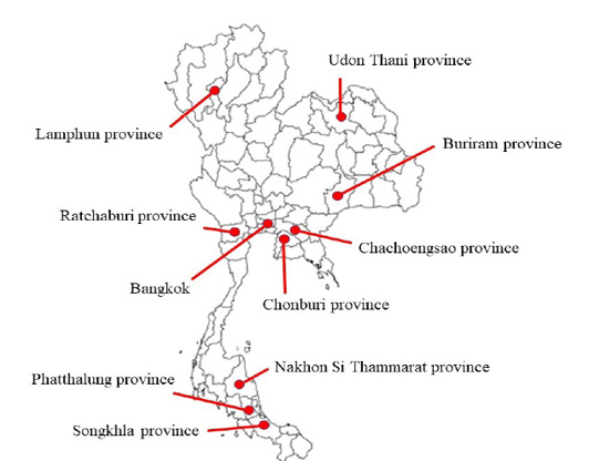
A serological survey was carried out during the year 2018-2021 to determine the seroprevalence of PCV2 in the domestic pigs of Thailand. This was done as part of health monitoring program of the farms. A total of 2,002 serum samples were collected from domestic pigs at different regions across the Thailand. Totally, 16 farms at dense areas of pig farming, located in 10 provinces scattered at different parts of Thailand were selected for sampling. Studied farms were categorized into3 groups based on number of pigs namely, small (50-500 sows), medium (501-5,000 sows) and large (>5,000 sows). Additionally, based on age the farms were categorized into 4 groups including nursery pig (2-10 weeks), fattening pig (10-26 weeks), gilt (>26 weeks, non-serviced) and sow (>26 weeks, serviced).There were 1 farm in northern (Lamphun province), 1 farm in central (Bangkok), 5 farms in eastern (Chachoengsao and Chonburi province), 4 farms in northeastern (Buriram and Udon Thani province), 1 farm in western (Ratchaburi province), and 4 farms in southern (Songkhla, Phatthalung and Nakhon Si Thammarat province) (Figure 1).
The blood samples were randomly collected for herd health monitoring regardless of with or without clinical signs, at different regions, farm sizes. In all farms, pigs have been immunized against PCV2 as routine measure, using different vaccine types and manufaturers according to own vaccination program of each farm. The collected samples were transported to the Laboratory of Molecular Genetics and Cellular Biotechnology (MGCB), Department of Animal Production Technology and Fisheries, School of Agricultural Technology, King Mongkut’s Institute of Technology Ladkrabang (KMITL) for serum separation, then processing and processed samples were stored at -20oC until further testing.
Serological Examination
The serum samples were investigated for IgG antibodies against PCV2 by indirect Enzyme-linked immunosorbent assay (ELISA) using PCV2 Antibody test kit (SK105; BioChek, Ascot, UK). The samples were tested according to the manufacturer’s instructions. Briefly, the serum was diluted according to the manual using provided diluent before adding into PCV2 antigen coated microtiter plate, then incubated at room temperature and washed. After that, conjugate (Anti-Swine: Alkaline Phosphatase in Tris buffer) was added and incubated again under room temperature. After second washing, pNPP (p-Nitrophenyl Phosphate) buffered substrate was added to each well. Finally, the reaction was terminated by adding stop solution after incubation at room temperature for 15 minutes. The optical density (OD) was determined using a monochromatic ELISA reader with a 405 nm wave length (AMR-100 Microplate Reader; Allsheng, Hangzhou, China). The result was calculated as S/P ratio. The cut-off was determined according to the criterias described in the instruction for positive and negative samples interpretation. Sample with an S/P ratio of 0.49 or less (titer ≤1,070) is considered as negative.
According to the study of Lin et al. (2020) which reported the serodynamics of pigs in PCV2 exposured farms measures using BioCheck ELISA kit, reveals that the S/P ratio of antibody to PCV2 will dramatically increased after exposed to PCV2 antigen, reaching between 2.0-2.5 in most pigs. Hence the authors have devided the positive samples in the present study into 2 categories regarding to the calculated S/P ratio. The S/P ratio which fall in the range 0.5-2.3 (titer between 1,071-5,766) are considered as low positive, while samples with an S/P ratio of 2.31 or greater (titer of ≥5,767) were determined as high positive.
Viral Genome Detection
Totally 914 serum samples were also tested for detection of PCV2 DNA by polymerase chain reaction (PCR). The DNA was extracted from serum using various genomic extraction kits, including TRIsureTM (Bioline, London, UK), ISOLATE II Genomic DNA Kit (Bioline, London, UK), dBIOZOL Genomic DNA Extraction (Bioer, Hangzhou, China), Biospin Whole Blood Genomic DNA Extraction Kit (Bioer, Hangzhou, China), MegaBio plus General Genomic DNA Purification Kit (Bioer, Hangzhou, China), and MegaBio plus Whole Blood Genomic DNA Purification Kit (Bioer, Hangzhou, China), according to the manufacturer’s instruction. The obtained DNA was measured for yield and purity using Nano-300 Micro-Spectrophotometer (Allsheng, Hangzhou, China) prior to PCR with primers targeting open reading frame 2 (ORF2) described by Cao et al. (2005), Chen et al. (2019) or by the real-time PCR (fluorescent probe) (LineGene K Plus; Bioer, Hangzhou, China) with the PCV II Real Time PCR Detection kit (Bioer, Hangzhou, China), which the specific primers and all reagents provided by manufacturer as commercial test kit. One of the above mentioned DNA extraction kits and PCR methods had been applied depending on the chemical set available in the laboratory when the samples were delivered to MGCB.
The study did not involve any animal experiments. All samples were submitted to the MGCB for diagnostic purposes. The permission of data using was granted by each farms and approved by The Animal Care and Use Committee of KMITL, approval number is CC-KMITL-/2021/021.
RESULTS
Serological Prevalence Of Pcv2
The seroprevalence of PCV2 in pigs revealed mainly low positive in all geographical regions, which was 81.17%. However, the proportion of low positive level varies according to location which were 77% in northern, 84.02% in northeastern, 82.26% in central, 81.33% in eastern, 70.63% in southern, and 68% in western. The high positive antibody against PCV2 was found in 10.29% of samples. There were 23% high positive in northern, 9.28% in northeastern, 4.71% in central, 14% in eastern, 18.25% in southern and 32% in western. Furthermore, seronegative groups revealed small numbers (8.54%) which fall in only 4 regions. There were 6.7% in northeastern, 13.03% in central, 4.67% in eastern and 11.11% in southern (Table 1).
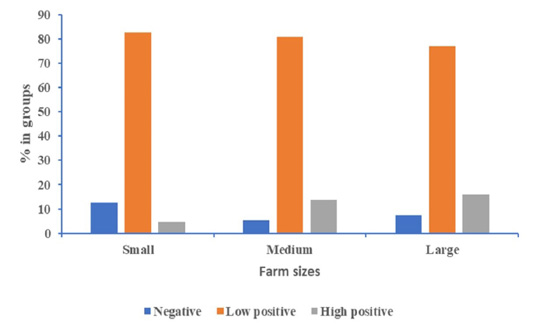
When the sizes of farm were considered, the results revealed that the most low positive number fall in small size farms, which was 82.69%. While medium and large size farms had 80.91% and 76.82% low positive antibody, respectively. The most high positive group was found in large farm size (15.88 %), while there were 13.89% and 4.6% were high positive in medium and small farms, respectively. Additionally, the most seronegative result was found in small farm size than medium and large farms, and those were 12.71%, 5.2% and 7.3%, respectively (Figure 2).
Table 1: Serological prevalence of PCV2 in different regions of Thailand.
Regions
% of sera at each level (No. of sera detected/ tested)
Negative1
Low positive2
High positive3
Northern
0 (0/100)
77 (77/100)
23 (23/100)
Northeastern
6.7 (26/388)
84.02 (326/388)
9.28 (36/388)
Central
13.03 (105/806)
82.26 (663/806)
4.71 (38/806)
Eastern
4.67 (26/557)
81.33 (453/557)
14 (78/557)
Southern
11.11 (14/126)
70.63 (89/126)
18.25 (23/126)
Western
0 (0/25)
68 (17/25)
32 (8/25)
Total
8.54 (171/2,002)
81.17 (1,625/2,002)
10.29 (206/2,002)
1Negative, S/P ratio ≤0.49 (titer ≤1,070)
2Low positive, S/P ratio 0.5-2.3 (titer 1,071-5,766)
3High positive, S/P ratio ≥2.31 (titer ≥5,767)
Table 2: Detection of PCV2 DNA in serum samples collected from different regions of Thailand.
Regions
No. of samples
% of positive by PCR (No. of sera detected/ tested)
Sero-Negative1
Low positive2
High positive3
Northern
55
0 (0/0)
0 (0/41)
0 (0/14)
Northeastern
388
0 (0/26)
0.92 (3/327)
0 (0/35)
Central
200
0 (0/26)
0 (0/172)
0 (0/2)
Eastern
120
0 (0/2)
2.78 (3/108)
20 (2/10)
Southern
126
0 (0/13)
0 (0/89)
4.17 (1/24)
Western
25
0/ (0/0)
0 (0/17)
0 (0/8)
Total
914
0 (0/67)
0.8 (6/754)
3.23 (3/93)
1Sero-Negative, S/P ratio ≤0.49 (titer ≤1,070)
2Low positive, S/P ratio 0.5-2.3 (titer 1,071-5,766)
3High positive, S/P ratio ≥2.31 (titer ≥5,767)
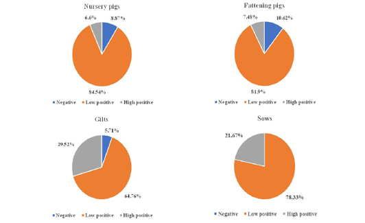
The low seropositive immune response to PCV2 was found mainly in all groups when categorized by age and displayed in Figure 3. There were 84.54% low positive in nursery
pigs, 81.9% in fattening pigs, 64.76% in gilts and 78.33% in sows. Meanwhile, the highest population that had high antibody level was in gilts (29.52%), followed by sows (21.67%), fattening pigs (7.48%) and nursery pigs (6.6%). Additionally, the most number of negative serum was found 10.62% in fattening pigs, followed by 8.87% and 5.71% in nursery pig and gilts, respectively. None of negative serum found in sows.
Orf2 Detection In Serum Samples
The results of PCR for ORF2 gene detection in 914 serum samples from various regions of Thailand is summarized in Table 2. The results showed that none of the detected PCV2 genetic material was derived from seronegative pigs. While some of low and high positive serum samples were detected as positive for PCV2 virus and those are only 0.8% and 3.23%, respectively.
DISCUSSION
The porcine circovirus associated disease (PCVAD) is now considered as one of the most important disease complexes in pigs worldwide. Since the first case of PCV2 infection in Thailand was confirmed in 1998, PCVAD then spread through the country ; ; . Although strict biosecurity measures to control the disease had been implemented in the field, the disease situation did not improve as expected. Thus the application of vaccine become an important tool to control the PCVAD. Vaccination with various types of vaccines against PCV2 has been applied and included in the routine programs of all commercial farms in Thailand. During two decades of vaccine use, there were few studies reported seroprevalence in pig population in Thailand, and even limited just in some local areas . So, the present study was taken to investigate the seroprevalence of PCV2 in pigs distributed in across the Thailand. This study also focused on prevalence in different farm sizes and ages of pigs.
The serological investigation results revealed that most pigs had seropositive at all regions of Thailand (91.46%). Among the seropositive population, most of them had low positive level of immunity against PCV2, distributed in all sizes of farms. The similar results had been displayed in all age categories. This was possibly caused by host response at early phase after immunization or in stage of seroconversion resulting from viremia. The interesting thing is the seronegative population is still remained, even after intensive vaccination in pig farms.
In this study, the result was represented as the samples derived from different regions of Thailand. Another perspective including farm sizes were also considered. Similar result was found as in the results enumerated in the regional aspect. All farm sizes revealed low positive immunity level as majority. The interesting thing is among farm size up to 50 sows and farms with more than 5,000 sows, have different levels of herd management, housing conditions, density as well as biosecurity system, also stress condition in captive pigs may affect the virus distribution. Nevertheless, our study showed that even with the differences in farm size, the antibody titer showed the similar level since PCV2 vaccines are applied in routine vaccination programs.
Among 4 age group categories of pigs, all of them illustrated that S/P ratio level mostly fall in range as displayed in the regions or farm sizes criterias. The interesting finding is no seronegative pig found in sow group. They were 100% seropositive, indicated that immunity against PCV2 had been established in all sows. This is very important key point of disease control measure. Because the maternal immunity will be delivered only through the seropositive sows. Then it plays important role in early protection for piglets prior to first vaccination. According to our research, in comparison to other age groups, gilts and sows had a higher of high positive rate. These results suggested that the presence of high levels of PCV2 maternal antibodies generally confer protection against PCV2 infection, but not total protection . When piglets are infected by contacting with the body fluids or vertical transmission of sows, the disease may develop after the maternal antibodies weaken or disappear. Therefore, it is essential to strengthening the control and detection of PCV2 in sows, which may play a role in reducing the economic loss caused by PCV2. López‐Soria et al. (2010) reported that sow and boar herds had pigs with medium to high titers, fattening pig herds had negative or low PCV2 antibody titers. The seroprevalence to PCV2 in sow herds ranged 93-100%, whereas it was 92-98% in fattening pig herds. Although these percentages were differcence comparable with the present study, but the same trend was found corresponding with our results.
Although the obtained results illustrated the seroprevalece of PCV2 of vaccinated pigs in Thailand, distributed in different regions, then enumerated into farm sizes and ages. The further testing had been processed, aimed to find out the viremia status hidden in serum samples. The collected serum then subjected to PCR testing for viral ORF2 gene detection. There is no viral genome detected in seronegative samples. While the positive PCR result was found in low positive immune level samples, but just in rare percentage (0.8%). This means among the normal herd immunity resulting from vaccination, the latent infection can still be found. The infection rate increased interestingly in high positive serum samples (3.23%), refers that highly immune response level may resulting from viremia. This means that the PCV2 can also outspread in vaccinated pigs and the PCV2 outbreak may repeat if preventive measures are not properly taken in the farms.
Currently, the usage of PCV2 vaccines in routine vaccination programs is adopted in all farms of Thailand. This high vaccination rate might also plays a role regarding the overall infection pressure and might explain the comparatively low amount of viremic status in our investigation. However, the PCVAD outbreaks in vaccinated pig herds can be occurred. In this case, antibody titer elicited by vaccination is assumed to be lower than by infection. Thus, our results showed that although PCV2 might be ubiquitous in the domestic pig population, PCV2 seroprevalence in Thailand seems to be lower than expected. A recent study showed that PCV2 DNA was detectable in sample materials not only from seropositive sows but also from seronegative sows . Therefore, seronegative sows can shed the virus to piglets. However, there was no viral gene detected in the serum of sows in the present study.
It should be annotated that great numbers of seropositive pigs can be found not only in farms with PMWS but also in farms without PMWS . However, the highest seroprevalence of PCV2 in pigs have been reported from farms with PMWS. . According to our findings, the geographical distribution of PCV2 infections in Thailand was intermittent, which may be due to differences in the denseness of farming, sanitation, farming techniques and environmental factors. The regional variation of PCV2 infection in Thailand may be related to feeding conditions, humidity, and local temperature. However, the results should be clarified with caution because there were few studies on seroprevalence rates of PCV2 in Thailand.
CONCLUSIONS AND RECOMMENDATION
Overall, PCVAD is an endemic disease in Thailand. The vaccines against PCV2 have been used routinely with several programs, depending on ages of pig, types of vaccine, or diseases status of each of the farms. The antibody levels in this study was accumulated from the farms disttibuted in dense areas of pig population across Thailand, proved that high prevalence of PCV2 antibodies was established in pig population in Thailand. It reveales the level of antibody titer elicited by vaccination fall in S/P ratio range 0.5-2.3 among most population. Although viremia status still occurred in seropositive pigs, but only few percentage can be found. It could be determined that the routine vaccination program used in farms can control PCV2 infection and distribution efficiently since long-term and continuing immunization. Therefore, the obtained data can be used as criterias for herd health management and determining for the herd immunity status, as well as the efficacy of PCV2 controlling measures in farms.
ACKNOWLEDGMENTS
The authors would like to express sincere appreciation to Union Agriphar Co., Ltd. (Thailand) for their assistance in blood sample collection and transportation to the laboratory.
CONFLICT OF INTEREST
The authors have no conflict of interest to declare.
NOVELTY STATEMENT
Since two decades after first PCV2 reported and the vaccines was included in routine programs of most farms in Thailand. There was no any nationwide serological survey in vaccinated pig herds, so far. The present study is the first report since long-term vaccines use in Thailand. It is revealed that the antibody levels against PCV2 elicited by vaccination, as well as the herd immunity status, elucidated in various aspects of population.
AUTHOR ’S CONTRIBUTION
All authors contributed to laboratory work, analysis and interpretation of the data, writing as well as editing the manuscript.
Figures
Figure 1. Figure 1.
The sampling areas located in 10 provinces of Thailand including Bangkok, Lamphun, Chachoengsao, Chonburi, Buriram, Udon Thani, Ratchaburi, Songkhla, Phatthalung and Nakhon Si Thammarat province.
Figure 2.
Serological prevalence of PCV2 in various farm sizes. The farms were categorized into 3 sizes, including small (50-500 sows), medium (501-5,000 sows) and large (>5,000 sows) sizes. The immune response levels are presented in three categories, negative (S/P ratio of 0.49 or less or the titer of ≤1,070), low positive (S/P ratio of 0.5-2.3 or the titer of 1,071-5,766) and high positive (S/P ratio of 2.31 or greater or the titer of ≥5,767).
Figure 3.
Serological prevalence of PCV2 in the different age categories of pigs. The pig populations were categorized into 4 groups, including nursery pig (2-10 weeks), fattening pig (10-26 weeks), gilt (>26 weeks, non-serviced) and sow (>26 weeks, serviced). The immune response levels are presented in three categoris, negative (S/P ratio of 0.49 or less or the titer of ≤1,070), low positive (S/P ratio of 0.5-2.3 or the titer of 1,071-5,766) and high positive (S/P ratio of 2.31 or greater or the titer of ≥5,767).
References
Afolabi KO, Iweriebor BC, Okoh AI, Obi LC (2017). Global status of porcine circovirus type 2 and its associated diseases in sub-Saharan Africa. Adv. Virol., 2017: 6807964. https://doi.org/10.1155/2017/6807964[CrossRef]
Cao S, Chen H, Zhao J, Lu J, Xiao S, Jin M, Guo A, Wu B, He Q (2005). Detection of porcine circovirus type 2, porcine parvovirus and porcine pseudorabies virus from pigs with postweaning multisystemic wasting syndrome by multiplex PCR. Vet. Res. Commun., 29: 263-269. https://doi.org/10.1023/B:VERC.0000047501.78615.0b[CrossRef]
Chen N, Huang Y, Ye M, Li S, Xiao Y, Cui B, Zhu J (2019). Co-infection status of classical swine fever virus (CSFV), porcine reproductive and respiratory syndrome virus (PRRSV) and porcine circoviruses (PCV2 and PCV3) in eight regions of China from 2016 to 2018. Infect. Genet. Evol., 68: 127-135. https://doi.org/10.1016/j.meegid.2018.12.011[CrossRef]
Chumsang S, Na Lampang K, Srikitjakarn L, Pringproa K (2021). Seroprevalence of the viral pig diseases among backyard pigs in Chiang Mai, Thailand. Prev. Vet. Med., 190: 105330. https://doi.org/10.1016/j.prevetmed.2021.105330[CrossRef]
Eddicks M, Koeppen M, Willi S, Fux R, Reese S, Sutter G, Stadler J, Ritzmann M (2016). Low prevalence of porcine circovirus type 2 infections in farrowing sows and corresponding pre-suckling piglets in southern German pig farms. Vet. Microbiol., 187: 70–74. https://doi.org/10.1016/j.vetmic.2016.03.010[CrossRef]
Ellis JA, Bratanich A, Clark EG, Allan G, Meehan B, Haines DM, Harding J, West KH, Krakowka S, Konoby C, Hassard L, Martin K, McNeilly F (2000). Coinfection by porcine circoviruses and porcine parvovirus in pigs with naturally acquired postweaning multisystemic wasting syndrome. J. Vet. Diagn. Invest., 12(1): 21–27. https://doi.org/10.1177/104063870001200104[CrossRef]
Franzo G, Segalés J (2018). Porcine circovirus 2 (PCV-2) genotype update and proposal of a new genotyping methodology. PLoS One, 13(12): e0208585. https://doi.org/10.1371/journal.pone.0208585[CrossRef]
Franzo G, Segalés J (2020). Porcine circovirus 2 genotypes, immunity and vaccines: multiple genotypes but one single serotype. Pathogens, 9(12): 1049. https://doi.org/10.3390/pathogens9121049[CrossRef]
Harms PA, Sorden SD, Halbur PG, Bolin SR, Lager KM, Morozov I, Paul PS (2001). Experimental reproduction of severe disease in CD/CD pigs concurrently infected with type 2 porcine circovirus and porcine reproductive and respiratory syndrome virus. Vet. Pathol., 38(5): 528–539. https://doi.org/10.1354/vp.38-5-528[CrossRef]
Jantafong T, Boonsoongnern A, Poolperm P, Urairong K, Lekcharoensuk C, Lekcharoensuk P (2011). Genetic characterization of porcine circovirus type 2 in piglets from PMWS-affected and negative farms in Thailand. Virol. J., 8:88. https://doi.org/10.1186/1743-422X-8-88[CrossRef]
Jittimanee S, Na Ayudhya SN, Kedkovid R, Teankum K, Thanawongnuwech R (2011). Genetic characterization and phylogenetic Analysis of porcine circovirus type 2 in Thai pigs with porcine circovirus associated diseases (PCVAD) during 2007-2010. Thai J Vet Med., 41(2): 163-169.
Klaumann F, Correa-Fiz F, Franzo G, Sibila M, Núñez JI, Segalés J (2018). Current knowledge on porcine circovirus 3 (PCV-3): a novel virus with a yet unknown impact on the swine industry. Front. Vet. Sci., 5:315. https://doi.org/10.3389/fvets.2018.00315[CrossRef]
Larochelle R, Magar R, D’Allaire S (2003). Comparative serologic and virologic study of commercial swine herds with and without postweaning multisystemic wasting syndrome. Can. J. Vet. Res., 67(2): 114-120.
Li X, Chen S, Niu G, Zhang X, Ji W, Ren Y, Zhang L, Ren L (2022). Porcine circovirus type 4 strains circulating in China are relatively stable and have higher homology with mink circovirus than other porcine circovirus types. Int. J. Mol. Sci., 23(6): 3288. https://doi.org/10.3390/ijms23063288[CrossRef]
Lin C-N, Ke N-J, Chiou M-T (2020) Cross-sectional study on the sero- and viral dynamics of porcine circovirus type 2 in the field. Vaccines, 8(2): 339. https://doi.org/10.3390/vaccines8020339[CrossRef]
López‐Soria S, Maldonado J, Riera P, Nofrarías M, Espinal A, Valero O, Blanchard P, Jestin A, Casal J, Domingo M, Artigas C, Segalés J (2010). Selected swine viral pathogens in indoor pigs in Spain, seroprevalence and farm‐level characteristics. Transbound Emerg Dis, 57(3): 171-179. https://doi.org/10.1111/j.1865-1682.2010.01135.x[CrossRef]
McKeown NE, Opriessnig T, Thomas P, Guenette DK, Elvinger F, Fenaux M, Halbur PG, Meng XJ (2005). Effects of porcine circovirus type 2 (PCV2) maternal antibodies on experimental infection of piglets with PCV2. Clin. diagn. lab. immunol., 12(11): 1347-1351. https://doi.org/10.1128/CDLI.12.11.1347-1351.2005[CrossRef]
Palinski R, Piñeyro P, Shang P, Yuan F, Guo R, Fang Y, Byers E, Hause BM (2017). A novel porcine circovirus distantly related to known circoviruses is associated with porcine dermatitis and nephropathy syndrome and reproductive failure. J. Virol., 91(1): e01879-16. https://doi.org/10.1128/JVI.01879-16[CrossRef]
Rodríguez-Arrioja GM, Segalés J, Calsamiglia M, Resendes AR, Balasch M, Plana-Duran J, Casal J, Domingo M (2002). Dynamics of porcine circovirus type 2 infection in a herd of pigs with postweaning multisystemic wasting syndrome. Am. J. Vet. Res., 63(3): 354-357. https://doi.org/10.2460/ajvr.2002.63.354[CrossRef]
Saporiti V, Franzo G, Sibila M, Segalés J (2021). Porcine circovirus 3 (PCV-3) as a causal agent of disease in swine and a proposal of PCV-3 associated disease case definition. Transbound Emerg Dis, 68(6): 2936-2948. https://doi.org/10.1111/tbed.14204[CrossRef]
Segalés J, Allan GM, Domingo M (2005). Porcine circovirus diseases. Anim. Health Res. Rev., 6(2): 119-142. https://doi.org/10.1079/ahr2005106[CrossRef]
Segalés J, Pastor J, Cuenca R, Domingo M. (2000). Haematological parameters in post-weaning multisystemic wasting syndrome affected pigs. Vet. Rec., 146(23): 675-676. https://doi.org/10.1136/vr.146.23.675[CrossRef]
Sibila M, Calsamiglia, M, Segalés J, Blanchard P, Badiella L, Le Dimna M, Jestin A, Domingo M (2004). Use of a polymerase chain reaction assay and an ELISA to monitor porcine circovirus type 2 infection in pigs from farms with and without postweaning multisystemic wasting syndrome. Am. J. Vet. Res., 65(1): 88-92. https://doi.org/10.2460/ajvr.2004.65.88[CrossRef]
Thangthamniyom N, Sangthong P, Poolpermc P, Thanantong N, Boonsoongnern A, Hansoongnern P, Semkum P, Petcharat N, Lekcharoensuk P (2017). Genetic diversity of porcine circovirus type 2 (PCV2) in Thailand during 2009–2015. Vet. Microbiol., 208: 239–246. https://doi.org/10.1016/j.vetmic.2017.08.006[CrossRef]
Tischer I, Gelderblom H, Vettermann W, Koch MA (1982). A very small porcine virus with circular single-stranded DNA. Nature, 295(5844): 64–66. https://doi.org/10.1038/295064a0[CrossRef]
Wang D, Mai J, Yang Y, Xiao C-T, Wang N (2022). Current knowledge on epidemiology and evolution of novel porcine circovirus 4. Vet. Res., 53:38. https://doi.org/10.1186/s13567-022-01053-w[CrossRef]
Wilfred E, Mutebi F, Mwiine FN, James OA, Lonzy O (2018). Porcine circovirus type 2 - systemic disease on pig farms and associated knowledge of key players in the pig industry in central Uganda. Int. J. Vet. Sci. Med., 6(2):178-185. https://doi.org/10.1016/j.ijvsm.2018.08.004[CrossRef]