
The avian lymphoid leukosis is caused by various avian retroviruses which induce a variety of benign and malignant neoplasm in domestic chickens with subsequent economic losses to poultry industry. Effected birds show lymphocyte tumor, abnormal proliferation and series of tumors in connective tissues and other visceral organs. To evaluate the status of lymphoid leukosis virussubgroup A (ALV-A) infection in free-range chickens in five different cities of Anhui province, a total of 732 serum samples were analyzed through enzyme-linked immuno sorbent assay (ELISA) in 2015 and 2016. The diagnosis of avian leukosis virus subgroup A (ALV-A) infection was confirmed by necropsy, histopathological examinations and PCR analysis. The results showed, 20 chickens as positive for ALV-A (2.73%, 95% CI 1.7-4.2) and these chickens were found to be positive for ALV-A with the further distribution of 2.91% (95% CI 1.0-6.7), 3.70% (95% CI 1.5-7.5), 2.48% (95% CI 0.5-7.1), 3.13% (95% CI 0.6-8.9) and 1.30% (95% CI 0.2-4.6) in Anhui province of Huaibei, Suzhou, Bozhou, Bengbu and Fuyang, respectively. The present survey showed that free-ranging chickens of Anhui province have been exposed to lymphoid leukosis virus subgroup A, indicating a high risk factor for poultry industry.
Research Article · Volume 49, Issue 3, 14 May 2017 · Open Access
Sero-prevalence and Pathological Examination of Lymphoid Leukosis Virus Subgroup A in Chickens in Anhui Province, China
· · · · · · ·
Affiliations & Notes ⌄
- 1College of Animal Sciences, Wenzhou Vocational College of Science and Technology, Wenzhou 325006, People‘s Republic of China 2College of Veterinary Medicine, Huazhong Agricultural University, Wuhan 430070, People‘s Republic of China , Hui Zhang and Yajing Wang made equal contributions to this article. , * Corresponding author: chviolet1984@sina.com
Article Info ⌄
- Journal:
- Pakistan Journal of Zoology
- Published:
- 14 May 2017

From the article
- The current result with a prevalence of 2.73% of ALV-A infection in Anhui province, which is significant lower than previous reports, but still indicates a high potential risk for poultry farmers in terms of morbidity and mortality.
- In the present study, pathological and histopathological findings suggested an ALV infection, as neoplastic lesions were found grossly and histologicaly.
- Only limited information is available about the prevalence and diagnosis analysis of ALV-A in chickens in China, however, our results suggests that ALV infection is prevailing among free-range chickens of Anhui province.
- In conclusion, we report the prevalence of ALV-A in Anhui province for the first time, and we also diagnosed the virus through necropsy, histopathological and PCR examinations.
Selected from the article's own text.
Abstract
INTRODUCTION
Avian leukosis (AL) is caused by avian leukosis virus (ALV) and supports the malignant proliferation in avian species ; . ALV is divided into 10 subgroups (A - J), based on the form of the virus neutralization reaction, host range, envelope glycoprotein types and other standards . A and B subgroups are common exogenous viruses that infect the layers by causing lymphoid leukemia . Subgroup C and D are rarely reported exogenous viruses ; however, subtype E is the lowest pathogenic or non-pathogenic of endogenous viruses . Moreover, Avian leukosis virus subgroup A (ALV-A) is the leading cause of benign and malignant neoplasms, neoplastic and reproductive disorders in poultry .
Economic losses caused by avian leukosis mainly have various aspects; i.e. tumors generally lead to the death of chickens and also enormous economic losses in terms of morbidity ; . ALV produce tumor syndrome, immunosuppression, retarded growth, less egg weight, decreased fertilization, decrease in egg production and hatching rate . The objectives of this survey was to evaluate the prevalence of the ALV-A in chickens kept under traditional free-ranging system and to determine the magnitude of ALV-A by providing reference for comprehensive prevention and control of Avian leukosis disease in Anhui Province, China.
MATERIALS AND METHODS
Serum and tissue samples preparation
A total of 732 blood samples were collected from different slaughterhouses of five cities (Huaibei, Suzhou, Bozhou, Bengbu and Fuyang) of Anhui province in 2015 and 2016 (Fig. 1). After collection, the serum was separated from each sample via centrifuge at 3000×g for 20 min and stored at -20°C, until subsequent use and further analysis.
Determination of antibodies against ALV-A by ELISA
The serum samples were used to determine the seroprevalence of ALV-A by using a commercially available ELISA kit (Bio-Swamp, Wuhan, China), according to the manufacturer’s instructions. The CUT OFF value was calculated based on the optical density (OD) values according to the following formula: CUT OFF= the average OD 450 of negative controls + 0.15. To ensure validity, the average OD 450 of negative controls was ≤0.10; and the average OD 450 value of positive was ≥ 1.00. The results were interpreted as negative if the OD 450 value of sample was < CUT OFF and considered positive if the OD 450 value of sample was ≥ CUT OFF.
Diagnostic analysis of ALV-A by necropsy, histopathology and PCR analysis
The positive samples were tested by ELISA and transported on ice to Huazhong Agricultural University, Wuhan for clinical and necropsy examination. The clinical signs and gross lesions were examined and then necropsy was performed. The specimens of intestine, heart, liver, and spleen were fixed in 10% formalin. After 72 h, the samples were dehydrated, embedded in paraffin, sectioned at 5 um thickness and stained with hematoxylin and eosin (Bio-Swamp, Wuhan, China). The tissue samples were used to extract ALV using the TIANamp Virus DNA/RNA Kit (TianGen, China). The PCR amplification approach was used to amplify a fragment (~200 bp) of the ALV-A gene. For this purpose, polymerase chain reaction primer forward: GGTTGGTCTAGACAGGAAGC and reverse CATTGCCACAGCGGTAC were used with the following cycling parameter, 35 cycles at 55 °C for 30 seconds, and 72 °C for 40 seconds and annealing at 56 °C for 40 seconds; and final extension at 72 °C for 10 min. PCR products were separated on agarose gel (1.5 %) along with ethidium bromide (at the rate of 0.5 μg/ml) following electrophoresis was performed in 0.5×TBE buffer at 5 V/cm for 60 min.
RESULTS
Seroprevalence of ALV-A
In present study, out of 732 serum samples only 20 samples (2.73%, 95% CI 1.7-4.2) were found to be positive for ALV-A with the further distribution of 2.91% (95% CI 1.0-6.7), 3.70% (95% CI 1.5-7.5), 2.48% (95% CI 0.5-7.1), 3.13% (95% CI 0.6-8.9) and 1.30% (95% CI 0.2-4.6) in Anhui regions of Huaibei, Suzhou, Bozhou, Bengbu and Fuyang, respectively (Table 1).
Table I.- Seroprevalence of ALV-A in free-ranging chickens of 5 different cities of Anhui province, China.
| Area | Samples | Positive serum | Sero-prevalence % (95% CI) |
| Huaibei | 172 | 5 | 2.91% (1.0-6.7) |
| Suzhou | 189 | 7 | 3.70% (1.5-7.5) |
| Bozhou | 121 | 3 | 2.48% (0.5-7.1) |
| Bengbu | 96 | 3 | 3.13% (0.6-8.9) |
| Fuyang | 154 | 2 | 1.30% (0.2-4.6) |
| Total | 732 | 20 | 2.73% (1.7-4.2) |
95% CI, confidence interval.
Necropsy examination
The visual examination revealed depressed and poor body condition of chickens with weakness, retarded growth, dehydration, emaciation, enlarged abdomen, pale violet comb and wattle. The gross lesions revealed the enlarged liver and diffused nodular tumor lesions (0.5 cm diameter) with perihepatitis (Fig. 2). The nodular lesions were also observed in the spleen and intestine (Fig. 2). Spleen revealed enlarged diffused tumors and damage to the normal structure of the spleen with hemorrhages. Grossly, other organs were also found affected; the carcass was condemned in the affected chicken. Spleen, kidney and other organs also showed different degree of swelling with gray nodules, glandular stomach mucosa, intestinal canal enlargement, and gray nodules, irregular and enlarged heart with firm and pale muscles. Myocardium was mildly to moderately discolored, and round to oval unstained basophilic inclusions were also found in the infected chicken.
Histopathological examination
Tissue samples were subjected to histopathological examination by H&E staining. Liver showed a massive intra and extra vascular accumulation of myeloblasts, and showed congestion in the portal vein with focal lymphoid cells (Fig. 3A). Spleen showed degenerative necrosis, hypertrophy with edema on the walls of follicular blood vessels and cells infiltration (Fig. 3B). Spleen tumor revealed large number of intensive lymphoblast with a consensus morphological structure, less cytoplasm, basophilic, enlarged thick dye nucleus and densely meshed chromatin (Fig. 3B). Lymphoid tumors in myocardial cells were composed of various tumor cells, the histopathological observation of cardiac muscle revealed the focal tumor lesions in the heart with mild myocardial necrosis, lymphocytic myocarditis, hyperplasia of myocyte nuclei, myocardial infiltration with rupture and myocarditis (Fig. 3C).
PCR identification of ALV-A gene
In this study, all the 20 (100%) ALV-A isolates were tested positive for the ALV-A gene by PCR (Fig. 4), confirming the results of ELISA.
DISCUSSION
Avian leukosis virus of chickens is divided into various subgroups and recently isolates from indigenous chicken breeds in East Asia were found to be clearly correlated with previously described subgroups . As the world’s largest broiler producer, China reported no ALV infections until 1999 . In recent years, ALV-A cases have been increasing in many regions of China, and becoming serious threat to the Chinese local chickens. Although many active measures have been adopted to prevent and control this disease, however, it is still the major infectious disease which arises as a big potential threat to the poultry industry. Previously, Yang et al. (2011) reported a prevalence of 9.29% of ALV-A in Shandong; Zhao and Li (2015) reported 16.7% of ALV-A in Guizhou. The current result with a prevalence of 2.73% of ALV-A infection in Anhui province, which is significant lower than previous reports, but still indicates a high potential risk for poultry farmers in terms of morbidity and mortality.
In present study, the seroprevalence of ALV-A was 2.73% which was obviously lower than that in Shandong and Guizhou province; however, it is obviously lower than that of ALV-A in chickens (1.24%) in Gansu .
In our study, we found depressed, emaciated chickens, with pale yellow comb, violet red wattles and swollen abdomen, grossly. Autopsy showed swelling with visible tumor tissues on liver, heart, spleen, intestine and other visceral organs. Tumor nodules were of different size and color (yellowish white, gray to yellowish white) using H&E staining and macroscopic findings. Despite these findings, enlarged and diffuse nodular tumors were also observed in liver, spleen, heart and intestine. Spleen also showed degenerative necrosis with infiltration, while the intestinal walls were found with nodular tumors and enlargement canal. The heart was found with lymphocytic inflammation and lymphocytic myocarditis. In the present study, pathological and histopathological findings suggested an ALV infection, as neoplastic lesions were found grossly and histologicaly. In chicken, a retrovirus strain has been reported causing myocarditis which is closely related to avian leukosis in chicken . Our pathological findings concomitant with the previous finding of Pandiri et al. (2009), where he found the tumor cells and the neoplastic cells within liver and spleen and other organs. The chicken with sexual maturity had the highest incidence of infection it causes chronic morbidity and mortality in adult chickens, the malignant lymphocytes spread and proliferate to other visceral organs to produce tumors . Only limited information is available about the prevalence and diagnosis analysis of ALV-A in chickens in China, however, our results suggests that ALV infection is prevailing among free-range chickens of Anhui province.
In conclusion, we report the prevalence of ALV-A in Anhui province for the first time, and we also diagnosed the virus through necropsy, histopathological and PCR examinations. Therefore, it is suggested that effective measures should be carried out to control and prevent the spread of virus.
ACKNOWLEDGMENT
This study was supported by the Wenzhou city public welfare science and technology plan project (N20140041) and Startup Project of Doctor scientific research of Wenzhou Vocational College of Science and Technology in 2016 (201604).
Conflict of interest statement
We declare that we have no conflict of interest.
Figures
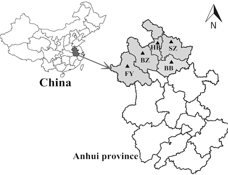
Collection sites of blood samples in Anhui province, China. (HB, Huaibei; SZ, Suzhou; BZ, Bozhou; BB, Bengbu; FY, Fuyang).
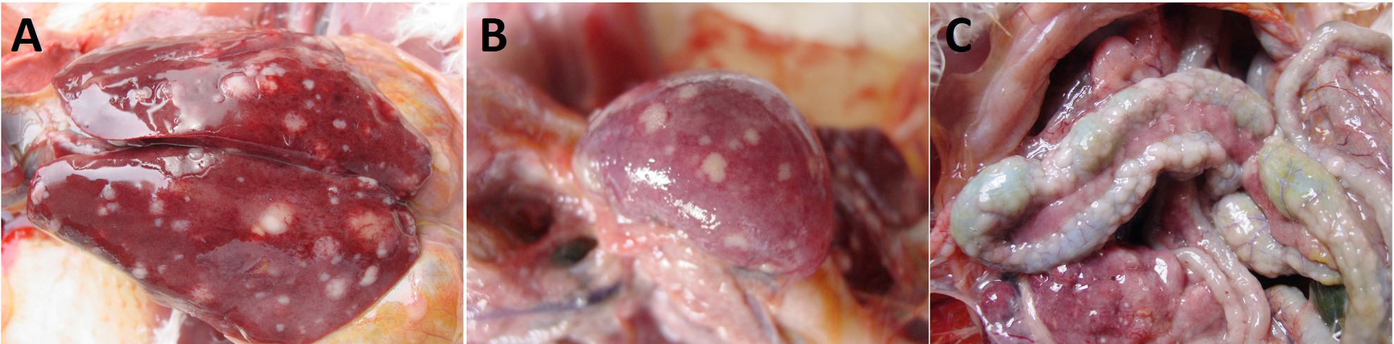
Morphological examination the enlarged liver and diffused nodular tumor lesions with perihepatitis (A), the nodular lesions in the spleen (B) and intestine (C).
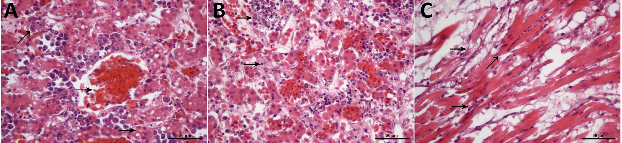
The pathological observation of the congestion in the portal vein with focal lymphoid cells in the liver (A), the degenerative necrosis, hypertrophy with edema on the walls of follicular blood vessels and cells infiltration in the spleen (B) and mild myocardial necrosis, lymphocytic myocarditis, hyperplasia of myocyte nuclei, myocardial infiltration with rupture and myocarditis in the heart (C). Stain, H&E; Magnification, 400x.
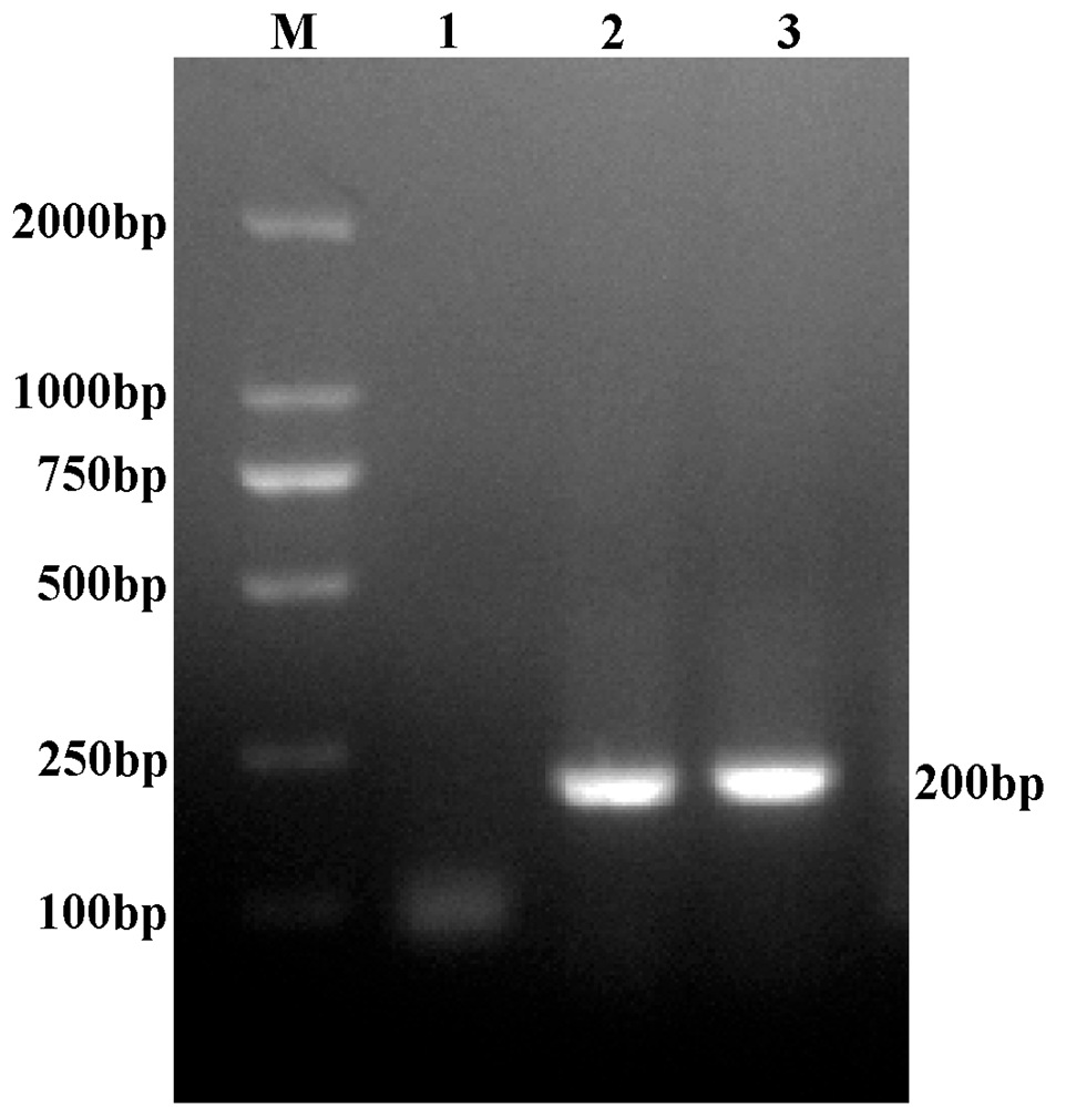
Specific PCR amplification of the ALV-A gene (200bp) on 1.5% agarose gel. M, bp DNA ladder.
References
- Crittenden, L.B., 1976. The epidemiology of avian lymphoid leukosis. Cancer Res., 36: 570-573.
- Cui, N., Su. S., Chen, Z., Zhao, X. and Cui, Z., 2014. Genomic sequence analysis and biological characteristics of rescued clone of avian leukosis virus strain JS11C1, isolated from indigenous chickens. J. Gen. Virol., 95: 2512-2522. https://doi.org/10.1099/vir.0.067264-0[CrossRef]
- Davidson, I., 2007. Avian oncogenic viruses: the correlation between clinical signs and molecular virus identification, knowledge acquired from the examination of over 1000 flocks. Israel J. Vet. Med., 62: 42-47.
- Du, Y., Cui, Z.Z. and Qin, A.J., 1999. Detected avian leukosis virus subgroup J from the market in commercial broiler (in Chinese). J. Chin. Poult., 3: 1-4.
- Gao, Y.N., Guan, X.L., Liu, Y.Z., Li, X.F., Yun, B.L., Qi, X.L., Wang, Y.Q., Gao, H.L., Cui, H.Y., Liu, C.J., Zhang, Y.P., Wang, X.M. and Gao, Y.L., 2015. An avian leukosis virus subgroup J isolate with a Rous sarcoma virus-like 59-LTR shows enhanced replication capability. J. Gen. Virol., 96: 150-158. https://doi.org/10.1099/vir.0.071290-0[CrossRef]
- Gilka, F. and Spencer, J.L., 1990. Chronic myocarditis and circulatory syndrome in a white leghorn strain induced by an avian leukosis virus: light and electron microscopic study. Avian Dis., 34: 174-184. https://doi.org/10.2307/1591349[CrossRef]
- Guan, H.W., Wu, R., Diao, X.L., Luo, P., Zhang, L., He, Z. and Ai, W.N., 2012. Epidemiological investigation of avian leukosis infection in avian in Lanzhou, China (in Chinese). Chin. Vet. Sci., 42: 1294-1301.
- Guo, F.X., Zhu, R.L., Cun, W.U., Zhao, X., Ting-He, Q.U. and Xiao-Hua, H.E., 2014. Effect of pathogenicity of bordetella aviumunder immunosuppression induced by avian leukosis virus in chickens. Chin. J. Vet. Sci., 34: 1072-1071.
- Liao, C.T., Chen, S.Y., Chen, W.G., Liu, Y., Sun, B.L., Li, H.X., Zhang, H.M., Qu, H., Wang, J., Shu, D.M. and Xie, Q.M., 2014. Single nucleotide polymorphism variants within TVA and TVB receptor genes in Chinese chickens. Poult. Sci., 93: 2482-2489. https://doi.org/10.3382/ps.2014-04077[CrossRef]
- Pandiri, A.R., Gimeno, I.M., Reed, W.M., Fitzgerald, S.D. and Fadly, A.M., 2009. Subgroup J avian leukosis virus-induced histiocytic sarcomatosis occurs only in persistently viremic but not immunotolerized meat-type chickens. Vet. Pathol., 46: 282-287. https://doi.org/10.1354/vp.46-2-282[CrossRef]
- Payne, L., Brown, S., Bumstead, N., Howes, K., Frazier, J.A. and Thouless, M.E., 1991. A novel subgroup of exogenous avian leukosis virus in chickens. J. Gen. Virol., 72: 801-807. https://doi.org/10.1099/0022-1317-72-4-801[CrossRef]
- Payne, L. and Nair, V., 2012. The long view: 40 years of avian leukosis research. Avian Pathol., 41: 11-19. https://doi.org/10.1080/03079457.2011.646237[CrossRef]
- Sandelin, K. and Estola, T., 1974. Occurrence of different subgroup of avian leukosis virus in Finnish poultry. Avian Pathol., 3: 159-168. https://doi.org/10.1080/03079457409353828[CrossRef]
- Smith, E.J., 1987. Endogenous avian leukosis viruses. Avian leukosis. Martinus Nijhoff, Boston, MA, pp. 101-120.
- Tomášek, O., Kvapil, P., Halouzka, R. and Kulíkova, L., 2005. Prevalence of tumours and antibodies against leukosis and sarcoma viruses from subgroups A and B in layers. Acta Vet. Brno, 74: 575. https://doi.org/10.2754/avb200574040575[CrossRef]
- Yang, F., Sun, H.L., Ni, W., Che, G.X. and Liu, S.D., 2011. Epidemiological study of avian leukosis on layer chickens with different stains in Shandong Province (in Chinese). Acta Vet. Zootech. Sin., 42: 698-703.
- Zhao, X.S. and Li, R.D., 2015. Epidemiological investigation, separation and identification of avian leukosis infection in chinckens in Guizhou, China (in Chinese). Shandong Anim. Husband. Vet., 8: 11-12.
Services
Article usage statistics
Loading…