
Intramuscular fat (IMF) content is one of the most important factors determining beef quality and price. Intramuscular adipocytes develop from mesenchymal stem cells (MSCs) in mesoderm. The mechanisms of preadipocytes differentiate into mature adipocytes to a great extent are clear, but the commitment of MSCs to preadipocytes is largely unknown. In this study, the Platelet-derived growth factor receptor α (PDGFRα) positive progenitor cells were isolated from the longissimus dorsi muscle (LM) of fetal bovine and induced adipogenesis. To optimize the in vitro IMF differentiation model, the effects of bone morphogenic protein 4 (BMP4) and rosiglitazone during differentiation were studied. Comparing with control group, progenitor cells treated with BMP4 or rosiglitazone accumulated more intracellular lipid. Furthermore, the mRNA expression level of adipocyte-specific genes also increased significantly in BMP4 or rosiglitazone treated cells. The result indicated that BMP4 and rosiglitazone could promote adipogenesis and be applied in adipogenic differentiation of fetal bovine derived progenitor cells.
Short Communication · Volume 53, Issue 1, 31 December 2020 · Open Access
BMP4 and Rosiglitazone Improves Adipogenesis of Bovine Fetal Muscle Derived Progenitor Cells
· · · · · · ·
Affiliations & Notes ⌄
- 1Key Laboratory of Animal Genetics Breeding and Reproduction, Ministry of Agriculture and Rural Affairs
- 2Institute of Animal Sciences, Chinese Academy of Agricultural Sciences, Beijing 100193, China 2Molecular and Cellular Biology, Gembloux Agro-Bio Tech, University of Liège, 5030 Gembloux, Belgium , * Corresponding author: zhanglupei@caas.cn
Article Info ⌄
- Journal:
- Pakistan Journal of Zoology
- Published:
- 31 December 2020

From the article
- In this study, the PDGFRα+ progenitor cells were isolated from the fetal LM and induced to differentiate into mature adipocytes in vitro.
- Previous reports have suggested that rosiglitazone enhance adipogenesis.
- In human MSCs, rosiglitazone was testified could counteract osteoblastogenesis and induce a preferential differentiation into adipocytes.
- There also has article showed that rosiglitazone induced adipogenic differentiation from mammalian MSC.
Selected from the article's own text.
Abstract
Materials and methods
Bovine fetuses between 120 to 150 days after conception were collected immediately after removal from uterus of slaughtered cows. LM was taken out from the fetus followed by isolating cells via enzyme digestion method. In virtue of PDGF Receptor α antibody (#5241, Cell Signaling Technology, USA) to filtrate PDGFRα⁺ cells and then incubated them at 37°C in 5% CO2-air. When the cells reached 80% confluence, the cells were digested with 0.25% trypsin (25200056, Gibco, USA) and subcultured into 12-well plates.
For adipogenic differentiation at 100% confluence, the cells were exposed to two types of differentiation medium (DM). One DM (CT3) contains growth medium (GM) with three inducers, 1 mM dexamethasone (D1756, Sigma, USA), 0.5 mM isobutyl-methylxanthine (I5879, Sigma, USA) and 3 μg/mL insulin (I5500, Sigma, USA). The other DM (CT4) contains an additional inducer, 1mM rosiglitazone (R2408, Sigma, USA). After 3 days, the medium was replaced every other day with maintenance medium (GM supplemented with 3 μg/mL insulin and 1mM rosiglitazone). Cells were treated with 10 ng/mL BMP4 during proliferation phase (from subculturing to 100% confluency), differentiation phase (after 100% confluency and induction) or both phases .
For oil red O staining after removing the culture medium, cells were washed once with phosphate-buffered saline (PBS) and fixed with 4% paraformaldehyde for 15 min at room temperature. Cells were washed and stained with 0.5% Oil Red O (O0625, Sigma, USA) for 15 min to assess intracellular lipid accumulation. Next, we measured the staining intensity of Oil Red O at 490 nm wavelength by microplate reader (Bio-Rad).
For quantitative real-time PCR total RNA was extracted using the TRIzol reagent (15596026, Invitrogen, USA) and reverse transcription was executed using PrimeScriptTM RT Master Mix (TaKaRa) following the manufacturer’s instructions. Quantitation of mRNA level by qRT-PCR was performed using ABI QuantStudio 7 Flex system to detect relative genes expression level. Primers used in qRT-PCR were list in Table I.
Six technical replicates were used for each group. Relative expression levels of each gene were calculated using the 2−ΔΔCt algorithm by normalizing to expression of 18S, and the data were presented as mean ± S.E.M. Results were analyzed using one-tailed Student’s t-test with GraphPad Prism software (GraphPad Prism, version 6.0). For BMP4 treating phase experiment, the significance between groups was determined by one-way ANOVA followed by tukey test for multiple comparisons. P < 0.05 was considered as significant difference.
Results and discussion
Skeletal muscle is a complex organ that composed of multiple types of cells, including several kinds of stem cells. One type of muscle-derived stem cells characterized with PDGFRα expression could differentiate into adipocyte . These adipogenic progenitor cells developed from non-myogenic lineage in mesoderm and are competent to differentiate into IMF . IMF can be detected at as early as 180 days of bovine fetus, but the adipogenic progenitor cells can be isolated at an earlier time using the PDGFRα as a positive cell surface marker . After inducing with adipogenic cocktail, the PDGFRα+ cells can differentiate into mature adipocytes . In this study, the PDGFRα+ progenitor cells were isolated from the fetal LM and induced to differentiate into mature adipocytes in vitro.
Previous reports have suggested that rosiglitazone enhance adipogenesis. In human MSCs, rosiglitazone was testified could counteract osteoblastogenesis and induce a preferential differentiation into adipocytes. There also has article showed that rosiglitazone induced adipogenic differentiation from mammalian MSC. Rosiglitazone is well-known as an insulin sensitizer, and PPARγ activator . Then, we compared the adipogenic efficiency of DM with or without rosiglitazone. We found that there were more mature adipocytes in CT4 group compared with CT3 group at day 9. (Fig. 1a and b). Next, we measured the staining intensity of Oil Red O at 490 nm wavelength and found the OD value was significantly higher in the group treated
Table I. Primers for real-time quantitative PCR.
| Name | Forward (5’-3’) | Reverse (5’-3’) |
| 18S ZNF423 PPARγ C/EBPα FABP4 LPL THRSP SFRP6 | CTAACCCGTTGAACCCCATT GGATTCCTCCGTGACAGCA TGGAGACCGCCCAGGTTTGC TGCGCAAGAGCCGGGACAAG GGATGATAAGATGGTGCTGGA TACCCTGCCTGAAGTTTCCAC AAGAGGCTGAGGAGGAGAGC CCTCCAGTGACCAAGATCTGTG | CCATCCAATCGGTAGTAGCG TCGTCCTCATTCCTCTCCTCT AGCTGGGAGGACTCGGGGTG ACCAGGGAGCTCTCGGGCAG ATCCCTTGGCTTATGCTCTCT CCCAGTTTCAGCCAGACTTTC GGACTGCCTTCTATCATGTGG TTCTTCATGTGCAGCACGAG |
with DM containing rosiglitazone(p<0.01) (Fig. 1c). To further study effects of rosiglitazone on adipogenesis, we analyzed adipocyte-specific genes expression level. The results showed that the mRNA expression of ZNF423, PPARγ, C/EBPα, FABP4, LPL, THRSP and SFRP6 genes were significantly higher in CT4 group than those of CT3 group (p<0.01) (Fig. 1d). These results indicated that rosiglitazone could improve adipogenesis of bovine fetal muscle derived progenitor cells.
Previous studies have shown that BMP4 can improve the commitment of C3H10T1/2 pluripotent stem cells to the adipocyte lineage (Tang et al., 2004a). Also, BMP4 could regulate preadipocyte commitment and PPARγ activation by modulating ZNF423 nuclear localization via WISP2 . In this study, to investigate the role of BMP4 in the adipogenesis of the PDGFRα+ progenitor cells, we treated the PDGFRα+ progenitor cells with BMP4 during proliferation phase and induced adipogenic differentiation with DM contains four kinds of inducers. After inducing the cells to adipogenic differentiation for 9 days, we observed that the cells treated with BMP4 appeared significantly more adipocytes compared with the control group. Furthermore, we found there were a larger staining area of those treated with BMP4 using ORO staining (Fig. 2a and b). Consistent with the result of ORO staining, the OD value was higher in the BMP4 group than that of the control group at day 9 (p<0.01) (Fig. 2c). Moreover, we also collected the cells at day 4 and 9 respectively to test the adipocyte-specific genes expression. The results showed that at both day 4 and 9 BMP4 could induce the higher expression level of relative genes than that of the control group (p<0.01) (Fig. 2d and 2e).
MSCs undergoes three stages to mature adipocytes, (a) adipocyte lineage commitment, (b) cell proliferation, (c) terminal differentiation to adipocytes (Tang et al., 2004a). In this study, we treated the cells with BMP4 in the proliferation phase to investigate the role of BMP4 in adipogenesis of the progenitor cells isolated from the LM of fetal bovine. Also, we compared the effects among groups treated with BMP4 during proliferation phase, differentiation phase and both proliferation and differentiation phase. The most accumulation of intracellular lipid as well as the highest OD value were found in the group treated only in proliferative phase (Fig. 2f). The increased expression of adipocyte-specific genes, especially ZNF423 in BMP4 group also provide the evidence for that BMP4 may mainly play a role before the differentiation phase. And the optimal treating period is consistent with a previous report (Tang et al., 2004a), in which they treated proliferating C3H10T1/2 cells with BMP4 followed by adipocyte differentiation, and the C3H10T1/2 pluripotent stem cells differentiation and expression of adipocyte protein marker were promoted.
Taken together, BMP4 is competent to trigger commitment in muscle derived progenitor cells to adipocyte lineage and promote adipogenesis in cattle. Furthermore, we also verified rosiglitazone could improve adipogenesis of bovine fetal muscle derived progenitor cells. In this work, we combined the two approaches in a novel control variate that should provide useful information for in vitro studies of bovine early IMF development.
Acknowledgement
Animal experiments were conducted according to the guidelines established by the Regulations for the Administration of Affairs Concerning Experimental Animals (Ministry of Science and Technology, China). All animal protocols were approved by Animal Ethics Committee of Institute of Animal Sciences, Chinese Acadamy of Agricultrual Sciences. Pregnant cows were raised in Inner Mongolia Aokesi Agriculture Co., Ltd (Wulagai, China). All efforts were made to minimize the cow’s suffering. This work was supported by National Natural Science Foundation of China (31672384), the Cattle Breeding Innovative Research Team (ASTIPIAS03 and ZDXT2018006).
Statement of conflict of interests
Authors have declared no conflicts of interest.
Figures
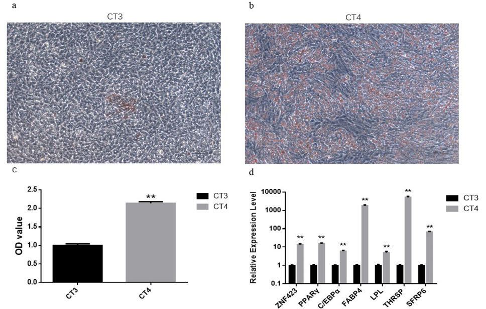
Rosiglitazone improved the efficiency of adipogenic differentiation of PDGFRα⺠cells. (a, b) ORO staining at day 9 after treating cells with CT3(a) or CT4(b). Scale bar, 100 μ m. (c) The OD value of ORO staining intensity of CT3 and CT4 group on day 9. (d) The relative genes expression level after treatment cells with different DM on day 9. Bars denote means ± SEM (n = 6 wells for each condition; **p < 0.01 versus control).
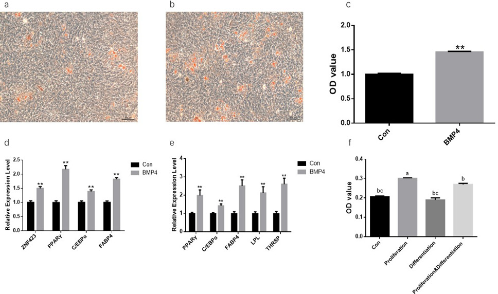
BMP4 promoted the efficiency of adipogenic differentiation of PDGFRα⺠cells. (a, b) ORO staining at day 9 after treating cells without (a) or with (b) BMP4 during proliferative phase. Scale bar, 100 μ m. (c) The OD value of ORO staining intensity of control and BMP4 group on day 9. (d, e) Relative genes expression of adipocyte-specific genes on day 4 (d) and day 9 (e). (f) The OD value of ORO staining intensity in different treatment strategies, treating with BMP4 during proliferation, differentiation and both proliferation and differentiation. The values with different superscript letters in a column are significantly different (p<0.05). Bars denote means ± SEM (n =6 wells for each condition; **p < 0.01 versus control).
References
- Attia, A.E.M. and EL-Sabagh, M.R., 2012. Am. J. Biochem. Biotechnol., 8: 171-178.
- Blazquez-Medela, A.M., Jumabay, M. and Bostrom, K.I., 2019. Obes. Rev., 20: 648-658. https://doi.org/10.1111/obr.12822[CrossRef]
- Bowers, R.R. and Lane, M.D., 2007. Cell Cycle, 6: 385-389. https://doi.org/10.4161/cc.6.4.3804[CrossRef]
- Dodson, M.V., Jiang, Z., Chen, J., Hausman, G.J., Guan, L.L., Novakofski, J., Thompson, D.P., Lorenzen, C.L., Fernyhough, M.E., Mir, P.S. and Reecy, J.M., 2010. J. Fd. Sci., 75: R1-8.
- Du, M., Huang, Y., Das, A.K., Yang, Q., Duarte, M.S., Dodson, M.V. and Zhu, M.J., 2013. J. Anim. Sci., 91: 1419-1427. https://doi.org/10.2527/jas.2012-5670[CrossRef]
- Du, M., Wang, B., Fu, X., Yang, Q. and Zhu, M.J., 2015. Meat Sci., 109: 40-47.
- Guan, L., Hu, X., Liu, L., Xing, Y., Zhou, Z., Liang, X., Yang, Q., Jin, S., Bao, J., Gao, H., Du, M., Li, J. and Zhang, L., 2017. Sci. Rep., 7: 43716. https://doi.org/10.1038/srep43716[CrossRef]
- Gupta, R.K., Arany, Z., Seale, P., Mepani, R.J., Ye, L., Conroe, H.M., Roby, Y.A., Kulaga, H., Reed, R.R. and Spiegelman, B.M., 2010. Nature, 464: 619-623. https://doi.org/10.1038/nature08816[CrossRef]
- Hammarstedt, A., Hedjazifar, S., Jenndahl, L., Gogg, S., Grunberg, J., Gustafson, B., Klimcakova, E., Stich, V., Langin, D., Laakso, M. and Smith, U., 2013. Proc. natl. Acad. Sci., 110: 2563-2568.
- Hong, J., Li, S., Wang, X., Mei, C. and Zan, L., 2018. Biosci. Rep., 38: BSR20181705. https://doi.org/10.1042/BSR20181705[CrossRef]
- Huang, H., Song, T.J., Li, X., Hu, L., He, Q., Liu, M., Lane, M.D. and Tang, Q.Q., 2009. Proc. natl. Acad. Sci., 106: 12670-12675.
- Huang, Y., Das, A.K., Yang, Q.Y., Zhu, M.J. and Du, M., 2012. PLoS One, 7: e47496.
- Kim, J., Kwak, H.J., Cha, J.Y., Jeong, Y.S., Rhee, S.D. and Cheon, H.G., 2014. J. biol. Chem., 289: 2755-2764. https://doi.org/10.1074/jbc.M113.493650[CrossRef]
- Knochel, S., Dillinger, K., Koster, M. and Knochel, W., 2001. Mech. Dev., 109: 79-82.
- Lee, Y.H., Petkova, A.P., Mottillo, E.P. and Granneman, J.G., 2012. Cell Metab., 15: 480-491. https://doi.org/10.1016/j.cmet.2012.03.009[CrossRef]
- Linhart, H.G., Ishimura-Oka, K., DeMayo, F., Kibe, T., Repka, D., Poindexter, B., Bick, R.J. and Darlington, G.J., 2001. Proc. natl. Acad. Sci., 98: 12532-12537.
- Mangino, M., Torrente, I., De Luca, A., Sanchez, O., Dallapiccola, B. and Novelli, G., 1999. J. Hum. Genet., 44: 76-77. https://doi.org/10.1007/s100380050113[CrossRef]
- Rosen, E.D. and MacDougald, O.A., 2006. Nat. Rev. Mol. Cell. Biol., 7: 885-896.
- Tang, Q.Q., Otto, T.C. and Lane, M.D., 2004a. Proc. natl. Acad. Sci., 101: 9607-9611.
- Tang, Q.Q., Zhang, J.W. and Daniel Lane, M., 2004b. Biochem. Biophys. Res. Commun., 319: 235-239.
- Uezumi, A., Fukada, S.-i., Yamamoto, N., Takeda, S.-i. and Tsuchida, K., 2010. Nature Cell Biol., 12: 143-152. https://doi.org/10.1038/ncb2014[CrossRef]
- Winnier, G., Blessing, M., Labosky, P.A. and Hogan, B.L., 1995. Genes Dev., 9: 2105-2116. https://doi.org/10.1101/gad.9.17.2105[CrossRef]
Services
Article usage statistics
Loading…