
To illustrate the protein composition of the Cryptocaryon irritans cyst wall, a shotgun-liquid chromatography-tandem mass spectrometry (LC-MS/MS) technique was applied and all identified proteins were categorised using a bioinformatics approach. The results showed that 3,472 proteins were identified with a theoretical molecular weight of 11.53–610.90 kDa and an isoelectric point of 4.13–11.77. A total of 63.07% or 50.09% proteins were annotated by Kyoto Encyclopaedia of Genes and Genomes (KEGG) or Gene Ontology (GO) terms, respectively. These results highlight that mass material is transported and catabolised and growth- and death-related proteins are highly expressed, including a number of vesicle-mediated transport, cytoskeletal and extracellular secretion-related proteins. These results provide a platform to study tomont cell characteristics and provide a further understanding of cyst wall formation and mechanisms.
Research Article · Volume 52, Issue 2, 27 January 2020 · Open Access
Proteomic Analysis by Shotgun LC-MS/MS of Cyst Wall Protein Components From the Protozoan Parasite Cryptocaryon irritans, the Causative Agent of White Spot Disease in Marine Fish
· · · ·
Affiliations & Notes ⌄
- 1Key Laboratory of Applied Marine Biotechnology, Ministry of Education, Collaborative Innovation Centre for Zhejiang Marine High-efficiency and Healthy Aquaculture, Ningbo University, Ningbo 315211, People’s Republic of China 2School of Marine Sciences, Ningbo University, 169 South Qixing Road, Ningbo 315832, People’s Republic of China , * Corresponding author: feige895@gmail.com
Article Info ⌄
- Journal:
- Pakistan Journal of Zoology
- Published:
- 27 January 2020

Abstract
Introduction
Cryptocaryon irritans, a parasitic ciliate that penetrates the epithelium of the gills, skin, and fins of marine fish, causes acute mortality in cultured fish. The tomont of C. irritans is an important stage for cell division and proliferation. Therefore, it is necessary to cut off the life cycle before the parasites proliferate and reinfect the host.
The cyst wall is the cell’s ‘armour’, which prevents a large number of drugs from penetrating the cell, which may otherwise cause damage to the protoplasm ; . Ultrastructural observations show that the thickness of the C. irritans cyst wall is about 4 μm, including a multi-layered structure composed of three regions with different densities but with similar forms . The chemical composition of the C. irritans cyst wall, however, is not known. The chemical composition of the protozoan cyst wall is mainly protein and polysaccharides . Furthermore, as the structural and regulatory proteins of the cyst wall of parasitic protozoa are often used as targets for screening and designing new anti-parasite drugs ; , identifying the C. irritans cyst wall proteins will be helpful to screen and develop drugs that inhibit cyst wall formation.
Shotgun liquid chromatography-tandem mass spectrometry (LC-MS/MS) is a technique used to determine proteins in complex systems. This method increases the probability that a protein of low abundance is identified. This technique has been widely used in many fields of life science , especially proteomics, including for large-scale screening of different proteins in Pseudourostyla cristata . In the present study, shotgun- LC-MS/MS and bioinformatics analysis techniques were used to identify the protein components of the C. irritans cyst wall.
Materials and methods
Cryptocaryon irritans cyst wall
C. irritans were isolated from naturally infected Larimichthys crocea, and the same species of fish with an average body mass of 100 g were used as animal models to establish the passage system . A large number of tomonts adhered to the bottom of the aquaria (1062-L, Фbottom=130 cm ×H=80 cm) four days after the infection. The fish were transferred to another clean aquarium, and residual tomonts were collected by carefully discarding the debris. The tomonts were incubated in a one liter beaker. The theronts were released about -four days later, and the empty cyst wall was harvested under a microscope. Approximately 1.5×10⁴ empty cyst walls were gathered and combined in a 1.5 ml centrifuge tube. The empty cyst walls were washed with three times in fresh seawater and clean empty cyst walls (Fig. 1) were stored at −80°C before the proteomic analysis.
Seawater salinity and temperature for aquaculture were 29%–31% and 28±1°C, respectively.
Protein extraction
The frozen clean empty cyst walls were transferred to tubes and ground with SDT lysis buffer (pH 8.5, 4% SDS, 100 mM Tris-HCl and 100 mM DL-dithiothreitol (DTT) using the MP FastPrep-24 5G homogeniser (MP Biomedical, Santa Ana, CA, USA). The homogenised cyst wall samples were incubated in a buffer of six M urea containing 2% SDS and 5 % β-mercaptoethanol at 100°C for 10 min to extract the cyst wall proteins. Subsequently, the cyst wall proteins were concentrated using an Ultrafree-0.5 centrifugal filter device with a high-flux Biomax-100 membrane (Millipore, Bedford, MA, USA). First, 500 μl of solubilised protein sample was added to the tube and centrifuged at 14,000×g for 30 min; the column was inserted into a new tube and centrifuged at 1,000×g for two min. Finally, 20 μl (20 mg/ml) of concentrated protein was obtained . The protein content in the supernatant was quantified by the Bradford method . Aliquots were stored at −80°C until use.
Cyst wall protein enzymolysis
The filter-aided proteome preparation method was used for enzymolysis of the cyst wall proteins. Total-protein samples (200 μg) diluted in 100 mM DTT solution to a concentration of 100 mg/mL were boiled for five min. The samples were left to cool to 25°C and were then loaded into a 10 kDa ultrafiltration tube (Sartorius, Goettingen Germany), followed by adding 200 μL UA buffer (8 M urea and 150 mM Tris-HCl pH 8.0) to the tube and centrifugation at 14,000×g for 15 min, followed by a wash in UA buffer. Then, 100 μL iodoacetamide (100 mM IAA in UA buffer) was added to block reduced cysteine residues, and the samples were incubated for 30 min in the dark. The filters were washed three times with 100 μL UA buffer and then twice with 100 μL 25 mM NH4HCO3 buffer. Finally, the protein suspensions were digested with 4 μg trypsin (Promega, Madison, WI, USA) in 40 μL of 25 mM NH4HCO3 buffer overnight at 37°C, and the resulting peptides were collected as the filtrate. The peptides in each sample were desalted on C18 Cartridges (Empore™ SPE Cartridges C18 (standard density), bed I.D. 7 mm, volume 3 mL; Sigma, St. Louis, MO, USA), concentrated by vacuum centrifugation, and reconstituted in 40 µL of 0.1% (v/v) formic acid. Finally, the concentration of the resulting peptide was analysed at OD280, and aliquots of the samples were freeze-dried for the proteomic analysis.
LC-MS/MS analysis
Peptide mixtures from the above extraction were separated by reverse phase-high-performance liquid chromatography (RP-HPLC) followed by tandem MS analysis. RP-HPLC was carried out using a surveyor EASY-nLC system (Thermo Finnigan, San Jose, CA, USA). The fractions were thawed and dissolved in buffer A (0.1% v/v formic acid in Milli-Q water). The column was equilibrated for 20 min with 95% (v/v) buffer A. The peptide mixtures were separated using a Thermo Scientific EASY column (2 cm × 100 μm 5 μm-C18) and a Thermo Scientific EASY column (75 μm × 100 mm 3 μm-C18) at a flow rate of 300 nL/min. The peptides were separated with solvent B [acetonitrile with 0.1% (v/v) formic acid] using a segmented gradient from 4–9% (v/v) in one min, from 9–50% in 54 min, from 50–90% (v/v) in one min and then at 90% (v/v) for 20 min.
A Q-Exactive (Thermo Finnigan) mass spectrometer was set to perform data acquisition in the positive ion mode, with a selected mass range of 300–1,800 mass/charge (m/z). Resolving power for the Q-Exactive was set to 70,000 for the MS scan and 17,500 for the MS/MS scan at m/z 200. MS/MS data were collected using the top 20 most abundant precursor ions with charge ≥ 2 as determined from the MS scan. The maximum ion injection times for the survey scan and the MS/MS scans were 50 and 60 ms, respectively, and the automatic gain control target values for both scan modes were set to 3 × 10⁶. These parameters were selected with an isolation window of 2 m/z and fragmented by higher-energy collisional dissociation with normalised collision energy of 27 eV. Dynamic exclusion for selected precursor ions was set to 60 s and the under ratio on the Q-Exactive was 0.1%.
Mass spectrometry data analysis and functional annotation of the proteins
Raw files were searched using MASCOT 2.2 software for database retrieval against the protein database (uniprot_Ciliophora_169050_20171127.Fasta, http://www.uniprot.org). The following search parameters were set: trypsin as the enzyme; missed cleavage sites were set to 2; fixed modification was carbamidomethyl (C); dynamic modification of setting oxidation (M). Blast2Go (https://www.blast2go.com/) software was used to screen the differentially expressed proteins in the Gene Ontology (GO) functional annotations. The expression levels of the proteins were annotated using a KO analysis (http://www.genome.jp/kegg/tool/map_pathway2.html). Kyoto Encyclopaedia of Genes and Genomes automatic annotation server (KASS) was used for the default parameters to perform the pathway annotation.
Results and discussion
Cyst wall proteins tandem mass spectrometric analysis
C. irritans cyst wall proteins were excised, digested and analysed by shotgun LC-MS/MS. The C. irritans cyst wall proteins were identified by searching the Ciliophora database at the NCBI; 3,472 proteins were identified with mascot scores ≥ 20, of which 1,194 proteins had a unique peptide value ≥ 2 (Supplementary Table I). All 3,472 proteins were distributed in a range of molecular weights of 5.72–998.30 kDa and isoelectric points of 4.13–11.77, according to the spectrometric analysis (Figs. 2, 3; Supplementary Table I).
GO and KEGG categories of C. irritans cyst wall proteins
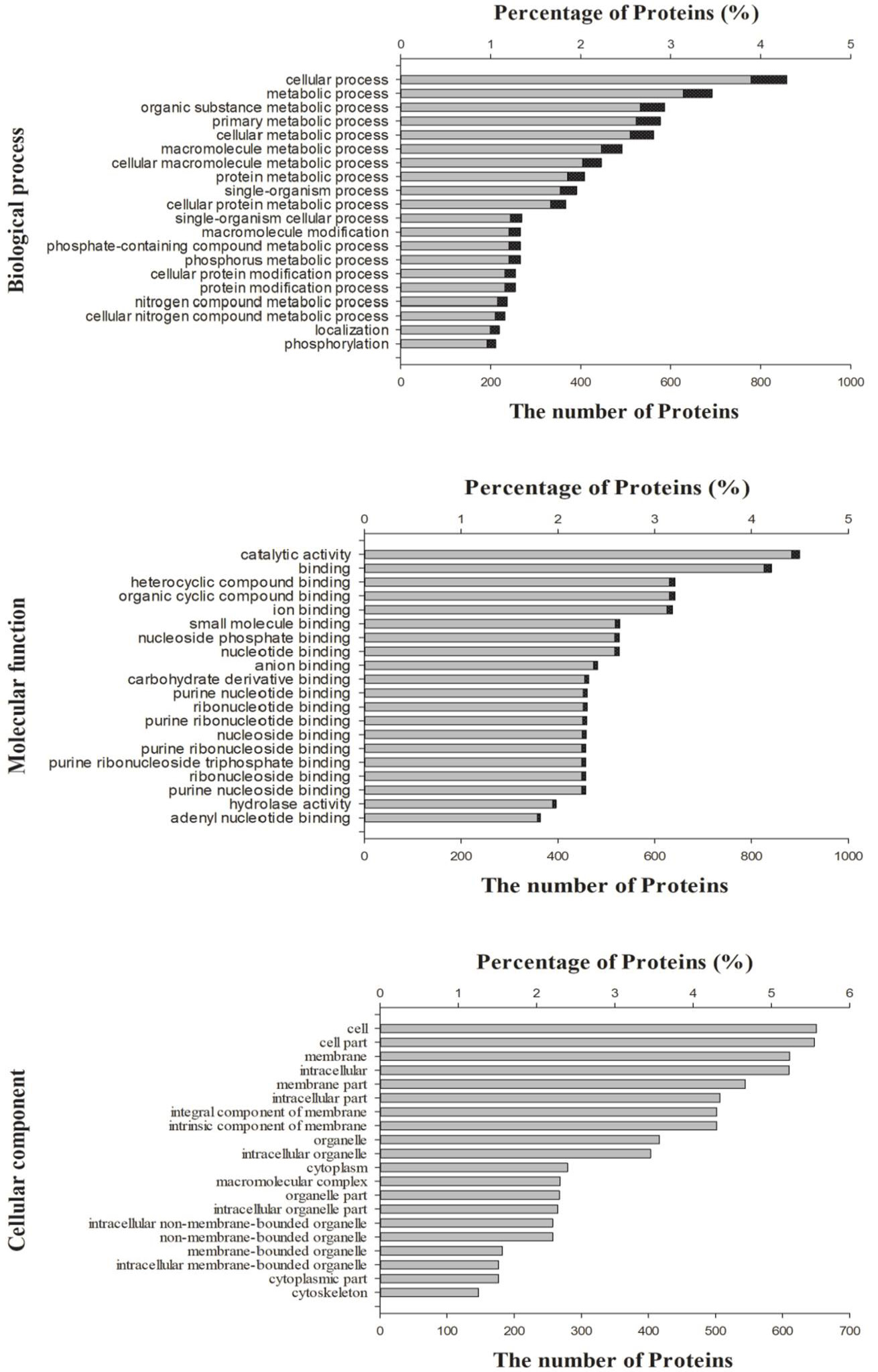
In this study, a total of 1,739 proteins (50.09 %) (Table I) were assigned predicted GO terms (Fig. 4, Supplementary Table II). These terms were summarized into 2167 sub-categories under three GO terms corresponding to the biological process (BP) category (1248), cellular component (CC) category (299), and molecular function (MF) category (620). In the BP category, the top 20 sub-categories were above 35 %, which proteins most enriched, included cellular process, several metabolic process, organism process, macromolecule modification, localization, and phosphorylation. In the MF category, catalytic activity, cyclic compound binding, ion binding, small molecular binding, nucleotide binding, and hydrolase activity were most enriched sub-categories, were above 52% of this part. In the CC category, the cell, memebrane, intracellular, organelle, cytoplasm, macromolecular complex, and cytoskeleton were the most enriched parts, were above 65%.
Table I.- Annotation of proteins of proteomic profiles of Cryptocaryon irritans cyst wall.
| Database | Number of annotated proteins | Percentage of annotated proteins in protein group |
| KO | 2,499 | 71.98% |
| GO | 1,739 | 50.09% |
| KEGG | 2,190 | 63.07% |
| Protein group (Total) | 3,472 | 100.00% |
The proteins were also categorized using the KEGG database to identify the biological pathways in C. irritans cyst wall proteins. A total of 2,190 proteins (63.07 %) (Table I) were further annotated by KEGG and classified into six categories with 47 subclasses (290 known KEGG pathways) (Fig. 5).
Studies on the components of the cyst wall have deepened. As early as the 1960s, researchers used cytochemical labelling to suggest that the Entamoeba cyst wall is composed of a carbohydrate-protein complex . In 1980, biochemical analysis, fluorescence labelling, ultrastructural observations and X-ray diffraction were combined to confirm that chitin is the main component of the E. invadens cyst wall . In recent years, studies have demonstrated that the cyst wall of protozoa is mainly composed of proteins and polysaccharides , and the protein components of the cyst walls of a variety of protozoa have been identified. For example, SDS-PAGE has been used to isolate small molecular proteins ; MS analysis of purified E. invadens cyst walls revealed chitinase and two lectins ; eight SDS-PAGE strips were analysed by MALDI-TOF MS, and 42 proteins were found in the E. encysticus cyst wall . In the present study, 3,472 proteins were identified by LC–MS/MS.
Table II.- The top 1% confident proteins in turn following unique peptide count.
| Reference | Unique pep count | |
| 1 | Agglutination/immobilization antigen OS=Cryptocaryon irritans PE=2 SV=1 | 22 |
| 2 | Agglutination/immobilization antigen OS=Cryptocaryon irritans PE=2 SV=1 | 20 |
| 3 | Surface antigen OS=Cryptocaryon irritans PE=2 SV=1 | 19 |
| 4 | Immobilization antigen (Fragment) OS=Cryptocaryon irritans PE=2 SV=1 | 17 |
| 5 | Agglutination/immobilization antigen OS=Cryptocaryon irritans GN=iAg PE=4 SV=1 | 14 |
| 6 | ADP/ATP carrier OS=Cryptocaryon irritans PE=2 SV=1 | 14 |
| 7 | Tubulin beta chain OS=Tetrahymena thermophila PE=1 SV=1 | 13 |
| 8 | Agglutination/immobilization antigen (Fragment) OS=Cryptocaryon irritans PE=2 SV=1 | 12 |
| 9 | Tubulin beta chain OS=Tetrahymena thermophila GN=BTU1 PE=1 SV=1 | 12 |
| 10 | Tubulin beta chain OS=Euplotes focardii PE=3 SV=1 | 11 |
| 11 | Immobilization antigen (Fragment) OS=Cryptocaryon irritans PE=2 SV=1 | 10 |
| 12 | Tubulin beta chain (Fragment) OS=Sorogena stoianovitchae GN=sbt PE=2 SV=1 | 10 |
| 13 | Tubulin beta chain OS=Euplotes crassus PE=3 SV=1 | 10 |
| 14 | Actin (Fragment) OS=Metopus es PE=3 SV=1 | 10 |
| 15 | Actin (Fragment) OS=Heterometopus palaeformis PE=3 SV=1 | 10 |
| 16 | Actin (Fragment) OS=Metopus es PE=3 SV=1 | 10 |
| 17 | Agglutination/immobilization antigen isoform 1 OS=Cryptocaryon irritans PE=2 SV=1 | 9 |
| 18 | Agglutination/immobilization antigen isoform 3 (Fragment) OS=Cryptocaryon irritans PE=2 SV=1 | 9 |
| 19 | Agglutination/immobilization antigen isoform 5 (Fragment) OS=Cryptocaryon irritans PE=2 SV=1 | 9 |
| 20 | Uncharacterized protein OS=Paramecium tetraurelia GN=GSPATT00035237001 PE=4 SV=1 | 9 |
| 21 | Tubulin beta chain (Fragment) OS=Chilodonella uncinata GN=btub PE=3 SV=1 | 9 |
| 22 | Tubulin beta chain (Fragment) OS=Colpoda sp. PE=3 SV=1 | 9 |
| 23 | Tubulin beta chain OS=Stylonychia lemnae GN=TUBB1 PE=3 SV=1 | 9 |
| 24 | Tubulin beta chain OS=Gastrostyla steinii PE=3 SV=1 | 9 |
| 25 | ATP synthase subunit alpha OS=Stentor coeruleus GN=SteCoe_10017 PE=3 SV=1 | 9 |
| 26 | ATP synthase subunit alpha OS=Paramecium tetraurelia GN=GSPATT00039376001 PE=3 SV=1 | 9 |
| 27 | Tubulin alpha chain OS=Tetrahymena thermophila PE=1 SV=1 | 9 |
| 28 | Tubulin alpha chain (Fragment) OS=Frontonia sp. PE=3 SV=1 | 9 |
| 29 | Tubulin alpha chain OS=Paramecium tetraurelia GN=alphaPT4 PE=3 SV=1 | 9 |
| 30 | ATP synthase subunit beta OS=Stylonychia lemnae GN=Contig4879.g5215 PE=3 SV=1 | 9 |
| 31 | Tubulin beta chain OS=Stentor coeruleus GN=BTU01 PE=3 SV=1 | 8 |
| 32 | Tubulin beta chain OS=Stentor coeruleus GN=BTU05 PE=3 SV=1 | 8 |
| 33 | Tubulin beta chain (Fragment) OS=Myrionecta rubra PE=2 SV=1 | 8 |
| 34 | Tubulin beta chain (Fragment) OS=Nyctotherus ovalis PE=3 SV=1 | 8 |
| 35 | Tubulin beta chain (Fragment) OS=Chilodonella uncinata GN=Btub PE=3 SV=1 | 8 |
If the unique peptide count was considered a reference indicator for the confidence of correctly identifying the proteins, the top 1% (35 in 3,472) of proteins were antigens, ATP/ADP transfer/creation-related proteins, and cytoskeleton-related proteins (tubulins and actin) (Table II). In addition, the top 100 proteins also included glyceraldehyde-3-phosphate dehydrogenase, heat shock proteins, ubiquitin, elongation factor (EF) 1-α, p-loop containing nucleoside triphosphate hydrolase, histone H4, calmodulin 1, fat domain-containing protein, transitional endoplasmic reticulum ATPase, histone variant H3.5 and preprotein translocase subunit-like protein (Supplementary Table I). It proved that the cytoskeleton-related proteins comprise the major components and protect against adverse environmental conditions in the resting cyst wall of E. encysticus . Agglutination/ immobilisation and surface antigens have been identified in all stages of the C. irritans life cycle ; . Fish immunised with C. irritans tomonts with a cyst wall produce specific antibody , suggesting that antigens are present in the cyst wall. Heat shock proteins are important housekeeping genes and are involved in encystation of parasitic protozoa when they encounter a hostile environment . ADP glycosylation factor-ARFs in Pseudvurostyla cristata cells are related to intracellular transport and extracellular polymerisation of cyst wall materials . The cyst wall protein-EF-Tu of C. cucullus is related to the formation of a fibrous or network-like structure of the scale layer of its cyst wall . Feeding RNAi to silence the EF-1α gene suppresses the formation of resting cysts in C. cucullus .
Structural and regulatory proteins of the cyst wall are often targets for screening and designing new drugs . For example, the formation of a cyst wall requires the synthesis of a large number of proteins, and puromycin is a protein synthesis inhibitor; a 50 μg mL−1 dose can cause failure of the C. cucullus cyst wall to form . β-1,3-glucan is a component of the oocyst wall of Toxoplasma and Eimeria, and biosynthesis of glucans is regulated by glucan-hydrolase/synthase. This enzyme is also a target of echinocandins, which inhibit formation of the cyst wall by interfering with glucan-hydrolase/synthase activity . Synthesis of E. invadens cysts relies on chitin synthase and chitinase, while inhibitors (poly D, nikkomycin and allosamidin) of these enzymes lead to failed cyst formation . Protein kinases, as an important drug target, are also widely regarded in the control of parasitic protozoa, and different protein kinases inhibitors (staurosporine, chelerythrine chloride and Calphostin C) exhibit an inhibitory effect on encystation . Human Glrac has been considered a target for designing anticancer drugs . Because Glrac regulates the trafficking and secretion of cyst wall proteins in the protozoa Giardia, it has also been used as a potential target to develop new anti-parasite drugs . In this study, eight nuclear transport-related proteins (40S ribosomal protein SA, GTP-binding nuclear protein, transmembrane proteins, N-acetyltransferase B complex non-catalytic subunit, GTP-binding nuclear protein ran and nucleoporin nup155) were found in the C. irritans cyst wall. Our previous study confirmed that the nuclear transporter inhibitor leptomycin B effectively induces C. irritans theronts and tomontsdeath . In addition, a number of cyst wall formation-related proteins, particularly some proteins peculiar to the tomonts of the C. irritans, were identified (Supplementary Table II) . These proteins will provide a basis for researchers to develop anti-C. irritans drugs.
Table III.- Vesicle-mediated transport-related proteins.
| Reference | MW (kDa) | PI | |
| 1 | Dynein heavy chain 1, axonemal protein OS=Tetrahymena thermophila (strain SB210) GN=TTHERM_00558640 PE=4 SV=2 | 487.70 | 5.79 |
| 2 | WD40-repeat-containing domain OS=Pseudocohnilembus persalinus GN=PPERSA_00474 PE=4 SV=1 | 222.44 | 8.9 |
| 3 | Zinc-binding dehydrogenase family oxidoreductase OS=Tetrahymena thermophila (strain SB210) GN= TTHERM_00384890 PE=4 SV=2 | 201.13 | 6.07 |
| 4 | Intraflagellar transport protein, putative OS=Ichthyophthirius multifiliis (strain G5) GN=IMG5_148350 PE=4 SV=1 | 198.15 | 6.55 |
| 5 | Clathrin heavy chain OS=Ichthyophthirius multifiliis (strain G5) GN=IMG5_187840 PE=3 SV=1 | 198.04 | 5.97 |
| 6 | Uncharacterized protein OS=Paramecium tetraurelia GN=GSPATT00020489001 PE=4 SV=1 | 133.07 | 7.81 |
| 7 | Transmembrane protein, putative OS=Tetrahymena thermophila (strain SB210) GN=TTHERM_00923100 PE=4 SV=3 | 113.30 | 8.2 |
| 8 | Adaptin amine-terminal region family protein OS=Tetrahymena thermophila (strain SB210) GN=TTHERM_00155580 PE=4 SV=3 | 113.00 | 5.73 |
| 9 | Phospholipase OS=Paramecium tetraurelia GN=GSPATT00019109001 PE=3 SV=1 | 112.86 | 8.67 |
| 10 | Coatomer subunit gamma OS=Tetrahymena thermophila (strain SB210) GN=TTHERM_00444320 PE=3 SV=1 | 105.39 | 5.47 |
| 11 | Adaptin amine-terminal region family protein OS=Tetrahymena thermophila (strain SB210) GN=TTHERM_00287890 PE=4 SV=2 | 100.45 | 5.8 |
| 12 | Coatomer subunit beta' OS=Stentor coeruleus GN=SteCoe_76 PE=3 SV=1 | 97.81 | 5.08 |
| 13 | Coatomer subunit beta OS=Pseudocohnilembus persalinus GN=PPERSA_03040 PE=4 SV=1 | 97.32 | 5.92 |
| 14 | Uncharacterized protein OS=Pseudocohnilembus persalinus GN=PPERSA_05656 PE=4 SV=1 | 91.97 | 5.3 |
| 15 | Phospholipid-transporting ATPase OS=Ichthyophthirius multifiliis (strain G5) GN=IMG5_078220 PE=3 SV=1 | 91.56 | 8.21 |
| 16 | Transport protein OS=Stylonychia lemnae GN=Contig1948.g2110 PE=4 SV=1 | 86.28 | 5.74 |
| 17 | Sec1-like protein OS=Pseudocohnilembus persalinus GN=PPERSA_07320 PE=3 SV=1 | 69.11 | 6.2 |
| 18 | Uncharacterized protein OS=Stentor coeruleus GN=SteCoe_14443 PE=3 SV=1 | 62.67 | 5.33 |
| 19 | Uncharacterized protein OS=Paramecium tetraurelia GN=GSPATT00026361001 PE=3 SV=1 | 50.81 | 8.54 |
| 20 | Mu1 adaptin OS=Oxytricha trifallax GN=OXYTRI_04462 PE=3 SV=1 | 50.28 | 8.65 |
| 21 | Uncharacterized protein OS=Paramecium tetraurelia GN=GSPATT00015008001 PE=4 SV=1 | 48.82 | 6.45 |
| 22 | Uncharacterized protein OS=Stentor coeruleus GN=SteCoe_5626 PE=4 SV=1 | 23.96 | 9.1 |
Acknowledgements
The authors gratefully acknowledge funding support from the Natural Science Foundation of Shanghai (Grant No. 17ZR1439600), the Natural Science Foundation of Zhejiang Province (Grant No. Y18C190005), and the K.C. Wong Magna Fund in Ningbo University.
Supplementary material
There is supplementary material (Tables I and II) associated with this article. Access the material online at: https://dx.doi.org/10.17582/journal.pjz/20190707110736
Statement of conflict of interest
The authors declare no conflict of interest.
Figures
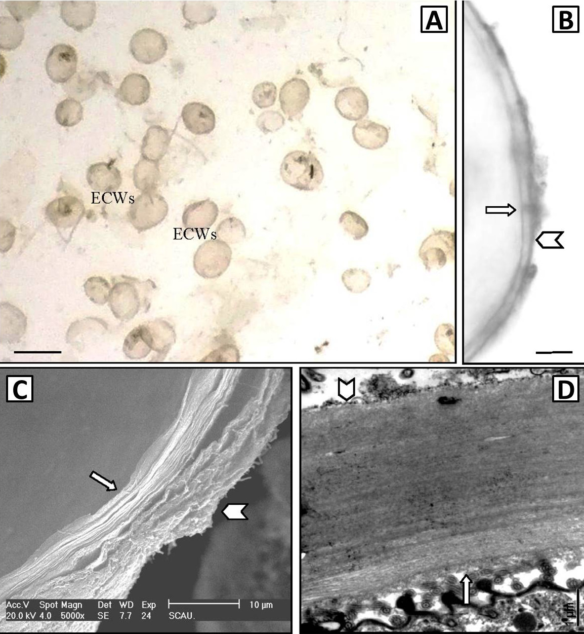
Cyst wall of Cryptocaryon irritans viewed by an optical microscope (A, B), SEM (C), and TEM (D). A, a holistic view of empty cyst walls; B, partial view of empty cyst walls; C, cyst wall stripped off in SEM sample preparation, showing the multi-layer structure of the thick cyst wall; D, sectional view of the cyst wall. ECWs represent empty cyst wall. Arrows indicate the inner surface of cyst wall, and Arrowheads indicate the outer surface of the cyst wall. Scale bars = 500 μm (A), 10 μm (B), 10 μm (C), and 1 μm (D).
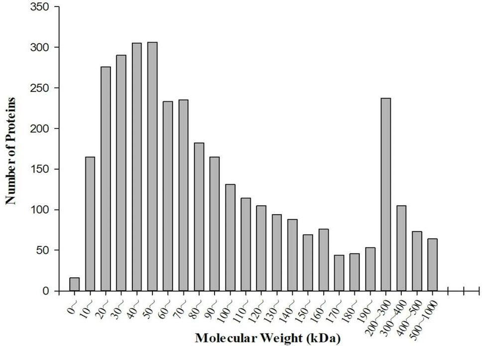
Distributions of molecular mass (MW) for cyst wall of Cryptocaryon irritans by the shotgun LC-MS/MS approach.
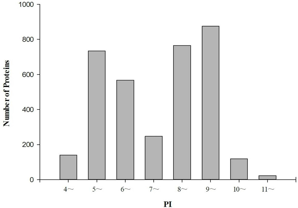
Distributions of isoelectric point (pI) for cyst wall of Cryptocaryon irritans by the shotgun LC-MS/MS approach.
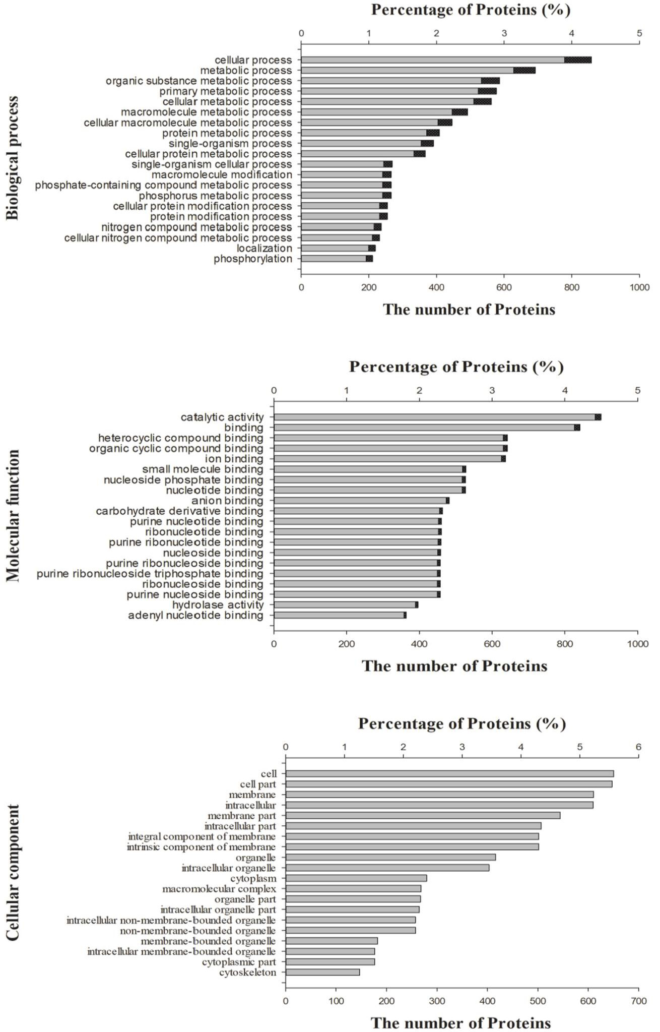
GO categories of Cryptocaryon irritans cyst wall proteins. These terms were summarized into 2167 sub-categories under 3 GO terms. Bar charts represented the top 20 sub-categories of the biological process (BP) category, cellular component (CC) category, and molecular function (MF) category, respectively. Grey bars showed the number of proteins, the black bars indicated the percentage of proteins.
Proteomic Analysis by Shotgun LC-MS/MS of Cyst Wall Protein Components From the Protozoan Parasite Cryptocaryon irritans, the Causative Agent of White Spot Disease in Marine Fish
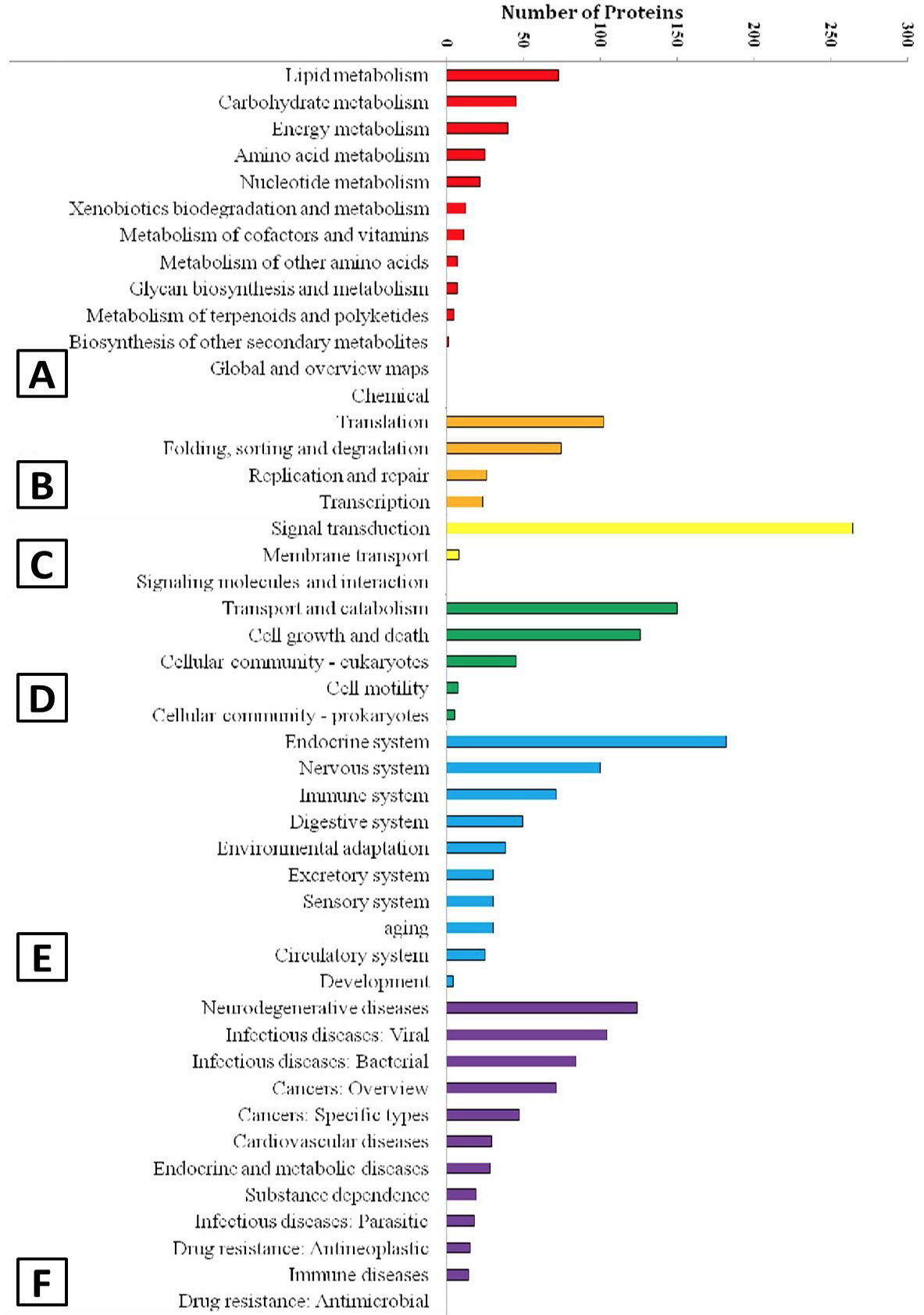
KEGG categories of Cryptocaryon irritans cyst wall proteins. A total of 2,190 proteins were annotated by KEGG and classified into 6 categories with 47 subclasses. The upper cases represent the title of 6 categories. A, metabolism; B, genetic information processing; C, environmental information processing; D, cellular processes; E, organismal systems; F, human diseases.
References
- Aggarwal, K., Choe, L.H. and Lee, K.H., 2006. Shotgun proteomics using the itraq isobaric tags. Brief. Funct. Genom., 5: 112-120. https://doi.org/10.1093/bfgp/ell018[CrossRef]
- Aguilar-Díaz, H., Carrero, J.C., Argüello-García, R., Laclette, J.P. and Morales-Montor, J., 2011. Cyst and encystment in protozoan parasites: Optimal targets for new life-cycle interrupting strategies? Trends Parasitol., 27: 450-458. https://doi.org/10.1016/j.pt.2011.06.003[CrossRef]
- Arroyo-Begovich, A., Carabez-Trejo, A. and Ruiz-Herrera, J., 1980. Identification of the structural component in the cyst wall of Entamoeba invadens. J. Parasitol., 66: 735-741. https://doi.org/10.2307/3280662[CrossRef]
- Aznar, S. and Lacal, J.C., 2001. Searching new targets for anticancer drug design: The families of ras and rho gtpases and their effectors. Prog. Nucl. Acid Res. Mol. Biol., 67: 193-234. https://doi.org/10.1016/S0079-6603(01)67029-6[CrossRef]
- Bai, J.S., Xie, M.Q., Zhu, X.Q., Dan, X.M. and Li, A.X., 2008. Comparative studies on the immunogenicity of theronts, tomonts and trophonts of Cryptocaryon irritans in grouper. Parasitol. Res., 102: 307-313. https://doi.org/10.1007/s00436-007-0766-6[CrossRef]
- Bradford, M.M., 1976. A rapid and sensitive method for the quantitation of microgram quantities of protein utilizing the principle of protein-dye binding. Anal. Biochem., 72: 248-254. https://doi.org/10.1006/abio.1976.9999[CrossRef]
- Bushkin, G.G., Motari, E., Magnelli, P., Gubbels, M.J., Dubey, J.P., Miska, K.B., Bullitt, E., Costello, C.E., Robbins, P.W. and Samuelson, J., 2012. Β-1,3-glucan, which can be targeted by drugs, forms a trabecular scaffold in the oocyst walls of Toxoplasma and Eimeria. MBio, 3: e00258-12. https://doi.org/10.1128/mBio.00258-12[CrossRef]
- Calvo, P., Fernandez-Aliseda, M.C., Garrido, J. and Torres, A., 2003. Ultrastructure, encystment and cyst wall composition of the resting cyst of the peritrich ciliate Opisthonecta henneguyi. J. Eukaryot. Microbiol., 50: 49-56. https://doi.org/10.1111/j.1550-7408.2003.tb00105.x[CrossRef]
- Dan, X.M., 2006. The propagation, preservation ofCryptocaryon irritans brown 1951 and control of cryptocaryonosis. Jinan University, Guangzhou, China.
- Funadani, R., Sogame, Y., Kojima, K., Takeshita, T., Yamamoto, K., Tsujizono, T., Suizu, F., Miyata, S., Yagyu, K.I. and Suzuki, T., 2016. Morphogenetic and molecular analyses of cyst wall components in the ciliated protozoan Colpoda cucullus NAG-1. FEMS Microbiol. Lett., 363: fnw203. https://doi.org/10.1093/femsle/fnw203[CrossRef]
- Gao, X.X., Chen, F.F., Niu, T., Qu, R.D. and Chen, J.W., 2015. Large-scale identification of encystment-related proteins and genes in pseudourostyla cristata. Scient. Rep., 5: 11360. https://doi.org/10.1038/srep14022[CrossRef]
- Ghosh, S.K., Field, J., Frisardi, M., Rosenthal, B., Mai, Z.M., Rogers, R. and Samuelson, J., 1999. Chitinase secretion by encysting Entamoeba invadens and transfected Entamoeba histolytica trophozoites: Localization of secretory vesicles, Endoplasmic reticulum, and Golgi apparatus. Infect. Immun., 67: 3073-3081.
- Huang, X.H., Sun, Z.Y., Guo, G.W., Zheng, C.F., Xu, Y., Yuan, L.P. and Liu, C., 2012. Cloning and characterization of a surface antigen cisa-32.6 from Cryptocaryon irritans. Exp. Parasitol., 130: 189-194. https://doi.org/10.1016/j.exppara.2012.01.012[CrossRef]
- Jarroll, E.L. and Sener, K., 2003. Potential drug targets in cyst-wall biosynthesis by intestinal protozoa. Drug Resist. Updates, 6: 239-246. https://doi.org/10.1016/S1368-7646(03)00065-7[CrossRef]
- Krtková, J., Thomas, E.B., Alas, G.C., Schraner, E.M., Behjatnia, H.R., Hehl, A.B. and Paredez, A.R., 2016. Rac regulates Giardia lamblia encystation by coordinating cyst wall protein trafficking and secretion. MBio, 7: e01003-16. https://doi.org/10.1128/mBio.01003-16[CrossRef]
- Lin, Q.Q., Yang, M., Huang, Z., Ni, W., Fu, G.L., Guo, G.W., Wang, Z.C. and Huang, X.H., 2013. Cloning, expression and molecular characterization of a 14-3-3 gene from a parasitic ciliate, Cryptocaryon irritans. Vet. Parasitol., 197: 427-435. https://doi.org/10.1016/j.vetpar.2013.07.028[CrossRef]
- Ma, R., Fan, X.P., Yin, F., Ni, B. and Gu, F.K., 2017. Ultrastructural features of the tomont of Cryptocaryon irritans (Ciliophora: Prostomatea), a parasitic ciliate of marine fishes. Parasitology, 144: 720-729. https://doi.org/10.1017/S0031182016002651[CrossRef]
- Mai, Y.Z., Li, Y.W., Li, R.J., Li, W., Huang, X.Z., Mo, Z.Q. and Li, A.X., 2015. Proteomic analysis of differentially expressed proteins in the marine fish parasitic ciliate Cryptocaryon irritans. Vet. Parasitol., 211: 1-11. https://doi.org/10.1016/j.vetpar.2015.05.004[CrossRef]
- McConnachie, E.W., 1969. The morphology, formation and development of cysts of Entamoeba. Parasitology, 59: 41-53. https://doi.org/10.1017/S003118200006981X[CrossRef]
- Rios, R.M., Sarmiento, R., Torres, A. and Fedriani, C., 2012. Solubilization and electrophoretic studies of cyst wall proteins of a hypotrichous ciliate. Biol. Cell, 67: 271-279. https://doi.org/10.1111/j.1768-322X.1989.tb00872.x[CrossRef]
- Samanta, S.K., Varghese, S.S., Krishnan, D., Baidya, M., Nayak, D., Mukherjee, S. and Ghosh, S.K., 2018. A novel encystation specific protein kinase regulates chitin synthesis in Entamoeba invadens. Mol. Biochem. Parasitol., 220: 19-27. https://doi.org/10.1016/j.molbiopara.2018.01.003[CrossRef]
- Samuelson, J. and Robbins, P., 2011. A simple fibril and lectin model for cyst walls of Entamoeba and perhaps Giardia. Trends Parasitol., 27: 17-22. https://doi.org/10.1016/j.pt.2010.09.002[CrossRef]
- Sogame, Y., Hori, M. and Matsuoka, T., 2016. Ef-1α silencing by feeding rnai suppresses resting cyst formation in Colpoda cucullus nag-1 strain. Inverteb. Surv. J., 13: 89-94.
- Wang, B.Z., Tao, N., Bhatti, M.Z., Chen, F.F., Lin, W. and Chen, J.W., 2017. Identification of cyst wall proteins of the hypotrich ciliate Euplotes encysticus using a proteomics approach. J. Microbiol., 55: 545-553. https://doi.org/10.1007/s12275-017-6422-7[CrossRef]
- Wang, Z.Q., Wang, L. and Cui, J., 2012. Proteomic analysis of Trichinella spiralis proteins in intestinal epithelial cells after culture with their larvae by shotgun lc-ms/ms approach. J. Proteom., 75: 2375-2383. https://doi.org/10.1016/j.jprot.2012.02.005[CrossRef]
- Yin, F., Liu, W.C., Bao, P.B., Jin, S., Qian, D., Wang, J.T. and Tang, B.J., 2018. Comparison of the susceptibility and resistance of four marine perciform fishes to Cryptocaryon irritans infection. Fish Shellf. Immunol., 77: 298-303. https://doi.org/10.1016/j.fsi.2018.03.052[CrossRef]
- Yin, F., Sun, P., Tang, B.J., Gong, H., Ke, Q.Z. and Li, A.X., 2016. Anti-parasitic effects of leptomycin b isolated from Streptomyces sp. CJK17 on marine fish ciliate Cryptocaryon irritans. Vet. Parasitol., 217: 89-94. https://doi.org/10.1016/j.vetpar.2015.12.034[CrossRef]
- Zahid, M.T., Shakoori, F.R., Zulfiqar, S., Al-Ghanim, K.A. and Shakoori, A.R., 2018. Growth characteristics, metal uptake and expression analysis of copper metallothionein in a newly reported ciliate, Tetrahymena farahensis. Pakistan J. Zool., 50: 1171-1181.
Supplementary material
Services
Article usage statistics
Loading…