but the lower mean weight was estimated at animals from Mymensingh (10.87±0.41). Narrower confidence intervals for all estimates were observed which might indicate an adequate sample size for the study.
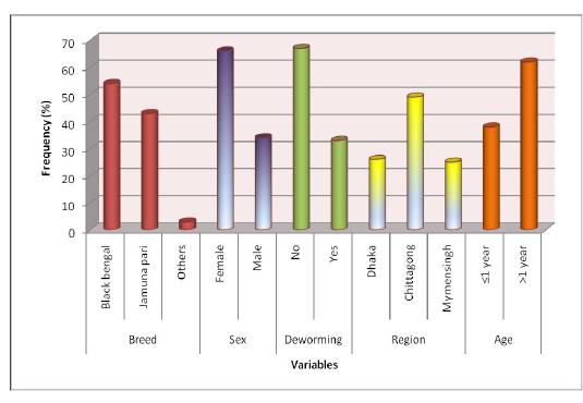
Figure 2: Descriptive statistics of the variables: breed, sex, deworming, region and age
Overall prevalence of gastrointestinal parasites: Nearly similar prevalence of gastrointestinal parasites in selected study areas was recorded (Figure 2). The highest prevalence of overall gastrointestinal parasitic infection in goats was recorded in samples from Central Veterinary Hospital, Dhaka (63.88%). A slightly lower prevalence was estimated in samples of Bangladesh Agricultural University Veterinary Hospital (Mymensingh) (62.13%). In S. A. Quaderi Teaching Veterinary Hospital overall prevalence was estimated as 59.43%. Around 40% goats were free from parasites in the study areas.
Prevalence on the basis of a type of parasite (trematodes, nematodes, and cestodes): Both nematodes (38%) and trematodes (40%) were highly prevalent in samples from Bangladesh Agricultural University Veterinary Hospital (Mymensingh). Samples from the other two study areas (Central Veterinary Hospital, Dhaka, and S. A. Quaderi Teaching Veterinary Hospital) were more positive for nematodes (66% and 49%, respectively) but the prevalence of trematodes was not remarkable (6% and & 2%, respectively) in the mentioned areas. Prevalence of cestode was nearly similar in all three study areas (lowest 3% and highest 8%) (Figure 4).
Prevalence of different genus of gastrointestinal parasites in goat: The current study revealed highest prevalence for Haemonchus (20.28%) and lowest prevalence for Toxocara (0.9%) in S .A. Quaderi Teaching Veterinary Hospital of Chittagong. In Central Veterinary Hospital, Dhaka highest prevalence was recorded for Haemonchus (39.81%) and lowest for Trichuris (0.01%). On the ot
er hand, in Bangladesh Agricultural University Veterinary Hospital (Mymensingh) the most prevalent parasite was Paramphistomum (24%) and lowest prevalence was estimated for Strongyloides and Cooperia (01%) (Figure 3).
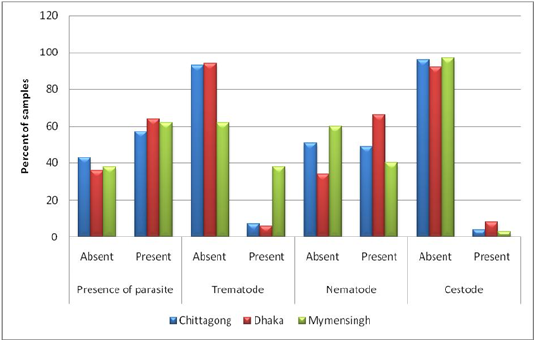
Figure 3: Prevalence on the basis of type of parasite (Trematodes, Nematodes and Cestodes) stratified by study areas.
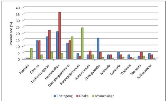
Figure 4: Prevalence of different genus of gastrointestinal parasites in goat
Considerably higher prevalence was recorded for Strongyloides (15.56%), Trichostrongylus (15.09%), Oesophagostomum (9.90%) and Eimeria (6.60%) than Paraphistomum, Bunostomum, Schistosoma and Moniezia infection in the population of S. A. Quaderi teaching Veterinary Hospital. Besides Haemonchus, a higher prevalence was estimated for Trichostrongylus (24.07%), Eimeria (13.80%), Oesophagostomum (13.80%) and Strongyloides (6.48%) than Paramphistomum, Bunostomum, Schistosoma, Moniezia and Toxocara infection in samples from Central Veterinary Hospital, Dhaka. On the other hand, Oesophagostomum (17%), Fasciola (8%) and Trichostrongylus (5%) were more prevalent in samples from Bangladesh Agricultural University Veterinary Hospital (Mymensingh) in comparison with the prevalence of Bunostomum, Haemonchus, Eimeria and Toxocara (Figure 4).
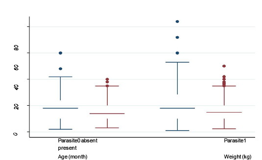
Figure 5: Box plot of continuous variables (age and body weight)
Median, maximum, minimum, 25th and 75th quartile values of continuous variables (age and body weight) stratified by presence and absence of parasite: box plot was created to show the difference in age and body weight in animals with and without parasitic infection
In the present study minimum age was 4 month and maximum age was 42 month for goats without parasitic infection, whereas, minimum and maximum age for the goats with parasitic infection was 3 months and 55 months, respectively.
Minimum and maximum body weights in both groups of goats (with and without parasitic infection) were nearly similar (4 kg and 38 kg in without infection and 4 kg and 39kg in with GI parasitic infection) (Figure 5). However, there were some outliers who had extreme age and body weight (shown as dots). Parasite-infected 25% goats were of 10 months old with 10 kg body weight and 75% goats
were of 30 months old with 20 kg body weight. Median age and median body weight for both groups of goats were nearly similar (Figure 4).
Table 2, We conducted a t-test analysis to identify if mean age and mean body weight was significantly different in groups of goats with and without gastrointestinal parasitic infection (Table 2). Mean age and body weight were slightly different in two groups of goats and the p-value of the t-test was not significant, indicates a less remarkable influence of age and body weight on the occurrence of parasitic infection in goat.
Table 2: comparison of means of continuous variables (age and weight) between the groups of animals with and without gastrointestinal parasitic infection using t-test
Relationship of different variables with a presence of gastrointestinal parasites in goats: Chi-square test was performed to identify the association of different variables with the presence of gastrointestinal parasites in goats. Prevalence was higher in females (63%) in comparison with males (56%), however, variables were not significantly associated (p-value = 0.16) (Table 3). It can be concluded that the prevalence was similar in different breeds as the slight difference among the breeds were not statistically significant (p-value = 0.42). A Higher prevalence (63%) was observed in a group of animals who were not dewormed in comparison with the dewormed animals. The highest prevalence was observed in Dhaka region (68%) followed by Mymensingh (62%) and Chittagong (56%), an association was not statistically significant though. After categorization, age was not significantly associated with the parasitic infection status of the animal, concordant with the previous analysis shown in Table 2.
We conducted Chi-square tests to assess the association of different variables with the presence of commonly isolated parasitic genus. The analysis revealed that there is a statistically significant varying prevalence of Haemonchus, Oesophagostomum, Paramphistomum, and Trichostrongylus according to different regions (Table 4). Very low prevalence of Haemonchus was recorded in Mymensingh (4%) in comparison with other two regions (21% in Chittagong and 36% in Dhaka). On the other hand, a very high prevalence of Paramphistomum was recorded in Mymensingh region (35%) in comparison to other regions (Table 4).
Prevalence of Haemonchus and Paramphistomum was significantly varied with breeds. High prevalence of Haemonchus was observed in Jamunapari (28%) but the prevalence of Paramphistomum was lowest in this breed (10%) in comparison with black Bengal and other non-descriptive breeds (Table 4).
Table 3: Relationship of different variables with the presence of gastrointestinal parasite
Table 4: Relationship of different variables with presence of Haemonchus, Oesophagostomum, Paramphistomum and Trichostrongylus