Research Article · Volume 12, 23 September 2025 · Open Access
Prevalence, Intensity and Risk Factors of Gastrointestinal Parasites in Cattle in The Northwest Region of Cameroon
· · · ·
Affiliations & Notes ⌄
1College of Technology, University of Bamenda, Bambili, Cameroon. *Correspondence | Julius Awah-Ndukum, College of Technology, University of Bamenda, Bambili, Cameroon
This study demonstrates a high prevalence and significant burden of both single and mixed gastrointestinal parasitic infections in cattle across the Northwest region of Cameroon.
Juvenile and old animals showed high and similar prevalence rates and intensities of GI parasite infections.
The study clearly indicates that season, location, breed, body condition score influenced the variations in GI parasite prevalence and intensity of infections.
Therefore, to mitigate the adverse effects of GI parasites, effective control measures that make use of the variations in GI parasite prevalence and intensity among the associated risk factors coupled with rational use of anthelmintics must be implemented.
Selected from the article's own text.
Abstract
Gastrointestinal (GI) parasites can cause severe impact to livestock health and productivity leading to reduced growth rates, delayed maturity, and reduced weight gains. There is dearth of information on the epidemiology of GI parasitic diseases in the major livestock production zones of Cameroon. The objective of this study was to determine the prevalence, intensity, and risk factors of GI parasite infections of cattle in the Northwest Cameroon. A cross-sectional study was conducted from November 2022 to October 2023 to detect helminth eggs and Eimeria oocysts cattle faecal samples based on morphological features using sedimentation and floatation techniques. The infection intensity was graded based on eggs per gram (EPG) into five categories; no infection (0 EPG), light (50–200 EPG), moderate (201–800 EPG), heavy (801–1200 EPG) or very heavy (>1200 EPG) infection. Data on breed, sex, age and body condition score were recorded prior to slaughter. Out of 1907 cattle examined, 1524 [79.92% (78.05–81.69)] were infected with one or more of the parasite species; 65.39% nematodes , 53.91% trematodes , Emeria spp. (10.23%), and 1.73% cestodes. Mixed infections (with two or more parasite species) (74.28%) were more common than single infections (25.72%) among the infected animals. The study showed high prevalence of GI parasitic infections in cattle in Northwest Cameroon. Season, location, breed, body condition score, mixed parasite and intensity of infections were significant risk factors (p<0.01). The significant impact of GI parasitism on cattle health and production, and challenges to the efforts of stakeholders to achieve self-sufficiency in food and animal-derived products were highlighted.
Cattle production in Cameroon is a major livestock enterprise and contributes to the country’s economic development, food and nutrition security, and poverty reduction ; FAO, 2000; , 2020). There are over 8 million cattle heads, with an annual growth rate of 1.97% and contributes about 16% of the national agricultural production and 61% of the national demand for meat in Cameroon . It also serves as the main source of exclusive livelihoods for over 35% of the rural communities in the country , 2014). Despite the vital importance of cattle husbandry, several challenges prevent breeders from benefiting on this enormous resource due to constraints. The challenges include poor nutrition, inadequate access to portable water, poor production systems, reproductive inefficiency, finance, management constraints, inadequate extension services, insufficient veterinary care, and disease being the most important (FAO, 2000; ; . Gastrointestinal (GI) parasites are among the most important diseases that limit ruminant production globally ; ; ; . Losses due to infection by GI parasites occur through mortalities, reduced production due to sub-clinical parasitism, and direct cost associated with control measures ; ; ; . GI parasites cause considerable economic losses, reduced weight gain, digestive disturbance, lowered production, impaired reproductive performance, condemnation of affected organs and mortality in infected animals ; FAO, 2000; ; ; .
GI parasite infections have been associated with several factors such as host age, sex, physiological status, body condition, breed, parasite species involved, and other epidemiological patterns such as husbandry practices and climate variables that determine the degree of infection ; ; ; ; ; ; ; . Environmental conditions such as temperature, rainfall and humidity provide suitable conditions for the development of nematode eggs and free-living stages ; ; ; . The contribution of GI parasitism to morbidity, mortality and production losses in cattle in Cameroon is not known. However, prevalence range of less than 10% to over 80% of GI parasites such as nematodes (Haemonchus spp., Oesophagostomum spp., Strongloides spp., Trichostrongylus spp., Bunostomum spp., Chabertia and Trichuris spp.); trematodes (Fasciola, Paraphistomum, Dicrocoelium spp.); cestodes (Monezia spp.); and protozoa (Eimeria spp.) have been reported in domestic ruminants in different parts of Cameroon ; , 2024; ; ; ; ; ; ; . The agro-ecological zones of Cameroon provide suitable environments for various livestock production. Domestic ruminants (Cattle, goats and sheep) are reared in traditional systems such as nomadic or pastoral, mixed farming and peri-urban systems , 2020). Grazing herds are normally fed on natural pastures, characterized generally by annual grass species. However, these traditional livestock systems cause stress and expose animals to several parasitic diseases, especially in areas with frequent rainfall, relatively high humidity, and mild temperatures , ; ; ; ; . Also, the occurrence of flooding, water pools and swamps are important habitats for the propagation of snail intermediate hosts of flukes and stages of other parasites ; ; ; ; .
Knowledge of cattle breeders on GI parasitic diseases , and different GI parasite species of cattle , 2024; ; ; ; ; as well as GI parasitic infections (57.4% in cattle) as the most prevalent infectious diseases of livestock have been reported in different parts of Cameroon. However, GI parasite species/burden may differ with agro-ecological zone, and there is dearth of information on the awareness of hazards caused by GI parasites on husbandry systems, and parasite distribution and abundance in the Cameroon. In addition, priority on infectious and contagious diseases of animals has been heightened in the country with limited attention on GI parasitic diseases. Understanding the epidemiology of GI parasite in a particular region is imperative in developing efficient and effective control programs . There is paucity of information on the epidemiology of GI parasites of cattle in Cameroon and the Northwest region. Therefore, this study aimed to determine the prevalence, fecal egg count intensity, and risk factors associated with GI parasitic infections in cattle in the Northwest region of Cameroon.
MATERIALS AND METHODS
Study area and animals
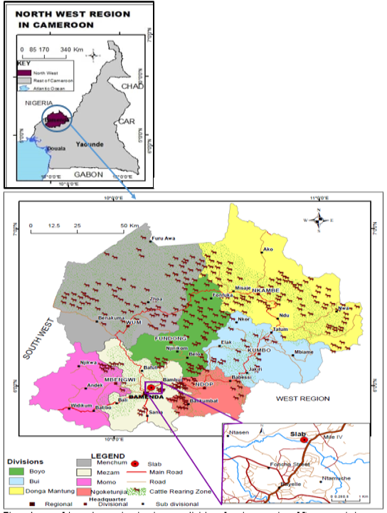
The study was carried out from November 2022 to October 2023 on cattle originating from the administrative divisions of the Northwest region of Cameroon (5o45”–9o9” N and 9o13”–11o13” E) destined for slaughter at the Bamenda municipal abattoir (Figure 1). The Northwest region of Cameroon is located at an altitude of 500 m–3000 m above sea level, and characterized by fertile volcanic soils. The choice of the study area was due to the following:
The Northwest region is ranked as a major livestock production area including cattle in the country , and has several grazing pasture and assembling sites for ranch farming, communal grazing, and other pastoral activities;
The study region has a subtropical type climate with average temperatures ranging from 21ºC–27ºC, annual rainfall of over 2259 mm, high humidity (75%–80%) and savannah vegetation with forest galleries. The region has two seasons: the rainy season from mid-March to mid-November and dry season from mid-November to mid-March ; ; ; . The warm humid subtropical climate and vegetation type of the study region ; ; ; provides suitable conditions for the growth and development of pathogens including the stages of GI parasites ; ; . There is dearth of information on the status of GI parasite infection and intensity in cattle in the Northwest region of Cameroon.
GI parasites have been detected in domestic ruminants (cattle, sheep and goats), and mostly in small ruminants (sheep and goats) in different parts of the country , 2024; ; ; ; ; ; ; . Additionally, there are many ethnic agropastoral communities with strong cultures of livestock rearing for livelihood with mix livestock husbandry being widely practiced in the region.
Sample collection
Determination of the prevalence and intensity of GI parasite infection in cattle was done at the Bamenda municipal abattoir. Selection of individual cattle at the abattoir during the study period was done using previously described systematic random sampling technique . An individual prevalence rate of 56.7% was used to estimate the sample size and animal selection sampling procedure was done as previously described . Briefly, based on a calculated sampling fraction of five (every fifth animal was sampled) for daily use, the first animal was selected by picking one animal by random generation method of the first five animals on the slaughter chain. Thereafter, every fifth animal (adding 5 to previous picked number) was chosen.
Faecal samples were collected per rectum into well-labelled sterile polythene bags and transported in ice packs to the Parasitology laboratory of the Northwest Regional Delegation for Livestock, Fisheries and Animal Industries, Bamenda.
Parasitological analysis
The sedimentation and floatation techniques described by Taylor et al. (2016), and Thienpont et al. (2003) were used to detect the presence of nematode, cestode and trematode eggs, and coccidian oocysts, while the McMaster technique was used to estimate the number of eggs per gram (EPG) of faeces as previously described. A sample was considered positive when a minimum of one GIP egg/oocyst was detected under the microscope. Eggs were identified based on their morphological features .
Given that the composition and pathogenicity of parasite populations may differ from area to area due to different climatic zones ; ; ; ; ; , the GI parasite infections was categorized based on faecal egg counts in mixed infections as previously described ; with slight modification. Briefly, the degree of infections was graded into five categories as no infection, light, moderate, heavy and very heavy infection according to their egg per gram of faeces (EPG) counts. Egg counts of ≤ 200 (50–200), 201–800, 801–1200 and > 1200 eggs per gram of faeces were considered as light, moderate, heavy and very heavy infections, respectively. Information related to breed, sex, age and body condition score of the sampled animals were noted prior to slaughter. Estimation of ages was done by dental inspection and examination of horn rings for animals without teeth (especially old/adult females) and classified according age at slaughter (young/immature ≤ 3 years, Adult/Matured 3 to 6 years, old > 6 years) while the breed of the animals was obtained as previously described ; ; ; . The body condition score was done by assessing the general appearance and palpation of the lumbar region of the animal on a scale of 1 to 5 and further classed into 3 categories: 1–2 (thin), 3 (moderate) and 4–5 (fat) as previously described .
Data analysis
All obtained data were initially entered into Excel 2010 and transferred to the Statistical Package for the Social Sciences (SPSS) version 20 (SPSS Inc. Chicago, IL, USA) for further analysis. Data were analyzed using descriptive statistics, chi-square test, odds ratios from logistic regression, and Student’s t-test in SPSS version 20. Significance was set at p<0.05. The univariate analysis (chi-square test) was used to assess the association between factors and logistic regressions (Odds-ratios) to determine the strength of association of different factors with the prevalence of GI parasite infection in cattle. The 95% confidence interval of proportions was calculated according to Wilson’s equation p±z√((p(1-p))/n), where p is the sample proportion, n is the sample size and z is 1.96, the z-score for a 95% confidence interval. The student t- test was used to compare mean intensity of GI parasites on the study animals. The statistical significance was set at P<0.05.
Ethical consideration
Risk assessments of the project were performed by the researchers to avoid hazards to all persons and animals involved in the study. Permission for the study and ethical approval were obtained from the required authorities in Bamenda-Cameroon [Ministry of Livestock, Fisheries and Animal Industries (MINEPIA) Ref No: MINEPIA/DREPIA/NW/107/40 of 02/11/2022] before carrying out the study. The purpose of the study was explained (with the assistance of resident veterinarians, local community leaders and or trusted intermediaries) to the targeted participants (owners, traders, and butchers) of the Bamenda municipal abattoir. An animal was included in the study after an informed verbal consent was given by the owner or trader-butcher. Apart from procedural restraining manipulations for safety purposes, the animals used in the present study were not subjected to suffering.
RESULTS
Prevalence and assessment of associated factors of gastrointestinal parasites in cattle slaughtered in Bamenda
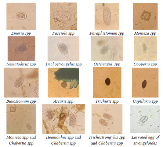
The cattle slaughtered at the Bamenda municipal abattoir were predominantly zebu breeds, with Red Fulani being the most common, followed by White Fulani and Gaudali, as well as crossbreeds (zebu and exotic). The results revealed that cattle from various administrative divisions within the Northwest region harboured different types of GI parasites, and the rate of infection was high (Figure 2). Out of the 1907 cattle examined, 1524 [79.92% (78.05–81.69] were infected with one or more of the following parasite species; 53.91 % trematodes [Fasciola (19.09%) and Paraphistomum (39.43%), and Dicrocoelium (0.16%)]; 65.39% nematodes [Ascaris (3.67%), Bunostomum (1.05%), Trichostrongylus (31.25%), Oesophagostomum (16.83%), Chabertia (6.19%), Haemonchus (16.10%), Cooperia (6.24%), Nematodirus (2.57%), Ostertagia (2.52%), Trichuris (13.48%), Dictyocaulus spp. (3.83%) and Capillaria spp. (0.05)]; 1.73% cestodes [Monezia (1.73%)], and protozoa, Emeria spp. (10.23%) (Figure 2 and Table 1).
Of the 1524 cattle infected, 392 (25.72%) had single infection and 1132 (74.28%) had mixed infections (Table 1). The study revealed that mixed infections (96.41%) were more common than single (3.59%) infections. Paramphistomum spp. (46.35%), Trichostrongylus spp. (41.69%), Fasciola spp. (24.25%), Oesophagostomum spp. (16.45%), Haemonchus spp. (15.78%), and Trichuris spp. (16.78%) were the most prevalent parasites in the two parasite mixed associations, and Paramphistomum spp. (60.00%), Trichostongylus spp. (54.88%), Haemonchus spp. (35.93%), Oesophagostomum spp. (34.39%), Trichuris spp. (23.41%) and Eimeria spp. (20.98%) were predominant in the three parasite mixed associations. For the more than three parasite mixed associations, Paramphistomum spp. (>83.12%), Trichostrongylus spp. (>62.50%), Fasciola spp. (>41.67%), Haemonchus spp. (>41.67%), Oesophagostomum spp. (>37.50%), Trichuris spp. (35.83%) and Eimeria spp. (39.17%) were the most frequently observed (Table 1).
Table 1: Prevalence and distribution of gastrointestinal parasites according to type of parasite associations in infected animals Cattle in Northwest region, Cameroon.
Gastrointestinal
parasite genera/ species
Total (N=1907); n (%)
Type of parasite associations GIP in infected cattle; n (%)
1 parasite
2 parasite associations
3 parasite associations
4 parasite associations
> 4 parasite associations
Overall
1524 (79.91)
392 (100)
602 (100)
410 (100)
105 (100)
15 (100)
Fasciola spp.
364 (19.09)
71 (18.11)
146 (24.25)
97 (5.09)
45 (42.86)
5 (33.33)
Paramphistomum spp.
752 (39.43)
130 (33.16)
279 (46.35)
246 (60.00)
83 (79.05)
14 (93.33)
Dicrcoelium spp.
3 (0.16)
1 (0.26)
1 (0.17)
1 (0.24)
0 (0)
0 (0)
Ascaris spp.
70 (3.67)
2 (0.51)
26 (4.32)
28 (6.83)
11 (10.48)
3 (20.00)
Bunostomum spp.
20 (1.05)
3 (0.77)
5 (0.83)
8 (1.95)
4 (3.81)
0 (0)
Trichostongylus spp.
596 (31.25)
45 (11.48)
251 (41.69)
225 (54.88)
66 (62.86)
9 (60.00)
Oesophgostomum spp.
321 (16.83)
36 (9.18)
99 (16.45)
141 (34.39)
36 (34.29)
9 (60.00)
Chabertia spp.
118 (6.19)
12 (3.06)
34 (5.65)
54 (13.17)
16 (15.24)
2 (13.33)
Haemonchus spp.
308 (16.15)
28 (7.14)
95 (15.78)
135 (35.93)
43 (40.95)
7 (46.67)
Cooperia spp.
119 (6.24)
16 (4.08)
41 (6.81)
42 (10.24)
15 (14.29)
5 (13.33)
Nematodirus spp.
49 (2.57)
7 (1.79)
15 (2.49)
18 (4.39)
8 (7.62)
1 (6.67)
Ostertagia spp.
44 (2.31)
2 (0.51)
20 (3.32)
15 (3.66)
6 (5.71)
1 (6.67)
Trichuris spp.
257 (13.48)
17 (4.34)
101 (16.78)
96 (23.41)
36 (34.29)
7 (46.67)
Dictyocaulus spp.
73 (3.83)
8 (2.04)
23 (3.82)
28 (6.83)
9 (8.57)
5 (33.33)
Capillaria spp.
1 (0.05)
0 (0)
1 (0.17)
0 (0)
0 (0)
0 (0)
Moneiza spp.
33 (1.73)
7 (1.79)
12 (1.99)
10 (2.44)
2 (1.90)
2 (13.33)
Eimeria spp.
195 (10.23)
7 (1.79)
55 (9.14)
86 (20.98)
40 (38.10)
7 (46.7)
N: total number of animals examined. n (%): number of infected animals (proportion of infected animal). GIP: Gastrointestinal parasite.
Table 2: Prevalence and assessment of potential risk factors of gastrointestinal parasites of cattle in northwest region, Cameroon.
Factors
Variables
Number infected
Prevalence
% (95%Cl)
P Value
(ꭓ2 value)
Odds ratio, (95%Cl)
P Value
(ꭓ2 Value)
Total (N=1907)
1524
79.92 (78.05–81.69)
Sex
Male (n=1450)
1152
79.45 (77.28–81.50)
0.364
(0.825)
1
Female (n=457)
372
81.40 (77.52–84.86)
1.13 (0.87–1.48)
0.364 (0.83)
Age (X)
X ≤ 3 years (n=240)
191
79.58 (73.92–84.50)
0.888
(0.021)
1
3 < X ≤ 6 years (n= 1361)
1088
79.94 (77.71–82.04)
1.02 (0.73–1.44
0.888 (0.02)
X > 6 years (n= 306)
245
80.07 (75.14–84.39)
1.03 (0.67–1.57)
0.888 (0.02)
Breed
White Fulani (n=598)
492
82.27 (78.97–85.25)
0.0150
(5.9143)
2.32 (1.61–3.34)
<0.0001 (20.96)
Red Fulani (n=1122)
891
79.41 (76.93–81.74)
1.93 (1.38–2.69)
<0.0001 (15.30)
Crossbreed (n=15)
13
86.67 (59.54–98.34)
3.25 (0.71–14.84)
0.0894*
Gaudali (n=172)
128
74.42 (67.22–80.76)
1
Body condition score
Thin (n=33)
31
93.94 (79.77–99.26)
0.0007
(14.466)
4.24 (1.01–17.81)
0.032 (4.61)
Medium (n=245)
214
87.35 (82.52–91.24)
1.89 (1.27–2.80)
0.001 (10.26)
Fat (n= 1629)
1279
78.51 (76.44–80.49)
1
Origin
Boyo (N=158)
127
80.38 (73.32–86.26)
0.0009
(11.106)
1.43 (0.89–2.28)
0.137 (2.21)
Bui (N=518)
423
81.66 (78.05–84.90)
1.55 (1.10–2.17)
0.011 (6.49)
Donga Mantung. (N=199)
169
84.92 (79.18–89.59)
1.96 (1.23–3.12)
0.004 (8.24)
Menchum (N= 363)
287
79.06 (74.51–83.13)
1.31 (0.92–1.88)
0.135 (2.23)
Mezam (N= 310)
230
74.19 (68.94–78.97)
1
Momo (N=245)
199
81.22 (75.76–85.91)
1.50 (1.0–2.27)
0.0497 (3.85)
Ngokentunjia (N= 114)
89
78.07 (69.35–85.28)
1.24 (0.74–2.07)
0.413 (0.67)
Season
Dry (n=533)
370
69.42 (65.31–73.31)
<0.0001
(50.79)
1
Rain (n=1374)
1154
83.99 (81.94–85.59)
2.13 (1.83–2.92)
<0.0001 (50.79)
Mixed infections (Number of parasites in infected animals) (N=1907)
No (Zero) infection
383
20.86 (18.31–21.95)
1 parasite
392
20.56 (18.76–22.44)
<0.00001
(902.382)
3.64 (2.93–4.51)
<0.0001 (152.27)
2 parasite associations
602
31.57 (29.49–33.71)
6.87 (5.58–8.46)
<0.0001 (396.92)
3 parasite associations
410
21.50 (19.67–23.41)
4.08 (3.29–5.06)
<0.0001 (184.29)
4 parasite associations
105
5.51 (4.48–6.53)
1
> 4 parasite associations
15
0.79 (0.39–1.18)
Category of intensity of infection (X EPG) (N=1907)
Light (X<800)
1218
63.87 (61.71–66.03)
<0.00001
(2033.49)
25.42 (20.72–31.19)
<0.00001 (1375.99)
Moderate (800≤X≤1200
124
6.50 (5.40–7.61)
1
0.0005 (11.95)
Heavy (X>1200)
182
9.54 (8.23–10.86)
1.52 (1.20–1.92)
* Fisher Exact Probability test, two tailed; N: total number of animals examined; n : number of animals examined; %: proportion of infected animal; EPG: eggs per gram.
The prevalence of GI parasites in cattle slaughtered in Bamenda according to the studied endogenous and exogenous factors is presented in Table 2. The results showed no significant difference (p>0.05) between the proportion of infected cattle based on sex or age. However, factors such as breed, body condition score (BCS), division of origin, co-infections, intensity of infection as well as season, significantly (p<0.01) influenced prevalence rates (Table 2). The prevalence rates were highest in crossbreeds [86.67% (59.54–98.34)], lean/thin animals [93.94% (79.77–99.26)], Donga Mantung [84.92% (79.18–89.59)] and during rainy season [83.99% (81.94–85.59)]. On the other hand, and lowest prevalence rates were recorded in Gaudali cattle [74.42% (67.22–80.76)], fat animals [78.51% (76.44–80.49)], and cattle from Mezam division [74.19% (68.94–78.97)] and during dry season [69.42% (65.31–73.31)]. The Red Fulani [OR=3.32 (95%CI; 1.61–3.34); χ2=20.96, P<0.0001] and White Fulani [OR=1.93 (95%CI; 1.38–2.69); χ2=15.30, P<0.0001] breeds, as well as lean/thin [OR=4.24 (95%CI; 1.01–17.81); χ2=4.61, P=0.032], and medium [OR=1.89 (95%CI; 1.27–2.80); χ2=10.26, P=0.001] animals were more likely to be infected with the GI parasites compared to the Gaudali breed, and fat animals, respectively. The highest prevalence rates were observed in cattle from Donga Mantung, followed by Bui, Momo and Boyo divisions, while the lowest rates were recorded in animals from Mentchum, Ngoketunjia, and Mezam divisions. In this light, cattle originating from Bui [OR=1.55 (95%CI; 1.10–2.17); χ2=6.49, P=0.011] and Donga Mantung [OR=1.96 (95%CI; 1.23–3.12); χ2=8.24, P=0.004] divisions were more likely to be infected compared to animals originating from Mezam division. Furthermore, infection rates were significantly higher during the rainy season [OR=2.13 (95%CI; 1.83–2.92); χ2=50.79, P<0.0001] than during the dry season (Table 2).
The infection rates were significantly (p<0.0001) different between mixed parasite infected animals and the category of intensity of infection. Co-infections with 2 or more parasites [59.36% (57.16–61.56)] were significantly higher [OR = 5.30 (4.60–6.10); χ2 = 574.18; p<0.0001)] than single (01) parasite infections [20.56% (18.76–22.44)]. Mixed parasite associations were widely observed, with significant differences (p<0.05) recorded among the various combinations in infected animals. Animals with 2 mixed parasite infection showed significantly higher (p<0.05) prevalence compared to animals with 3, 4 and more than 4 mixed parasite co-infections (Table 2).
In the present study, animals were more likely infected with single parasite [OR=3.64 (95%CI; 2.93–4.51); χ2=152.27, p<0.0001)] and double combinations [OR=6.87 (95%CI; 5.58–8.46); χ2=396.92, p<0.0001], and triple [OR=4.08 (95%CI; 3.29–5.06); χ2=184.29, p<0.0001] parasite combinations compared to animals with more than 3 mixed parasite combinations (Table 2).
The results revealed that sex, age, breed, body condition score (BCS), location of origin, and season significantly (p<0.0001) influenced prevalence rates of GI parasites based on types of mixed parasite association (Table 3) and the category of intensity of infection (Table 4). The Red Fulani, White Fulani, male, 3 to 6 years old, and fat (good body condition) cattle showed the highest (p<0.05) prevalence rates regardless of the type of parasite association (Table 3) and or the category of infection intensity (Table 4), when breed, sex, age group and body condition scores were considered. Similarly, the highest (p<0.05) prevalence rates, irrespective of mixed parasite association (Table 3) and infection intensity (Table 4) were recorded in animals from Bui, Menchum and Mezam divisions, as well as during the rainy season compared to other divisions and the dry season, respectively.
Monthly and seasonal prevalence (%) of gastrointestinal parasites in cattle in the northwest region of Cameroon
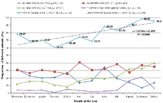
Gastrointestinal (GI) parasitic infections, including single parasite and co-infections were recorded throughout the study period (Figure 3). The monthly prevalence ranged from 60.83–98.57% for infections with at least 1 parasite type. Infection rates varied as follows; 7.14–34.36%, 25.71–40.91%, 8.57–40.25% and 1.99–21.43% for infections with single parasite types, two, three and more than three parasite associations, respectively.
The detection rates of GI parasites varied widely between season with significant differences (p<0.05) in the seasonal and monthly rates, and with several fluctuating peaks observed during the months of November, April, June and September irrespective of the type of parasite association, for infections involving at least 1 parasite type (Figure 3). Furthermore, regression analysis of the overall infection rates recorded during the study period revealed a slope with a positive gradient (Y = 2.6785X + 61.403; R2 = 0.6095) suggesting an increase in the trend from dry season [(69.42%; 95% CI: 65.31–73.31)] to rainy season [(83.99%; 95% CI: 81.94–85.59)] in GI parasites detection rates in cattle slaughtered at the Bamenda municipal abattoir (Figure 3).
Intensity of gastrointestinal parasite infections in cattle slaughtered in Bamenda
The distribution of the relative abundance of GI parasites based on worm egg count (or oocysts count for Emeria spp.) per gram of faeces (EPG) according to associated risk factors, mixed parasite associations in infections, and category of intensity of infection are shown in Tables 5 and 6, respectively. Calculation of the EPG data obtained showed significant variations about the average EPG values among infected animals in the study. Though the average EPG obtained in the study based on the intrinsic (breed, sex, age, BCS) and extrinsic (location, Season) factors were < 800, EPG suggesting light (EPG ≤ 200) intensity of GI infections, moderate (200 < EPG ≤ 800), heavy (800 < EPG ≤ 1200) and very heavy (EPG > 1200) intensity GI infections were also frequently observed across all factors
Table 3: Prevalence of gastrointestinal parasite associations of cattle in northwest region, Cameroon according to breed, sex, age, body condition score and season.
Factors
Variables
Type of gastrointestinal parasite associations in infected animals
Single parasite
2 parasites
3 parasites
More than 3 parasites
Number infected
Preva-lence
% (95% Cl)
Number infected
Preva-lence
% (95%Cl)
Number infected
Preva-lence
% (95% Cl)
Number Infected
Preva-lence
% (95% Cl)
Total (N=1907)
392
20.56 (18.74-22.37)
602
31.57 (29.48-33.65)
410
21.50 (19.66-23.34)
120
6.29 (5.20-7.38)
Sex
Male (n=1450)
304
20.97 (18.87-23.06)
451
31.10 (28.72-33.49)
297
20.48 (18.41-22.56)
100
6.90 (5.59-8.20)
Female (n=457)
88
19.26 (15.64-22.87)
151
33.04 (28.73-37.35)
113
24.73 (20.77-28.68)
20
4.38 (2.50-6.25)
Age (X)
X ≤ 3 years (n=240)
49
20.42 (15.32-25.52)
72
30.00 (24.20-35.80)
58
24.17 (18.75-29.58)
12
5.00 (2.24-7.76)
3 < X ≤ 6 years (n= 1361)
284
20.87 (18.71-23.03)
428
31.45 (28.98-33.91)
288
21.16 (18.99-23.33)
88
6.47 (5.16-7.77)
X > 6 years (n= 306)
59
19.28 (14.86-23.70)
102
33.33 (28.05-38.62)
64
20.92 (16.36-25.47)
20
6.54 (3.77-9.31)
Breed
White Fulani (n=598)
137
22.91 (19.54-26.28)
194
32.44 (28.69-36.19)
129
21.57 (18.28-24.87)
32
5.35 (3.55-7.15)
Red Fulani (n=1122)
227
20.23 (17.88-22.58)
347
30.93 (28.22-33.63)
238
21.21 (18.82-23.60)
79
7.04 (5.54-8.54)
Crossbreed (n=15)
4
26.67 (4.29-49.05)
6
40.00 (15.21-64.79)
3
20.00 (0-40.24)
0
0 (0-0)
Gaudali (n=172)
24
13.95 (8.78-19.13)
55
31.98 (25.01-38.95)
40
23.26 (16.94-29.57)
9
5.23 (1.90-8.56)
Body Con-dition Score
Thin (n=33)
5
15.15 (2.92-27.38)
13
39.39 (27.72-56.07)
9
27.27 (12.08-42.47)
4
12.12 (0.99-23.26)
Medium (n=245)
57
23.27 (17.97-28.56)
81
33.06 (27.17-38.95)
52
21.22 (16.10-26.34)
24
9.80 (6.07-13.56)
Fat (n= 1629)
330
20.26 (18.31-22.21)
508
31.18 (28.94-33.43)
349
21.42 (19.43-23.42)
92
5.65 (4.53-6.77)
Origin
Boyo (n=158)
37
23.42 (16.81-30.02)
52
32.91 (25.58-40.24)
28
17.72 (11.77-23.68)
10
6.33 (2.53-10.13)
Bui (n=518)
97
18.73 (15.37-22.09)
152
29.34 (25.42-33.26)
125
24.13 (20.45-27.82)
49
9.46 (6.94-11.98)
Donga Mantung. (n=199)
50
25.13 (19.10-31.15)
74
37.19 (30.47-43.90)
41
20.60 (14.98-26.22)
4
2.01 (0.06-3.96)
Menchum (n= 363)
69
19.01 (14.97-23.04)
121
33.33 (28.48-38.18)
78
21.49 (17.26-25.71)
19
5.23 (2.94-7.53)
Mezam (n= 310)
53
17.10 (12.91-21.29)
92
29.68 (24.59-34.76)
66
21.29 (16.73-25.85)
19
6.13 (3.46-8.80)
Momo (n=245)
55
22.45 (17.22-27.67)
78
31.84 (26.00-37.67)
53
21.63 (16.48-26.79)
13
5.31 (2.50-8.11)
Ngokentunjia (n= 114)
31
27.19 (19.02-35.36)
33
28.95 (20.62-37.27)
19
16.67 (9.83-23.51)
6
5.26 (1.16-9.36)
Season
Dry (n=533)
124
23.26 (19.68-26.85)
154
28.89 (25.04-32.74)
75
14.07 (11.12-17.02)
16
3.00 (1.55-4.45)
Rain (n=1374)
268
19.51 (17.41-21.60)
448
32.61 (30.13-35.08)
335
24.38 (22.11-26.65)
104
7.57 (6.17-8.97)
n: number of animals examined; %: proportion of infected animal; 95%Cl : 95% Confidence Interval.
Table 4: Prevalence of categories of intensity of gastrointestinal parasite infection of cattle in northwest region, Cameroon according to breed, sex, age, body condition score and season.
Factors
Variables
Category of Gastrointestinal parasite intensity in infected animals
Light (X ≤ 200)
Moderate (200<X≤800)
Heavy (800<X≤1200)
Heavy (X>1200)
Number Infected
Preva-lence
% (95% Cl)
Number Infected
Preva-lence
% (95% Cl)
Number Infected
Preva-lence
% (95% Cl)
Number Infected
Preva-lence
% (95% Cl)
Total (N=1907)
651
34.14 (32.01– 36.27)
544
28.53 (26.50– 30.55)
149
7.81 (6.61-9.02)
180
9.44 (8.13-10.75)
Sex
Male (n=1450)
494
34.07 (31.63– 36.51)
421
29.03 (26.70– 31.37)
110
7.59 (6.22-8.95)
127
8.76 (7.30-10.21)
Female (n=457)
158
34.57 (30.21– 38.93)
122
26.70 (22.64– 30.75)
39
8.53 (5.97-11.10)
53
11.60 (8.66-14.53)
Age (X)
X ≤ 3 years (n=240)
80
33.33 (27.37– 39.30)
72
30.00 (24.20– 35.80)
21
8.75 (5.18-12.32)
18
7.50 (4.17-10.83)
3 < X ≤ 6 years (n= 1361)
456
33.50 (31.00– 36.01)
372
27.33 (24.97– 29.70)
115
8.45 (6.97-9.93)
145
10.65 (9.01-12.29)
X > 6 years (n= 306)
116
37.91 (32.47– 43.34)
99
32.35 (27.11– 37.59)
13
4.25 (1.99-6.51)
17
5.56 (2.99-8.12)
Breed
White Fulani (n=598)
231
38.63 (34.73– 42.53)
169
28.26 (24.65– 31.87)
38
6.35 (4.40-8.31)
54
9.03 (6.73-11.33)
Red Fulani (n=1122)
373
33.24 (30.49– 36.00)
312
27.81 (25.19– 30.43)
98
8.73 (7.08-10.39)
108
9.63 (7.90-11.35)
Crossbreed (n=15)
6
40.00 (15.21– 64.79)
7
46.67 (21.42– 71.91)
0
0 (0-0)
0
0 (0-0)
Gaudali (n=172)
42
24.42 (18.00– 30.84)
55
31.98 (25.01– 38.95)
13
7.56 (3.61-11.51)
18
10.47 (5.89-15.04)
Body Con-dition Score
Thin (n=33)
14
42.42 (25.56– 59.29)
14
42.42 (25.56– 59.29)
0
0 (0-0)
3
9.09 (0-18.90)
Medium (n=245)
117
47.76 (41.50– 54.01)
63
25.71 (20.24– 31.19)
8
3.27 (1.04-5.49)
26
10.61 (6.76-14.47)
Fat (n= 1629)
521
31.98 (29.72– 34.25)
466
28.61 (26.41– 30.80)
141
8.66 (7.29-10.02)
151
9.27 (7.86-10.68)
Origin
Boyo (n=158)
59
37.34 (29.80– 44.88)
45
28.48 (21.44– 35.52)
6
3.80 (0.82-6.78)
17
10.76 (5.93-15.59)
Bui (n=518)
177
34.17 (30.09– 38.25)
133
25.68 (21.91– 29.44)
43
8.30 (5.93-10.68)
70
13.51 (10.57-16.49)
Donga Mantung. (n=199)
87
43.72 (36.83– 50.61)
55
27.64 (21.42– 33.85)
18
9.05 (5.06-13.03)
9
4.52 (1.64-7.41)
Menchum (n= 363)
135
37.19 (32.22– 42.16)
93
25.62 (21.13– 30.11)
28
7.71 (4.97-10.46)
31
8.54 (5.66-11.41)
Mezam (n= 310)
74
23.87 (19.13– 28.62)
96
30.97 (25.82– 36.11)
32
10.32 (6.94-13.71)
28
9.03 (5.84-12.22)
Momo (n=245)
85
34.69 (28.73– 40.65)
81
33.06 (27.17– 38.95)
14
5.71 (2.81-8.62)
19
7.76 (4.41-11.10)
Ngokentunjia (n= 114)
35
30.70 (22.23– 39.17)
40
35.09 (26.33– 43.85)
8
7.02 (2.33-11.71)
6
5.26 (1.16-9.36)
Season
Dry (n=533)
194
36.40 (32.31– 40.48)
121
22.70 (19.15– 26.26)
29
5.44 (3.52-7.37)
26
4.88 (3.05-6.71)
Rain (n=1374)
458
33.33 (30.84– 35.83)
422
30.71 (28.27– 33.15)
120
8.73 (7.24-10.23)
154
11.21 (9.54-12.88)
n: number of animals examined; %: proportion of infected animal; 95%Cl : 95% Confidence Interval.
Table 5: Intensity of categories of intensity of gastrointestinal parasite infection of cattle in northwest region, Cameroon according to breed, sex, age, body condition score and season.
Factors
Variables
Category of intensity of GI infection (egg per gram of faeces; mean±STD)
Total
Light (X≤200)
Moderate (200<X ≤800)
Heavy (800<X≤1200)
Heavy (X>1200)
Total (N=1907)
93.01± 47.57
450.55± 154.67
974.50±130.70
3389.44±2843.17
556.35± 1295.56
Sex
Male (n=1450)
91.80± 46.90
455.34± 154.92
972.27±131.78
3062.60±2734.49
505.48± 1166.40
Female (n=457)
97.47± 50.09
436.07± 152.43
980.77±129.07
4172.64±2970.13
717.72± 1630.40
Age (X)
X ≤ 3 years (n=240)
90.00± 45.98
461.11± 160.81
983.33±117.62
3258.33±3005.15
498.75± 1163.37
3 < X ≤ 6 years (n= 1361)
92.76± 48.06
459.95± 156.81
972.61±133.81
3319.31±2869.92
592.62± 1358.33
X > 6 years (n= 306)
93.98± 47.68
410.10± 134.20
991.67±15.83
4126.47±2463.87
440.20± 1085.96
Breed
White Fulani (n=598)
93.51± 47.33
432.54± 150.02
1003.95±126.48
3384.26±2517.89
527.76± 1203.53
Red Fulani (n=1122)
93.16± 49.33
462.18± 156.31
969.39±129.56
3535.65±3094.86
584.49± 1389.31
Crossbreed (n=15)
91.67± 37.64
350.00± 95.74
-
-
200.00± 162.57
Gaudali (n=172)
91.67± 36.49
457.27± 156.47
926.92±142.33
2527.78±2035.29
503.20± 986.83
Body condition score
Thin (n=33)
110.71± 56.09
432.14± 133.89
-
6433.33±3733.07
815.15± 2039.97
Medium (n=245)
99.15± 46.54
405.56± 140.88
1056.25±111.60
5032.69±3931.36
720.20± 1962.66
Fat (n= 1629)
91.36± 46.54
457.73± 155.94
969.86±130.50
3046.03±2472.37
526.46± 1140.03
Origin
Boyo (n=158)
95.76± 52.76
447.78± 178.04
1083.33±68.31
2252.94±1706.37
446.84± 872.49
Bui (n=518)
94.92± 48.58
464.66± 150.60
962.79±127.75
3917.86±3222.62
761.10± 1739.99
Donga Mantung (n=199)
95.40± 49.79
412.73± 121.42
955.56±141.31
5005.56±3880.76
468.59± 1291.45
Menchum (n= 363)
97.78± 46.06
440.32± 157.12
998.21±154.85
3646.77±3047.73
537.60± 1325.94
Mezam (n= 310)
88.51± 44.99
461.98± 155.72
948.44±126.03
2712.50±2010.28
507.10± 965.77
Momo (n=245)
88.24± 46.71
442.59± 156.95
967.86±101.16
2626.32±1979.29
435.92± 875.31
Ngokentunjia (n= 114)
78.57± 42.51
477.50± 162.89
1031.25±92.34
2266.67±1140.03
383.33± 591.78
Season
Dry (n=533)
90.72± 45.99
432.23± 137.37
965.52±134.37
2453.85±1565.63
303.38± 647.72
Rain (n=1374)
94.21± 48.44
456.40± 158.73
976.67±130.28
3547.40±2980.42
654.48± 1460.48
n : number of animals examined; mean±STD : mean EPG ± Standard deviation.
(breed, sex, age, BCS, location and season) (Table 5). Among the infested cattle in the study, the relative average GI parasite intensity was comparable or similar between age groups and breeds except for crossbreeds which had revealed lower intensities (Table 5). However, the average GI parasite intensity seemed higher (P>0.05) in female animals and during the rainy season compared to male animals and the dry season, respectively. Poor body condition score favoured higher GI parasite intensity with lean/thin animals showing the highest intensities followed by medium and fat animals. The average GI parasite intensity was higher in animals from Bui division and lower (p>0.05) in those from Ngokentunjia division respectively, compared to those from the other divisions (Table 5).
The present results showed average GI parasite intensities (EPG) of 93.01±47.57, 450.55±154.67, 974.50±130.70 and 3389.44±2843.17 for light (EPG < 200), moderate (200 < EPG ≤ 800), heavy (800 < EPG ≤ 1200) and very heavy (EPG > 1200) infection intensity categories respectively. While the GI parasite intensity for single (182.02±495.05 EPG) and 2 mixed parasite (488.12±882.05 EPG) infections revealed light and moderate intensities category, heavy intensity of infection was seen in 3 mixed parasite (1143.17±1756.16 EPG) infections, and very heavy intensity for more than 3 mixed parasite (1890.83±2661.86 EPG) infections (Table 5). The average GI parasite intensity increased with the number of parasites in the type of parasite association, reaching its highest level in animals that harboured more than 3 GI parasite associations, followed by those with 3, 2, and single parasites infections. Heavy and very heavy intensities were also observed in animals infected with 3 and more parasite associations, except for crossbreeds (Table 5).
Overall, light intensity-infections (EPG < 200) were observed among individual parasite in the study, among which Oesophagustomum spp. (140.14 ± 623.57), Trichostrongylus spp. (158.73±540.89), Haemoncus spp. (112.77 ± 550.96) and Trichuris spp. (52.75 ± 237.66) showed higher EPG values compared to the other GI parasites.
Table 6: Intensity of gastrointestinal parasite associations of cattle in northwest region, Cameroon according to breed, sex, age, body condition score and season.
Factors
Variables
Mixed GI parasite infections (egg per gram of faeces; mean±STD)
Total
Single GI parasite
2 GI parasite associations
3 GI parasite associations
More than 3 GI parasite associations
Total (N=1907)
182.02±495.05
488.12± 882.05
1143.17± 1756.16
1890.83±2661.86
556.35± 1295.56
Sex
Male (n=1450)
157.89±317.90
454.77± 781.38
948.48± 1293.66
1980.00±2808.45
505.48± 1166.40
Female (n=457)
265.34±860.48
587.75± 1127.93
1654.87± 2544.56
1445.00±1740.46
717.72± 1630.40
Age (X)
X ≤ 3 years (n=240)
95.92±143.55
611.11± 1391.20
1031.03± 1574.77
925.00±608.84
498.75± 1163.37
3<X≤6 years (n=1361)
206.69±554.08
473.60± 743.94
1219.27± 1816.30
2203.98±3942.77
592.62± 1358.33
X > 6 years (n= 306)
134.75±354.44
462.25± 957.52
902.34± 1628.77
1092.50±1672.00
440.20± 1085.96
Breed
White Fulani (n=598)
233.58±731.36
478.61± 1029.28
1196.12± 1873.42
1137.50±930.92
527.76± 1203.53
Red Fulani (n=1122)
156.61±311.90
508.21± 852.48
1103.99± 1716.31
2291.77±3152.59
584.49± 1389.31
Crossbreed (n=15)
125.00±119.02
233.33± 112.55
366.67± 189.30
-
200.00± 162.57
Gaudali (n=172)
137.50±144.65
422.73± 447.82
1263.75± 1685.69
1050.00±694.62
503.20± 986.83
Body condition score
Thin (n=33)
170.00±268.33
623.08± 1355.95
1444.44± 3453.39
1237.50±1553.69
815.15± 2039.97
Medium (n=245)
155.26±670.13
416.67± 843.36
1458.65± 2869.65
2416.67±3603.79
720.20± 1962.66
Fat (n= 1629)
186.82±462.38
496.06± 874.74
1088.40± 1457.21
1782.07±2408.30
526.46± 1140.03
Origin
Boyo (n=158)
155.41±331.41
390.38± 351.19
825.00± 701.26
2145.00±2441.59
446.84± 872.49
Bui (n=518)
141.24±311.67
588.49± 1139.99
1474.00± 2327.12
2178.57±2990.82
761.10± 1739.99
Donga Mantung (n=199)
122.00±191.44
368.92± 437.28
906.10± 1163.52
5675.00±6601.07
468.59± 1291.45
Menchum (n= 363)
315.22±973.75
470.66± 1107.13
1281.41± 2128.87
868.42±513.76
537.60± 1325.94
Mezam (n= 310)
203.77±326.09
547.83± 770.97
1034.09± 1300.95
1457.89±1787.40
507.10± 965.77
Momo (n=245)
176.36±398.36
428.36± 709.24
766.98± 751.85
1773.08±2393.45
435.92± 875.31
Ngokentunjia (n= 114)
114.52±117.75
486.36± 483.04
807.89± 430.52
1458.33±1513.80
383.33± 591.78
Season
Dry (n=533)
196.77±391.56
422.08± 730.12
781.33± 1025.58
821.88±569.49
303.38± 647.72
Rain (n=1374)
175.19±536.77
510.83± 928.21
1224.18± 1872.86
2055.29±2816.76
654.48± 1460.48
n: number of animals examined; mean±STD: mean EPG ± standard deviation, GI: Gastrointestinal.
Discussion
Gastrointestinal parasites are among the major parasitic diseases that affect cattle productivity in the Northwest region of Cameroon. These parasites, especially parasitic worms cause significant economic losses due to, anemia, diarrhea, poor growth, low weight gain, decreased reproductive effectiveness, condemnation of affected organs, and mortality in infected animals. In addition, some of these parasites, such as Trichostrongylus and Fasciola spp. have zoonotic potential, posing risks to human health. Gastrointestinal parasites which cause direct and indirect production losses include; Nematodes, Trematodes, Cestodes and Protozoa have been widely reported in cattle in tropical and subtropical regions ; ; ; ; ; . The prevalence and intensity of GI parasites are influenced by several factors, including variations in animal immunity, environmental factors such as climate, as well as feeding and management conditions of the animals.
Gastrointestinal helminthes and Eimeria spp. infections have been widely reported in cattle, and differences in prevalence rates and infection intensity attributed to variations in immunity of the animals, environmental factors such as climate, as well as feeding and management conditions of the animals. The present study revealed an overall prevalence of gastrointestinal parasitic infection was over 79% with an average infection intensity of about 560 EPG. This study provides valuable information on the burden of GI parasites among cattle in the Norwest region of Cameroon, as the animals slaughtered in this abattoir are representative of the cattle population across the entire region. Nematode and trematode infections were particularly high and similar to previous reports of prevalence for nematode (71.7%, 32.68%), trematode (26.5%, 11.22%) and cestodes (2.01%, 0.49%) in Nigeria ; , respectively. The present study also agrees with those of Hamid et al. (2016) who found that infections with nematodes were common especially Strongyles (54%) than other types of worms, followed by trematodes with Paramphistomum spp. being the most prevalent (39.43%), followed by Fasciola spp. (19.09%). High nematode and trematode infections have significant negative impact on livestock productivity, leading to reduction in production potentials such as decreased growth rate, weight loss in young calves, delayed maturity of the animals, decreased milk production, and reduced endurance in working animals ; ; . Cestodes (1.73%) and Emeria (10.23%) infections were less common. However, cestode and trematodes are of significant are of zoonotic and public health concern. Fasciola spp. is a zoonotic liver trematode notable for causing huge economic losses and health problems ; , 2024; ; FAO, 2000; ; .
The overall prevalence of GIPs reported in this study was similar with findings in other regions, such as, 81.4% in Douala Abattoir Cameroon , 80.17% in Bangladesh , 80.71% in Nigeria and 95.5% in Southern Ghana . However, it was higher than 56.7% reported in Jakiri, Bui Division, Cameroon , 65.96% in Central Java Indonesia and 41-67% in different parts of Nigeria ; ; ; ; . On the contrary, it was lower than 95.12% reported in Abeokuta Nigeria . Several factors could be attributed to these observed differences in GI parasite prevalence rates including; the periods or seasons in which the studies were conducted, location or source/origin of cattle sampled, differences in geographical and climatic conditions as well as environmental and ecological factors ; ; ; ; ; ; ; ; .
The GI parasite species identified in this study aligns with those previously identified in cattle ; ; ; ; ; and goats ; ; ; in different parts of Cameroon. Mixed livestock husbandry which includes domestic ruminants (Cattle, goats and sheep) is usually practiced using traditional systems such as nomadic or pastoral farming, mixed farming and peri-urban systems. Management systems range from free-range or extensive systems on natural grazing in less populated areas, to year-round confinement and cut-and-carry feeding in densely populated areas , 2020; . The findings of this study suggests that the observed similarities in GI parasite profiles could be attributed to the free movement and exposure of the animals to common conditions, such as ecology, pasture quality and humidity which are prevalent in the agro-ecological zones of Cameroon, and provides suitable environments for various livestock production and occurrence of GI parasite. In this study, the sampled cattle were sourced from across the entire Northwest region (seven administrative divisions) of Cameroon before being transported to the Bamenda Municipal abattoir.
In the present study, more male animals than females were sampled. Although GI parasite intensity seemed to be higher in females than in males, male animals were observed to be more aggressive during feeding and heavier feeders than females and thus more likely picked up worm eggs/ova and coccidia oocytes while grazing. However, the findings revealed that neither sex nor age group significantly influenced the prevalence of GI parasites in cattle, and both the male and female animals and all age groups (≤ 3 years, 3 to 6 years and > 6 years old) of animals had an equal likelihood of being infected with GI parasites. While some studies have reported no influence of sex on GI parasite infection rates ; ; ; , others have observed significant differences . Previous reports have suggested that male animals were more susceptible to GI parasite infections than the female animals due to hormones debilitating immune functions, which favour growth and spread of parasites in the GI tracks of male animals ; ; . Contrarily, female animals were more likely to be infected during pregnancy due to stress and decreased immune competence . Furthermore, poor feeding practices, and inadequate veterinary care which play major role in the susceptibility of animals to GI parasite infections as well as the slaughter of pregnant , 2018) were commonly observed in the study region and abattoir.
Breed, body condition score (BCS), division of origin, co-infections, infection category, and season significantly influenced prevalence rates of GI parasites. These findings agree with previous studies that reported similar influences due to these factors on GI parasites infection of cattle ; ; ; . The differences in the prevalence could be due to the presence of favourable environmental conditions necessary for the survival and development of GI parasite life stages ; ; ; . Lean-thin and moderate animals showed higher prevalence and harboured more GI parasites in this study contrary to Adedipe et al. (2014) who reported that cattle with moderate body condition score had higher prevalence of GI helminths compared to those that were emaciated. The management system of animals , and other factors such as better immunity and nutritional status of medium and fat animals (good BCS) in this study provided better protection and/or tolerance against GI parasite infections. Consequently, these animals often remain asymptomatic during the study period ; . On the other hand, malnutrition contributes to the lack of or weakened immunity and increases susceptibility of animals to both single and mixed parasite infections ; ; . In agreement with previous works ; ; ; ; ; ; , mixed GI parasite infections, which is defined as the presence of two or more GI parasite species, were more prevalent than single infections in this study. Mixed GI parasite infections cause stress leading to immunosuppression of the host immune system which increases host susceptibility to diseases including GI parasites , resulting in severe morbidity and reduced production in livestock ; . Additionally, factors such as differences in geographic location, climatic conditions, disease resistance, and grazing behavior have been associated with mixed GI parasite infections .
In this study, significant variations were observed around the average egg per gram (EPG) values in infected animals. The average EPG, based on intrinsic factors (breed, sex, age, body condition score [BCS]) and extrinsic factors (location, season), were below 800 EPG, indicating light to moderate intensities of GI parasite infections. However, heavy (800 < EPG ≤ 1200) and very heavy (EPG > 1200) intensities of infections were also widely recorded across all factors (breed, sex, age, BCS, location, and season), including in animals with two or more mixed GI parasite associations. Heavy mixed gastrointestinal helminth infections in cattle can cause significant health and gross economic impact. These infections can result in reduced growth rates, decreased productivity, poor reproductive performance, increased susceptibility to other diseases and even death ; ; ; FAO, 2000; ; .
The low prevalence (10.23%) and average intensity (<100 oocysts per gram [OPG]) of Eimeria spp. observed in this study suggest that adult cattle, which are predominantly brought for slaughter, still harbour coccidia infections, and that infection with coccidia occurred in adult cattle. Adult cattle often develop immunity and become asymptomatic carriers due to repeated exposure, serving as reservoirs of infection for more susceptible juvenile animals ; due to low levels of immunity. Subclinical coccidial infections caused by one or more Eimeria species and a low prevalence (20.90%) of Eimeria spp. in cattle have also been reported previously. However, the high prevalence and intensities of GI parasite infections recorded in juvenile animals in the present study was associated to failure in separating young animals from the adults and older animals in the various husbandry systems, overgrazing of infested pastures as well as inappropriate and inadequate use of anthelmintics by farmers. Numerous studies have documented wide variations in GI parasite intensity around average EPG values ; ; , and mixed GI parasite infections in ruminants ; ; ; ; ; ; ; ; ; ; with varying infection intensity categories. Therefore, farmers should be educated on the importance of using their own pasture, rotation of grazing pastures and having feed reserves to ensure safe feed for their cattle during periods of scarcity. Also, anthelminthic treatment of animals by farmers at every 3–4 months and inter-seasonal periods to reduce infection rates and the risk of re-infection is recommended.
The animals used in this study were destined for slaughter and human consumption and selected from the farms located in the administrative divisions of the Northwest region of Cameroon. They were apparently in clinical healthy states and transported usually by trekking (50–80 km for 2–3 days) to the Bamenda municipal abattoir and judged fit for slaughter following standard antemortem inspection procedures as described by the Cameroon veterinary services and supported by manuals on routine meat inspection (FAO, 1994; . However, the rates and intensity of GI parasite infections obtained in the present study could be under-estimations of the exact situation in the region. Animals that were in poor health and unfit for transportation to the Bamenda municipal abattoir were not selected in the farms around at the level of the divisions. Also, the evolution of GI parasites in infected animals was not investigated since repeat sampling and analysis of the studied animals was not done.
Conclusion
This study demonstrates a high prevalence and significant burden of both single and mixed gastrointestinal parasitic infections in cattle across the Northwest region of Cameroon. Juvenile and old animals showed high and similar prevalence rates and intensities of GI parasite infections. The study clearly indicates that season, location, breed, body condition score influenced the variations in GI parasite prevalence and intensity of infections. GI parasitic diseases severely affect animal health and production, leading to reduction in performance and productivity, including slower growth rates, delayed maturity, and reduced weight gains. These challenges hinder the efforts of stakeholders to achieve self-sufficiency in food and animal-derived products. Therefore, to mitigate the adverse effects of GI parasites, effective control measures that make use of the variations in GI parasite prevalence and intensity among the associated risk factors coupled with rational use of anthelmintics must be implemented. Therefore, the assistance of the veterinary services to educate livestock farmers on the importance of using their own pasture, pasteur rotation and having feed reserves to ensure safe feed for their cattle during periods of scarcity; and to strategically deworm their animals with broad spectrum anthelmintics especially at the beginning and end of rainy seasons should be emphasised. Further investigation should be done on the zoonotic potentials GI parasites of cattle and its public health significant in concerned communities. Investigation of the evolution of GI parasites in infected animals would be vital to better understand the degree of influence of risk factors and development of control measures including deworming protocols.
ACKNOWLEDGEMENT
The authors are grateful to the Delegation for Livestock, Fisheries and Animal Industries of Northwest Region Cameroon for allowing the collection and analysis of faecal samples from abattoir cattle. The authors also appreciated the generous cooperation of the cattle professionals of the Bamenda municipal abattoir, Cameroon.
NOVELTY STATEMENT
This study has highlighted the high prevalence, intensity and significant impact of single and mixed gastrointestinal (GI) parasite infections in cattle in the Northwest region of Cameroon.
AUTHOR’s CONTRIBUTION
MCK, BKN and JAN conceived, designed and coordinated the study. MCK, BKN, PYL and JAN designed data collections tools, methodology and implementation. MCK, PYL, BKN, and PJD contributed materials and carried out field investigation and data entry. MCK and JAN supervised the field and laboratory work as well as data entry. MCK, BKN, PYL and JAN were involved in data validation, statistical analysis and interpretation. MCK and BKN drafted the original manuscript which was reviewed and edited by JAN. All authors participated in preparation and critical reviewed of the manuscript. All authors have read and approved the final version of the manuscript.
Funding
No specific funding was received for this study.
Generative AI and AI-assisted technology statement
The authors declare that they have not used generative AI and AI-assisted technologies in the drafting, reviewing and editing process of this article before submission.
Conflict of interest
The authors have declared no conflict of interest.
Figures
Figure 1:.
Map of Cameroon showing the seven administrative divisions of the Northwest Region of Cameroon, cattle production villages and location of Bamenda municipal abattoir.
Source: Adapted from NIS, (2015) with information about cattle distribution from the Northwest Regional Delegation of Livestock, Fisheries and Animal Industry.
Figure 2:.
Images of Emeria spp. Oocyst and helminth eggs detected in Cattle from Northwest region, Cameroon slaughtered in the Bamenda municipal Abattoir.
Figure 3:.
Monthly prevalence (%) of mixed gastrointestinal parasite infections of cattle in northwest region, Cameroon.
References
Abass CG, Mamoudou A, Pagou KH, Abah S, Woi M, Mpoame M (2020). Hepatic and Rumenal Worms Infestations of Cattle in Vina Division (Adamawa–Cameroon). Int. J. For. Anim. Fish. Res., 4: 14-28. https://doi.org/10.22161/ijfaf.4.1.2[CrossRef]
Abdoulmoumini M, Pierre FS, Ebene NJ (2015). Trypanosomes and helminths infections in Mayo Rey Division of Cameroon and impact of concurrent infections on cattle. J. Vet. Med. Anim. Health, 7: 215-220. https://doi.org/10.5897/JVMAH2015.0373[CrossRef]
Abebe Y, Befikadu U, Getachew A (2019). Prevalence and risk factors of bovine schistosomiasis in Northwestern Ethiopia. BMC Vet. Res., 15: 12. https://doi.org/10.1186/s12917-018-1757-9[CrossRef]
Adedipe OD, Emmanuel CU, Victor OA, Oyeduntan AA, Simeon IBC (2014). Gastrointestinal helminths in slaughtered cattle in Ibadan, South-Western Nigeria. J. Vet. Med., Article ID 923561, 923566 pages. https://doi.org/10.1155/2014/923561[CrossRef]
Akter, S, Prank MR, Islam S, Akter S, Hasnine I, Ahmed MU, Faruk MSA (2023). Prevalence of gastrointestinal parasite of cattle in Ullapara Upazila, Bangladesh. J. Adv. Parasitol., 10: 1-7. https://doi.org/10.17582/journal.jap/2023/10.1.7[Full text][CrossRef]
Apio A, Plath M, Wronski T (2006). Patterns of gastrointestinal parasitic infections in the bushbuck Tragelaphus scriptus from the Queen Elizabeth National Park, Uganda. J. Helminthol., 80: 213-218. https://doi.org/10.1079/JOH2006343[CrossRef]
Atanga HM, Ngum HN, Oumar M (2024). Prevalence of bovine fasciolosis and direct financial losses in cattle slaughtered in Bamenda and Bafoussam abattoirs, Western Highlands, Cameroon. Vet. Med. Sci., 10: e70002. https://doi.org/10.1002/vms3.70002[CrossRef]
Atanga MHN, Keambou C, Kimbi HN (2019). Bovine fasciola infection: Prevalence and intensity among cattle slaughtered in Bamenda Abattoir. Int. J. Biomed. Res., 10: e4981.
Awa DN, Mbunkah DA (2009). Review of livestock pathology in the central African region: epidemiological considerations and control strategies. In: Review of livestock pathology in the central African region: epidemiological considerations and control strategies, eds. L. Seiny-Boukar and P. Boumard, Savanes africaines en développement : innover pour durer, 20 - 23 avril 2009, 13p. Garoua, Cameroun; Prasac, N’Djaména, Tchad: Cirad, Montpellier, France, cédérom.
Belem AMG, Ouinoaga PO, René B (2001). Gastro-intestinal nematodes and cestodes of cattle in Burkina Faso. Biotechnol. Agron. Soc. Environ., 5(1): 17–21.
Biffa D, Jobre Y, Chakka H (2006). Ovine helminthosis, a major health constraint to productivity of sheep in Ethiopia. Anim. Health Res. Rev., 7. https://doi.org/10.1017/S1466252307001132[CrossRef]
Blench R (1999). Traditional livestock breeds: Geographical distribution and dynamics in relation to the ecology of West Africa. In: Traditional livestock breeds: Geographical distribution and dynamics in relation to the ecology of West Africa, 67. London, UK: Overseas Development Institute.
Chollet JY, Martrenchar A, Bouchel D, Njoya A (1994). Epidemiology of digestive parasitic diseases of young cattle in northern Cameroon. Revue d’elevage et de medecine veterinaire des pays tropicaux 47: 365-374. https://doi.org/10.19182/remvt.9073[CrossRef]
Ebangui AL, Erasmus GL, Mbah D, Tawah CL, Messine O (2001). Prediction of maternal performance from preweaning weight traits in Gudali beef in a tropical environment. Rev. Portug. Zoot. AnoVII: 33 - 42.
Ebene NJ, Njonka D, Onyali IO, Manchang TK, Ikpeze OO (2022). Survey of gastrointestinal and pulmonary parasitism in two local cattle breeds in the high guinea savannah, Cameroon. Bioscientist, 10: 223-243.
Edosomwan EU, Shoyemi OO (2012). Prevalence of gastrointestinal helminth parasites of cattle and goats slaughtered at abattoirs in Benin City, Nigeria. Afr. Sci., 13: 109–114.
Ekong PS, Juryit R, Dika NM, Nguku P, Musenero M (2012). Prevalence and risk factors for zoonotic helminth infection among humans and animals-Jos, Nigeria, 2005–2009. Pan Afr. Med. J., 12.
Elele K, Owhoeli O, Gboeloh LB (2013). Prevalence of species of helminths parasites in cattle slaughtered in selected abattoirs in Port Harcourt, south-south, Nigeria. Int. Res. Med. Sci., 1(2): 10–17. https://doi.org/10.1155/2014/435913[CrossRef]
FAO (1994). Manual for meat inspection for developing countries (by Herenda D, Chambers P.G., Ettriqui A., Seneviratna P., and T.l.P. da Silva and reprinted in 2000). Food and Agriculture Organization of the United Nations, Rome, Italy. https://www.fao.org/4/t0756e/t0756e00.htm
FAO (2000). Distribution and impact of helminth diseases of livestock in developing countries. In: Corporate document repository- agriculture and consumer protection. In Distribution and impact of helminth diseases of livestock in developing countries. Food and Agricultural Organisation, Rome, Italy.
Fassi FA (2006). Collecte et maturation des ovocytes bovins: Effet de l’état nutritionnel sur le rendement et la qualité des ovocytes, Thèse de Doctorat d’Etat Es-Sciences Biologiques, Institut Agronomique et Vétérinaire Hassan II.
Grist A (2011). Ovine Meat Inspection: Anatomy, Physiology, and Disease Conditions (2nd ed.). Nottingham University Press, Nottingham, UK. pp. 335.
Hakoueu NBF, Hassanu FM, Wuld DPM, Ambrose KL, Elvis NN, Gilbert AN, Mouafo CLM, Isabelle LN, Munji VN (2021). Reproductive status of cows slaughtered at the Bamenda city slaughter house, Cameroon. EAS J. Vet. Med. Sci., 3: 50-54.
Hakoueu NBF, Gilbert N, Hassan FM, Fadila NW, Isabelle LW, Victorine NM, Pougueu HDB (2018). Incidence of foetal wastage in cattle slaughtered and its economic implications at the Bamenda city slaughter house, Cameroon. Int. J. Vet. Sci. Anim. Husb., 3: 14-18.
Hamid PH, Yuli PK, Joko P, Liliana MRS (2016). Gastrointestinal parasites of cattle in central java. Am. J. Anim. Vet. Sci., 11: 119-124. https://doi.org/10.3844/ajavsp.2016.119.124[CrossRef]
Hansen J, Perry BD (1994). The epidemiology, diagnosis and control of helminth parasites of ruminants. A handbook. Printed by the International Livestock Centre for Africa Addis Ababa, Ethiopia: Published by the International Laboratory for Research on Animal Diseases, P.O. Box 30709, Nairobi, Kenya.
Hughes S, Kelly P (2006). Interactions of malnutrition and immune impairment, with specific reference to immunity against parasites. Parasit. Immunol., 28: 577-588. https://doi.org/10.1111/j.1365-3024.2006.00897.x[CrossRef]
Kumsa B, Wossene A (2007). Abomasal nematodes of small ruminants of Ogaden region, Eastern Ethiopia prevalence,worm burden and species composition. Rev. Med. Vet., 157: 27-32.
Kumsa B, Tadesse T, Sari T, Duguma R, Hussen B (2011). Helminths of sheep and goats in Central Oromia (Ethiopia) during the dry season. J. Anim. Vet. Adv., 10: 1845-1849. https://doi.org/10.3923/javaa.2011.1845.1849[CrossRef]
Luka J, Ologunja JA, Ngozi PC, James OOB, James TT (2018). Gastrointestinal parasites of cattle and sheep slaughtered at Gombe abattoir, Gombe State, north-eastern Nigeria. Bull. Anim. Health Prod. Afr., 66: 101-109.
Mengoue LET, Rostand JAT, Pythagore SF, Estelle MM, Arnol AZT, Rolin MNK, Gustave S (2025). Schistosome infections and other gastrointestinal parasites in cattle of the Magba sub-division in the West region of Cameroon. Vet. Parasitol. Region. Stud. Rep., pp. 101199. https://doi.org/10.1016/j.vprsr.2025.101199[CrossRef]
Menkir S, Uggla A, Waller PJ (2007). Prevalence and seasonal incidence of nematode parasites and fluke infections of sheep and goats in eastern Ethiopia. Trop. Anim. Health Prod., 39. https://doi.org/10.1007/s11250-007-9035-z[CrossRef]
MINEPIA. (2000). Law NO. 2000/017 of 19 December 2000 to Regulate Veterinary Health Inspection. MINEPIA, Article 7. MINEPIA, Yaoundé, Cameroun
MINEPIA (2002). La stratégie sectoriel de l’élevage, des peches et industries animales. In: Cabinet Management 2000 MINEPIA. ed. A. Doufissa. Yaounde, Cameroon: Ministry of Livestock, Fisheries and Animal Industries, Yaounde.
MINEPIA (2014). Annuaire des statistiques du sous-secteur élevage, pêche et industries animales 2013, rapport EPIA STAT N° 002 89P. In Annuaire des statistiques du sous-secteur élevage, pêche et industries animales 2013, rapport EPIA STAT N° 002 89P. Yaoundé-Cameroun.
MINEPIA (2020). Situation des productions et des importations du sous-secteur élevage, Pêches et industries animales en 2019. In Situation des productions et des importations du sous-secteur élevage, Pêches et industries animales en 2019. Division des Etudes, de la Planification, de la Coopération et des Statistiques.: MINEPIA, Yaounde, Cameroon.
Modupe CA, Akinseye V, Cadmus E, Awosanya E, Popoola OA, Akinyemi OO, Perrett L, Stack AT, Moriyon I, Cadmus I (2017). Prevalence of bovine brucellosis in slaughtered cattle and barriers to better protection of abattoir workers in Ibadan, South-Western Nigeria. Pan Afr. Med. J., 28: 68. https://doi.org/10.11604/pamj.2017.28.68.10925[CrossRef]
Mohamed HI, Waleed MA, Khaled ME-D (2023). Prevalence and associated risk factors of gastrointestinal helminths and coccidian infections in domestic goats, Capra hircus, in Minya, Egypt. Beni-Suef Univ. J. Basic Appl. Sci., 12: 29. https://doi.org/10.1186/s43088-023-00369-6[CrossRef]
Mollah MRR, Islam AWMS, Islam MK (1996). Epidemiology of abomasal helminth of black Bengal goats in Bengladash. Indian J. Vet. Med., 16: 29-31.
Molua EL, Cornelius ML (2006). Climate, hydrology and water resources in Cameroon. CEEPA, Pretoria 37.
Molua EL (2006). Climatic trends in Cameroon: implications for agricultural management. Clim. Res., 30: 255-262. https://doi.org/10.3354/cr030255[CrossRef]
Moreau E, Chauvin A (2010). Immunity against helminths: Interactions with the host and the intercurrent infections. J. Biomed. Biotechnol., 9 pages. https://doi.org/10.1155/2010/428593[CrossRef]
Mouiche MMM, Eugenie ENW, Serge EM, Frédéric M, Jean MKF, Arouna NN, Youssouf MM, Claude S, Mamoudou A (2023). Zoo-Sanitary situation assessment, an initial step in country disease prioritization process: Systematic review and meta-analysis from 2000 to 2020 in Cameroon. Pathogens, 12: 1076. https://doi.org/10.3390/pathogens12091076[CrossRef]
Nack J, Carine CY, Georges F (2024). Diversity, prevalence and intensity of gastrointestinal helminths parasitizing goats in abattoir of the Central Market of Douala. Moroccan J. Agric. Sci., 5: 45-50.
Natumanya R, Owiny D, Kugonza R (2008). The potential of Ankole cattle abattoir ovaries for in vitro embryo production. Asian J. Anim. Biomed. Sci., 3: 1819-4214.
Ng’ang’a CJ, Maingi N, Kanyari PWN, Munyua WK (2004). Development, survival and availability of gastrointestinal nematodes of sheep on pastures in a semi-arid area of Kajiado District of Kenya. Vet. Res. Com. 28: 491-501. https://doi.org/10.1023/B:VERC.0000040246.22919.cd[CrossRef]
Ntonifor HN, Ndaleh WN (2012). Prevalence of liver fluke infections and other gastrointestinal tract parasites in slaughtered cattle in Douala, Cameroon. Bull. Anim. Hlth. Prod. Afr., 60: 439-445.
Ntonifor HN, Shei SJ, Ndaleh NW, Mbunkur GN (2013). Epidemiological studies of gastrointestinal parasitic infections in ruminants in Jakiri, Bui Division, North West Region of Cameroon. J. Vet. Med. Anim. Health, 5: 344-352.
Nwigwe JO, Njoku OO, Odikamnoro OO, Uhuo CA (2013). Comparative study of intestinal helminths and protozoa of cattle and goats in Abakaliki metropolis of Ebonyi State, Nigeria. Adv. Appl. Sci. Res., 4: 223–227.
Pamo ET, Awah-Ndukum J, Boukila B, Kana JR, Tedonkeng F, Essie MN (2006). A study on the anthelmintic property of fresh cassava (Manihot esclenta) leaves incorporated in the diet of West African Goats. Bull. Anim. Health Prod. Afr., 54: 230-233. https://doi.org/10.4314/bahpa.v54i3.32773[CrossRef]
Pfukenyi DM, Willingham A, Mukaratirwa S, Monrad J (2007). Epidemiological studies of parasitic gastrointestinal nematodes, cestodes and coccidia infections in cattle in the highveld and lowveld communal grazing areas of Zimbabwe. Onderstepoort J. Vet. Res., 74(2): 129-142. https://doi.org/10.4102/ojvr.v74i2.132[CrossRef]
Qadeer MA, Terry TL, Abduljalil FU (2018). Prevalence and predilection sites of gastro-intestinal parasites among slaughtered cattle in yola modern abattoir. Adamawa State Univ. J. Sci. Res., 6: 94-99.
Rafiullah TA, Sajid A, Shah SR, Ahmad S, Shahid M (2011). Prevalence of gastrointestinal tract parasites in cattle of Khyber Pakhtunkhwa. ARPN J. Agric. Biol. Sci., 6: 1-15.
Regassa F, Sori T, Dhuguma R, Kiros Y (2006). Epidemiology of gastrointestinal parasites of ruminants in Western Oromia, Ethiopia. Int. J. Appl. Res. Vet. Med., 4: 51-57.
Rossanigo CE, Gruner L (1995). Moisture and temperature requirements in faeces for the development of free-living stages of gastrointestinal nematodes of sheep, cattle and deer. J. Helminthol., 69: 357–362. https://doi.org/10.1017/S0022149X00014954[CrossRef]
Schutz JS, Carroll JA, Gasbarre LC, Shelton TA, Nordstrom ST, Hutcheson JP, Van Campen H, Engle TE (2012). Effects of the gastrointestinal parasites on parasite burden, rectal temperature, and antibody titer responses to vaccination and infectious bovine rhinotracheitis virus challenge. J. Anim. Sci., 90: 1948-1954. https://doi.org/10.2527/jas.2011-4059[CrossRef]
Sedrique ZT, Nfor JT (2021). Rainfall variability and quantity of water supply in Bamenda I, Northwest Region of Cameroon. In: African Handbook of Climate Change Adaptation, eds. Nicholas Oguge, Desalegn Ayal, Lydia Adeleke and Izael da Silva, pp. 713-733. Cham: Springer International Publishing. https://doi.org/10.1007/978-3-030-45106-6_139[CrossRef]
Squire SA, Amafu-Dey H, Beyuo J (2013). Epidemiology of gastrointestinal parasites of cattle from selected locations in Southern Ghana. LRRD, 25: 29-34.
Swai ES, Mtui PF, Mbise AN, Kaaya E, Sanka P, Loomu PM (2006). Prevalence of gastro intestinal parasite infections in Maasai cattle in Ngorongoro District, Tanzania. Livest. Res. Rural Dev., 18.
Tadesse G, Mengistie A (2016). Challenges, opportunities and prospects of dairy farming in Ethiopia: A review. World J. Dairy Food Sci., 11: 01 - 09.
Takang E, LeBreton M, Ayuk C, MacLeod E (2020). A socio-economic study of Fasciola infections in cattle and sheep at the Etoudi slaughterhouse, Yaoundé, Cameroon. J. Helminthol., 94. https://doi.org/10.1017/S0022149X19000890[CrossRef]
Takeet MI, Badru OB, Olubgbogi E, Abakpa SAV (2016). Prevalence of gastrointestinal parasites of cattle in Abeokuta, Ogun State, Nigeria. Niger. J. Anim. Sci., 18: 458-465.
Taylor MA, Coop RL, Wall RL (2016). Veterinary parasitology. 4th ed.; John Wiley & Sons, Ltd, The Atrium, Southern Gate, Chichester, West Sussex, UK
Tchoumboue J, Awah-Ndukum J, Tong JC (2000). A survey of gastrointestinal parasites in sheep and goats of the Western Highlands of Cameroon. Bull. Anim. Health Prod. Afr., 48: 250–253.
Tembely S (1998). Development and survival of infective larvae of nematode parasites of sheep on pasture in a cool tropical environment. Vet. Parasitol., 79: 81-87. https://doi.org/10.1016/S0304-4017(98)00108-3[CrossRef]
Tembely S, Lahlou-Kassi A, Rege JEO, Sovani S, Diedhiou ML, Baker RL (1997). The epidemiology of nematode infections in sheep in a cool tropical environment. Vet. Parasitol., 70: 129-141. https://doi.org/10.1016/S0304-4017(96)01144-2[CrossRef]
Terfa W, Kumsa B, Ayana D, Bekele H, Maurizio A, Tessarin C, Cassini R (2023). Epidemiology of gastrointestinal parasites of sheep in three agroecological zones in West Shewa Zone, Oromia State, Central Ethiopia. J. Vet. Med. Res., 10: 1241. https://doi.org/10.3390/ani13020285[CrossRef]
Thienpont D, Rochette F, Vanparijs O (2003). Diagnosing helminthiasis through coprological examination. Janssen Research Foundation, Beerse, Belgium.
Thrusfield M (2007). Veterinary epidemiology. Oxford, UK: Blackwell Science Ltd, a Blackwell publishing company.
Tiele D, Sebro E, HMeskel D, Mathewos M (2023). Epidemiology of gastrointestinal parasites of cattle in and around hosanna town, Southern Ethiopia. Vet. Med. Res. Rep., pp. 1-9. https://doi.org/10.2147/VMRR.S389787[CrossRef]
Tume SJP (2021). Rainfall seasonality and standardized precipitation valuation of water resources susceptibility to climate variability on the Bui Plateau, Northwest Region, Cameroon. Adv. Hydrol. Meteorol., 1: 1-16. https://doi.org/10.33552/AHM.2021.01.000502[CrossRef]
Turton J (1999). How to estimate the age of cattle Onderspoort, South Africa: National Department of Agriculture, ARC- Onderspoort Veterinary Institute.
Umar YA, Babayo SA, Mao PS (2018). Gastrointestinal helminths of slaughtered cattle in Bauchi Central Abattoir, Bauchi state, Nigeria. GSC Biol. Pharma. Sci., 4(2). https://doi.org/10.30574/gscbps.2018.4.2.0036[CrossRef]
Verocai GG, Umer NC, Manigandan L (2020). Diagnostic methods for detecting internal parasites of livestock. Vet. Clin. Food Anim. Pract., 36: 125-143. https://doi.org/10.1016/j.cvfa.2019.12.003[CrossRef]
Vlassoff A, Leathwick DM (2001). The epidemiology of nematode infections of sheep. N. Z. Vet. J., 49: 213-221. https://doi.org/10.1080/00480169.2001.36235[CrossRef]
Waller PJ (2006). Sustainable nematode parasite control strategies for ruminant livestock by grazing management and biological control. Anim. Feed Sci. Technol., 126(3-4): 277-289. https://doi.org/10.1016/j.anifeedsci.2005.08.007[CrossRef]
Waruiru R, Nansen P, Kyvsgaard N, Thamsborg S, Munyua W, Gathuma J, Bøgh H (1998). An abattoir survey of gastrointestinal nematode infections in cattle in the central highlands of Kenya. Vet. Res. Commun., 22: 325-334. https://doi.org/10.1023/A:1006164805185[CrossRef]
Zachée B, Mahamat O, Saboun M, Awah-Ndukum J (2020). Prevalence, intensity and risk factors of tick infestation of cattle in N’djamena Chad. Int. J. Vet. Sci. Anim. Husb., 5: 139-146.