
Pigeons (Columba livia), the members of Columbidae family are domesticated all around the globe in the villages, towns and cities. Jhang is a distant and underdeveloped district of Punjab province, Pakistan. The purpose of the present study was the determination of the prevalence of gut parasites in domestic pigeons through routine laboratory methods in district Jhang. Fresh fecal samples (n = 1800) of pigeons were gathered in plastic bags from district Jhang home fanciers and local retailers. Smearing, sedimentation, and flotation procedures were among the various parasitological methods used to analyze all of the samples that were gathered. Herein, the prevalence of coccidiosis was 70.7% and that of capillariasis was 15.7% whereas, mixed infection was 13.4%. Eggs per gram (EPG) were recorded as 750 and 700 for Coccidia and Capillaria, respectively. This study recommends the proper management and deworming strategies to reduce the risk of economic losses due to parasitic infections. This was the first study to determine the incidence of these parasitic infections in pigeons of district Jhang. However, further studies are necessary to confirm the reported incidence in the current investigation by means of advanced molecular techniques.
Research Article · Volume 69, Issue 2, 22 December 2023 · Open Access
Prevalence of Gastro-Intestinal Parasites in Pigeons of District Jhang-Pakistan
· · · · · ·
Affiliations & Notes ⌄
- 1Department of Pathobiology, University of Veterinary and Animal Sciences, Lahore sub-campus CVAS, Jhang-35200, Pakistan
- 2Department of Zoology, Government College University, Lahore-54000, Pakistan
- 3Department of Basic Sciences, University of Veterinary and Animal Sciences, Lahore Sub-Campus CVAS, Jhang-35200, Pakistan. , *Correspondence | Muhammad Fiaz Qamar, Department of Pathobiology, University of Veterinary and Animal Sciences, Lahore sub-campus CVAS, Jhang-35200, Pakistan
- 4Email: fiaz.qamar@uvas.edu.pk
Article Info ⌄
- Journal:
- Biologia (Lahore)
- Published:
- 22 December 2023
From the article
- The proportion of the infections in the pigeons were 51% for Eimeria spp. and 46% for Capillaria spp. Different researchers have reported different prevalence rates of coccidia and capillaria spp. in pigeons.
- The present study revealed that pigeons were infected with various kinds of endo-parasites along with mixed infections.
- This was a preliminary study for identification and prevalence of parasites in the pigeons.
- The prevalence of these infections in pigeons would be better identified by sophisticated molecular techniques.
Selected from the article's own text.
Abstract
Introduction
Domestic pigeons (Columba livia domestica) are kept by humans all around the globe . The population density of feral pigeon is about one pigeon per 10-20 city dwellers while the estimated worldwide population of the pigeons ranges between 165 to 330 million pigeons heads . Pigeons are not only reared as pets but also for food, ornamental and recreational purposes. That’s why they become pets in domestic, commercial and public places . However, their faces constitute the main cause of environmental pollution . A single pigeon may produce about 10-12kg of feces annually on public places including historic buildings causing health issues to humans and livestock (Haag-Wackernagel, 2005; .
Although, pigeon is not considered an endangered bird species, however its excessive hunting might produce a risk of its extinction . Pigeons feed on grains but also eat insects and worms . So, they can never be neglected as a potential carrier of different protozoa and helminths . Pigeons do not relocate, however if they are permitted, they may return from longer distances to their nests due to their home loving nature. So, they might be infected by numerous pathogens and parasites that may be transmitted to their flocks . These birds are the main cause of zoonotic infections closely associated with humans whereas figuring out how common intestinal parasites are in these birds could be crucial to enhancing public health . Nevertheless, these birds are often neglected in terms of disease investigation and associated further research . Exposure to huge burden of parasites may cause severe health issues or mortalities in affected birds .
Avian coccidiosis is the most significant protozoal disease of birds around the world that is caused by one or more of the seven birds-infecting Eimeria species ; . Eimeria species survive for longer time in the oocysts as oocysts are source of infection when excreted in the feces of infected bird , so its utmost important to control this disease. Capillaria is also an important infection of pigeons . Different endoparasites including coccidia are usually present in their droppings . Gastrointestinal parasites are under-investigated in birds in which capillaria species was the most prevalent parasite, with these, Ascaridia species and coccidial oocysts are also reported . Hence, such investigations offer the essential implications for managing the risk of zoonotic disease transmission among birds and its effects on veterinary practitioners, birds, and the general public’s health . Therefore, the current study was performed to identify the fecal parasites of pigeons in Jhang district through different simple laboratory methods. The reported prevalence of parasitic diseases would greatly help in the improvement of possible control strategies. Investigation on the distributions of the different parasitic diseases will support the clinicians to properly treat and give awareness to the fanciers about suitable control measures.
Materials and Methods
Collection and examination of fecal samples
Fresh fecal samples (n = 1800) of pigeons were collected in the plastic bags from the local shops and home fanciers of district Jhang. Two hundred collected samples were thrown out because of an artifact that occurred during transportation. All the remaining viable samples (n = 1600) were analyzed through different parasitological techniques including direct smear inspection, sedimentation and flotation techniques.
For direct smear analysis, 0.5 gram of fecal samples were mixed in 10 ml of normal saline. Following that, the debris was removed by filtering the suspension through a sieve. Then, a thin smear was made by the strained material (two drops) using glass slide and a coverslip. The slide was observed under the light microscope (Nikon, Model Eclipse E200LED; Nikon Corporation, Tokyo Japan) for the identification of oocysts . The samples positive by direct smear investigation were further recognized through sedimentation and flotation methods.
Sedimentation method was done as proposed by in which 3 grams of fecal sample were mixed in distilled water and subsequently filtered. Thereafter, centrifugation of the filtrate was done for 2 minutes at 1500 rpm (251.55 x g). Then, we discarded the supernatant and one drop of the sediments was placed on the clean glass slides to detect the parasitic oocysts under the light microscopy setup. The flotation method was carried out as proposed by . Counting of eggs per gram (EPG) was done by means of McMaster method .
Statistical analysis
Statistical analysis was accomplished using Quantitative Parasitology 1.0 (QP1.1) software . We calculated the prevalence values (percentage), mean abundance and mean intensity following previously employed statistical methods .
Results and Discussion
Overall, 890 samples were positive out of total 1600 analyzed pigeon samples. One protozoan (Eimeria spp.) and one Helminth (Capillaria spp.) parasites were recorded in the examined bird samples. Moreover, the prevalence of Coccidia (70.7%) was higher than that of Capillaria spp. (15.7%). While, the prevalence of mixed infection (Eimeria spp. + Capillaria spp.) prevalence was (120/890) 13.4% (Figure 1 and Table 1). Besides, the EPG of Coccidia and Capillaria were estimated as 750 and 700, respectively.
Poultry birds are reared at poultry farms with all the hygienic measures and proper vaccination against different infections. However, the pigeons are inhabited in the open environment, their feed is unhygienic and there is no vaccination plan available for these birds. Several protozoa can potentially infect the digestive system of the pigeons . There are different endopartasites i.e., nematodes, cestodes and protozoa that might cause the reduced growth, less egg production, increased susceptibility to different diseases and high mortality in the pigeons. Therefore, this study was designed to diagnose the parasitic infections in pigeons through fecal examination. The results obtained in this study indicated the incidence of Eimeria and Capillaria spp. in pigeons. The proportion of the infections in the pigeons were 51% for Eimeria spp. and 46% for Capillaria spp.
Different researchers have reported different prevalence rates of coccidia and capillaria spp. in pigeons. Akram et al. (2019) reported 100% prevalence of Eimeria spp. in pigeons also reported high prevalence of Eimeria spp. in pigeons that correlates with the present study. Herein, EPG was recorded at a moderate level of infection (700 for Eimeria spp. and 600 for Capillaria spp.) because of moderate temperature and humidity in November, those are not very favorable for parasitic infections. Similar findings were reported by . In this present study, the findings of temperature and humidity with relation to level of infection are similar to the findings of , who reported that nematodal infection was found at moderate level when the temperature was low. Whereas, high level of infection was attributed to monsoon weather (June to September) and the lower infection rates were recorded during summer season (February to May). Moreover, the prevalence and level of parasitic infection is influenced by the epidemiological factors such as sex, age, and breed .
Coccidia are the common pathogenic parasites in pigeons . Dovč et al. (2004) examined 139 pigeons for the most common parasitic diseases. Eimeria and Capillaria spp. were documented in 71.9% and 26.6% of the examined dropping samples. Hence, the prevalence of both coccidia and capillaria spp. is comparable to the present study. In contrast, found coccidia oocysts in 40.0% and Ascaridia columbae in 30.0% of the examined fecal samples that is very high prevalence in comparison with the current study.
Table 1: Values of fecal parasites in sampled pigeons (Columba livia).
| Parasite species in pigeons | Total infected birds | Infected birds | Not infected birds | *Percentage prevalence infected bird | **Mean intensity | ***Mean abundance |
| Eimeria spp. | 890 | 630 | 710 | 70.7 | 1.41 | 0.39 |
| Capillaria spp. | 140 | 15.7 | 6.35 | 0.08 | ||
| (Mixed infection) Eimeria spp. + Capillaria spp. | 120 | - | 13.4 | 7.41 | 0.07 |
*Calculations of Percentage Prevalence of infected bird. No. of birds (infected)/total no. of birds examined (infected and non-infected) × 100. **calculation of mean intensity. Total no. of parasites within infected host/total no. of infected birds with that parasite. ***calculation of mean abundance. Total no. of individual parasites in infected host/total no. of hosts examined (infected and non-infected).
Moreover, protozoa (mostly Eimeria sp.) were noticed in 86.05% of the free-living pigeons while 20.93% of the birds were infected by mixed parasites that were almost similar as detected herein .
In another prior study of 132 fecal samples of pigeons, overall prevalence of gastrointestinal parasites was 72.7% including Ascaridia colombae (33.3%), Eimeria spp. (31.0%), Capillaria colombae (17.4%), Raillietina (9.0%) and mixed infections (31.8%) . This study coincides with the findings of capillaria spp. and mixed infection reports of the current investigation. While, the prevalence of coccidia spp. was low as compared to the present findings. However, the low prevalence of Capillaria spp. was recorded by that is analogous to the present study. Furthermore, the previously reported prevalence of 26% for Capillaria spp. is also consistent with our studies.
Conclusions and Recommendations
The present study revealed that pigeons were infected with various kinds of endo-parasites along with mixed infections. This was a preliminary study for identification and prevalence of parasites in the pigeons. Our findings have showed that the parasitic infestations might vary from area to area and species to species as the temperature and environment have the significant role for the parasitic infections. The prevalence of these infections in pigeons would be better identified by sophisticated molecular techniques. Necessary prevention and control programs are necessary to alleviate the risk of infection in pigeons. Furthermore, proper management practices and public awareness are the other strategies to stop the spread of pathogens from pigeons to the other livestock.
Acknowledgement
The authors greatly acknowledge the assistance of the Parasitology Lab staff for the accomplishment of these experiment.
Novelty Statement
This initial investigation was carried out in a specific location to determine the gut parasite frequency in pigeons in the district of Jhang. The findings will help to improve management of certain parasitic disorders, public education, and the application of practical control measures.
Author’s Contribution
MFQ supervised the entire work and wrote manuscript. ARA, FY, TB and TH helped in planning of the experiment. IL, ARA helped in the analysis and refining the manuscript. IL, FY, and TB helped in statistical analysis of data. TH helped in collection of data.
Conflict of interest
The authors have declared no conflict of interest.
Figures
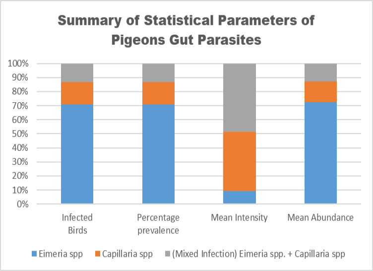
Summary of statistical parameters of gut parasites in pigeons.
References
- Adang, K., S. Oniye, A. Ezealor, P. Abdu, O. Ajanusi and K. Yoriyo. 2009. Ectoparasites and intestinal helminths of speckled pigeon (columba guinea hartlaub and finsch 1870) in Zaria, Nigeria. Sci. World J., 4(2): 1-5. https://doi.org/10.4314/swj.v4i2.51836[CrossRef]
- Akram, M.Z., M.A. Zaman, H. Jalal, S. Yousaf, A.Y. Khan, M.Z. Farooq, A.S. Tauseef-ur-Rehaman, M.F. Qamar and D.D. Bowman. 2019. Prevalence of gastrointestinal parasites of captive birds in Punjab, Pakistan. Pak. Vet. J., 39(1): 132-134. https://doi.org/10.29261/pakvetj/2018.123[CrossRef]
- Al-Barwari, S. and I. Saeed. 2012. The parasitic communities of the rock pigeon Columba Livia from Iraq: Component and importance. Türkiye Parazitol. Derg., 36(4): 232. https://doi.org/10.5152/tpd.2012.56[CrossRef]
- Bogach, M., 2021. Parasites of domestic and wild pigeons in the south of Ukraine. Biosyst. Divers, 29(2): 135-139. https://doi.org/10.15421/012118[CrossRef]
- Dar, J., T. Syed, S. Dar and J. Kuchai. 2013. First report of Capillaria anatis (nematoda: Capillariidae) from corvus species of Kashmir-India. Glob. Vet., 10(4): 467-471.
- Djelmoudi, Y., M. Daoudi-Hacini and S. Doumandji. 2014. Common endoparasites of wild rock pigeon (columba livia livia) and wood pigeon (columba palumbus) in the Algiers Sahel, Algeria. Int. J. Zoo, 4(3): 99-106.
- Dovč, A., O. Zorman-Rojs, A.V. Rataj, V. Bole-Hribovšek, U. Krapež and M. Dobeic. 2004. Health status of free-living pigeons (columba livia domestica) in the city of Ljubljana. Acta Vet. Hung., 52(2): 219-226. https://doi.org/10.1556/avet.52.2004.2.10[CrossRef]
- Faust, E.C., J.S. D’Antoni, V. Odom, M.J. Miller, C. Peres, W. Sawitz, L.F. Thomen, J. Tobie and J.H. Walker. 1938. A critical study of clinical laboratory technics for the diagnosis of protozoan cysts and helminth eggs in feces. Am. J. Trop. Med., 1(2): 169-183. https://doi.org/10.4269/ajtmh.1938.s1-18.169[CrossRef]
- Foreyt, W.J., 2013. Veterinary parasitology reference manual. John Wiley and Sons.
- Ghosh, K.K., M.S. Islam, S. Sikder, S. Das, S. Chowdhury and M.A. Alim. 2014. Prevalence of ecto and gastrointestinal parasitic infections of pigeon at Chittagong metropolitan area, Bangladesh. J. Adv. Parasitol., 1(1): 9-11. https://doi.org/10.14737/journal.jap/2014/1.1.9.11[Full text][CrossRef]
- Greiner, E.C., 2008. Isospora, atoxoplasma, and sarcocystis. Parasitic diseases of wild birds. pp. 108-119. https://doi.org/10.1002/9780813804620.ch5[CrossRef]
- Haag-Wackernagel, D. and A.J. Bircher. 2010. Ectoparasites from feral pigeons affecting humans. Dermatology, 220(1): 82-92. https://doi.org/10.1159/000266039[CrossRef]
- Haag‐Wackernagel, D., 2005. Parasites from feral pigeons as a health hazard for humans. Ann. App. Biol., 147(2): 203-210. https://doi.org/10.1111/j.1744-7348.2005.00029.x[CrossRef]
- Haug, A., P. Thebo and J.G. Mattsson. 2007. A simplified protocol for molecular identification of Eimeria species in field samples. Vet. Parasitol., 146(1-2): 35-45. https://doi.org/10.1016/j.vetpar.2006.12.015[CrossRef]
- Jha, B., 2017. Gastrointestinal parasites of feral pigeon (columba livia gmelin, 1789) at two temples of Kathmandu valley. Tribhuvan University Kirtipur, Kathmandu Nepal, 10: Batch: 2070/71.
- Kumar, S., R. Garg, A. Moftah, E.L. Clark, S.E. Macdonald, A.S. Chaudhry, O. Sparagano, P.S. Banerjee, K. Kundu and F.M. Tomley. 2014. An optimised protocol for molecular identification of Eimeria from chickens. Vet. Parasitol., 199(1-2): 24-31. https://doi.org/10.1016/j.vetpar.2013.09.026[CrossRef]
- Latif, A.A., S. Fazal, F. Manzoor, A. Maqbool, S. Asghar, I. Wajid and A. Ashraf. 2016. A comparative study on prevalence of coccidian parasites in broiler chicken (gallus gallus domesticus), Japanese quail (Coturnix coturnix japonica) and wild pigeon (Columba livia). Pak. J. Zool., 48(1).
- Mushi, E., M. Binta, R. Chabo, R. Ndebele and R. Panzirah. 2001. Seroprevalence of toxoplasma gondii and chlamydia psittaci in domestic pigeons (Columba livia domestica) at sebele, gaborone, botswana. Onderstepoort J. Vet. Res., 68(2): 159-161.
- Opara, M., I. Ogbuewu, C. Iwuji, E. Ihesie and I. Etuk. 2012. Blood characteristics, microbial and gastrointestinal parasites of street pigeons (Columbia livia) in Owerri, Imo state, Nigeria. Sci. J Anim. Sci., 1(1): 14-21.
- Parsani, H., R. Momin, A. Lateef and N. Shah. 2014. Gastro-intestinal helminths of pigeons (columba livia) in gujarat, india. Egypt. J Biol., 16(1): 63-71. https://doi.org/10.4314/ejb.v16i1.9[CrossRef]
- Qamar, M., A. Butt, S. Ehtisham-ul-Haque and M. Zaman. 2017. Attributable risk of Capillaria species in domestic pigeons (Columba livia domestica). Arq. Bras. Med. Vet. Zoot., 69: 1172-1180. https://doi.org/10.1590/1678-4162-7829[CrossRef]
- Qamar, M.F., R.Z. Abbas, M.A. Zaman, K. Ali, F.A. Atif, T. Hussain, I. Liaqat, M. Kiran, Y.A. Fadladdin and A. Bibi. 2021. Molecular detection of fancy birds parasites for clinical diagnosis and epidemiology-A review.
- Qi, M., R. Wang, C. Ning, X. Li, L. Zhang, F. Jian, Y. Sun and L. Xiao. 2011. Cryptosporidium spp. in pet birds: Genetic diversity and potential public health significance. Exp. Parasitol., 128(4): 336-340. https://doi.org/10.1016/j.exppara.2011.04.003[CrossRef]
- Reddy, B.S., S. Sivajothi and V. Rayulu. 2015. Clinical coccidiosis in adult cattle. J. Parasit. Dis., 39(3): 557-559. https://doi.org/10.1007/s12639-013-0395-1[CrossRef]
- Reiczigel, J., M. Marozzi, I. Fábián and L. Rózsa, 2019. Biostatistics for parasitologists–a primer to quantitative parasitology. Trends Parasitol., 35(4): 277-281. https://doi.org/10.1016/j.pt.2019.01.003[CrossRef]
- Rózsa, L., J. Reiczigel and G. Majoros, 2000. Quantifying parasites in samples of hosts. J. Parasitol., 86(2): 228-232. https://doi.org/10.1645/0022-3395(2000)086[0228:QPISOH]2.0.CO;2[CrossRef]
- Salem, H.M., N. Yehia, S. Al-Otaibi, A.M. El-Shehawi, A.A. Elrys, M.T. El-Saadony and M.M. Attia. 2021. The prevalence and intensity of external parasites in domestic pigeons (Columba livia domestica) in Egypt with special reference to the role of deltamethrin as insecticidal agent. Saudi J. Biol. Sci., pp. 1-7. https://doi.org/10.1016/j.sjbs.2021.10.042[CrossRef]
- Sari, B., B. Karatepe, M. Karatepe and M. Kara, 2008. Parasites of domestic (Columba livia domestica) and wild (Columba livia livia) pigeons in Nigde, Turkey. Bull. Vet. Inst. Pulawy, 52: 551554.
- Sivajothi, S. and B.S. Reddy. 2015. A study on the gastro intestinal parasites of domestic pigeons in Ysr Kadapa district in Andhra Pradesh, India. J. Dairy Vet. Anim. Res., 2(6): 10.15406. https://doi.org/10.15406/jdvar.2015.02.00057[CrossRef]
- Tietz Marques, S., R. Marinho De Cuadros, C. Jardim Da Silva and M. Baldo. 2007. Parasites of pigeons (Columba livia) in urban areas of lages, southern Brazil. Parasitol. Latinoamericana, 62(3-4): 183-187. https://doi.org/10.4067/S0717-77122007000200014[CrossRef]
- Toure, A., I.A. Konan, O.A. Etchian and A. Gogbe. 2023. Parasites pathogens and prevalence of the green pigeon (Treron calvus, TEMMINCK, 1811) in the South-East of Côte d’Ivoire: Histopathological Assessment. J. Biosci. Med., 11(11): 45-54. https://doi.org/10.4236/jbm.2023.1111005[CrossRef]
- Vidyanagar, V., 2020. Density assessment of totally urbanized bird species feral pigeons and their relationship with humans a dissertation thesis submitted to. Sardar Patel University, pp. 84.
Services
Article usage statistics
Loading…