Special Issue: Emerging and Re-Emerging Animal Health Challenges in Low and Middle-Income Countries · Volume 13, Issue 1, 14 October 2025 · Open Access
Physiological and Histological Effects of Aqueous Extract of Cinnamon and Frankincense on Albino Rats with Induced Diabetes
· ·
Affiliations & Notes ⌄
1Department of Plant Protection, Agriculture Engineering Sciences College, University of Baghdad, Baghdad, Iraq
2Department of Biology, College of Science for Women, University of Baghdad, Baghdad, Iraq
3Biotechnology Research Center, Al-Nahrain University, Iraq. *Correspondence | Majeed Hameed Nawar, Department of Plant Protection, Agriculture Engineering Sciences College, University of Baghdad, Baghdad, Iraq
Data of the body weight showed a significant increase of the control animals on the last day of the experiment, when the final weight was 179.67 ±3.18 compared to the initial weight of the same group (160.30±3.09 g) (Table 1).
In addition, a significant reduction was shown in the final weight of the diabetic categories (140.30±1.63)g compared with the initial weight of (168.22±2.09g) the same group.
While a significant increase in the final body weight was shown in the diabetic group treated with the aqueous extract of Cinnamon and Frankincense scoring 167.32±2.35 g compared with the initial weight (157.10 ±3.64 g).
Selected from the article's own text.
Abstract
One of the major causes of inability in human body to properly use or produce insulin is diabetes mellitus. Cinnamon is a food flavoring produced from the bark of the Cinnamon cassia tree. It contains many compounds including cinnamic acid, cinnamon anhydride, tannin, and methyl-hydroxychalcone polymer (MHCP) etc. Frankincense (FRN) is an aromatic resin extracted from tree species belonging the genus Boswellia within the family Burseraceae family. This study was initiated to evaluate the physiological activities of Cinnamon and Frankincense aqueous extracts, which included blood sugar level, B. urea, lipid profiles, TNF-α, LDH, creatinine, ALT, AST, MDA, CAT, and SOD. Thirty albino rats were used in this experiment, divided into three groups(10/group). The first was considered a negative control group, the second was considered a diabetic group without treatment, and the third was a diabetic group treated with the aqueous extract of the of aqueous extract of Cinnamon and Frankincense (250g/kg). A significant increase in both blood sugar levels, cholesterol, triglyceride, LDL, VLDL, Urea, creatinine, ALT, AST, MDA.CAT, TNF-α, LDH and SOD was shown in the diabetic group without treatment. The diabetic group treated with aqueous extract of Cinnamon and Frankincense showed a clear significant decrease in the above values at the end of experiment.Based on these finding, it can be concluded that using the aqueous extract of cinnamon and frankincense can lower the blood sugar level and reduce the negative side effects of diabetes and offer animals as good model to study impact of compounds.
Humans use plant extracts for various purposes to protect their food and health. They serve to protect plants from insect and other pest infestation and to protect food sources from infection (Nawar et. al., 2024; . The failure of pancreatic glands, to produce or proper use insulin by human body is termed to as diabetes mellitus (DM). Characterization of this chronic endocrine disorder can be through a high concentration of blood glucose resulted from an absolute or relative insulin deficiency, associated with insulin resistance ; . DM can be a silent killer affecting millions of people worldwide . DM as a common metabolic disease referred to as hyperglycemia as well, may occur from one or both the insufficient insulin sensitivity and failure of insulin secretion, causing the loss of glucose homeostasis. DM can impact almost every organ system of human body, but the damage level can be mainly related to the disease severity and duration ; .
Diabetes mellitus can be grouped into four categories: Diabetes Type-1: Insulin dependent diabetes mellitus or also termed hyperplasia because it is characterized by an absolute insufficiency of insulin. Beta cells are destroyed because of invasion by virus, action of chemical toxins or due to action of autoimmune antibodies . This beta cell necrosis is causing insulin deficiency and caused Type-1 diabetes .
Diabetes Type-2: Non-insulin dependent diabetes mellitus occurs when target organ insulin resists the boundary responsiveness to both exogenous and endogenous insulin . Diabetes Type-3: This type is caused by chronic drug treatment with glucocorticoids, growth hormone thiazids and diuretics . Diabetes Type- 4: This type of diabetes presented in about 4-5% of total pregnancies, can caused by placental hormones that develop insulin resistance .
Most medicinal plants comprise potentially useful chemical compounds that can be utilized as basic materials for the synthesizing contemporary medications . Royal jelly also works as an antioxidant in addition to being an anti-diabetic . Cinnamon is a common spice produced from the bark of the Cinnamon cassiae, contains many compounds including cinnamic acid, cinnamon anhydride, methyl-hydroxychalcone polymer (MHCP) and tannins. Historically, it has been prescribed as an anti-diabetic spice but results from trials involving cinnamon supplementation were inconstant . Studies showed that cinnamon extract could cause a mild effect through minimizing the fasting plasma glucose concentrations in diabetic patients suffering poor control of glycaemia .
Frankincense (FRN) an aromatic resin can be extracted from tree species belonging to the genus Boswellia within the family Burseraceae. Traditionally, Boswellia sacra can be discovered and used in numerous geographical regions, possessing a range of pharmacological properties, including curing activities as anti-hyperglycemic, antioxidant and anti-inflammation ; . Formulations containing FRN extract can decrease blood sugar in STZ-induced DM ; and alloxan-induced DM . FRN curing activity made it a potential candidate for many studies to investigate this resin as an antioxidant on DM. Thus, the current study was designated to investigate the antioxidant activities of FRN on RBC’s against the initiation of DM on experimental rats.
This study was conducted to evaluate the physiological activities of Cinnamon and Frankincense aqueous extracts on blood sugar level and the negative side effects of diabetes.
Experimental design
Albino male rats (n=30) weighing 155-170 grams were selected for treatments and divided into three groups (10 rats/group). G1: negative control, G2: diabetic group without treatment (positive group), G3: diabetic group orally treated with 250 mg/kg of Cinnamon and Frankincense aqueous extracts as a single dose/day for 30 days. The rats were obtained from the Animal House of the Center for Biotechnology at the University of Nahrain ; .
Blood collection
Blood samples, collected under anaesthetized conditions, were immediately centrifuged for 10 min, at 3000 rpm ; . The resultant serum was used for further analyses including, ALT, AST, MDA, CAT, SOD, creatinine, blood urea, lipid profile, TNF-α, CRP and LDH .
Lipid profile
Th concentration of serum cholesterol was estimated using a commercial diagnostic kit (Biolab, USA) following the manufacturer`s standard protocol. Spectrophotometry was applied to estimate the OD of the dyed complex. The absorbance values at a wavelength 500 nanometer were measured.
Triglyceride
Triglycerides concentration was estimated using a commercial diagnostic kit supplied by (Biolab, USA) of Fossati. The absorbance values were measured at a wavelength of 500 nm using a spectrophotometer.
High density lipoprotein (HDL)
This test was performed using a commercial diagnostic kit provided by (Biolab, USA). A colorimetric method was performed to measure the final reaction produced at a wavelength of 505 nm using a spectrophotometer .
Low density lipoprotein (LDL)
It is measured according to the following equation:
It was measured according to the following equation
Concentration of very low-density protein fats = concentration of triglycerides/5
ALT (Rndox/British), AST (Rndox/British), ALP (Biolabo/France). Enzymatic and colorimetric methods (Spinreact/Spain), (Cat) and Biovision-USA kits and protocols were used to measure urea and serum Creatinine in bloods of treated animals.
Diabetes induction
Diabetes in male rats was induced experimentally through a single intraperitoneal (IP) injection of alloxan (Sigma Chemical Co.) freshly dissolved in normal saline at a dose of 150 mg /kg body wt. To prevent hypoglycemia that might be resulted from alloxan week later after injection, experimental animals were let to feed overnight on glucose solution (5%) w/v. For blood glucose determination, Alloxan–treated rats were fasted for 12 hours, and blood samples were collected from the tail venous. Rats in diabetic group suffered blood sugar levels more than 250 milligram/dl were considered diabetic and chosen for additional studies (Salah et al., 2020).
Results
Data of the body weight showed a significant increase of the control animals on the last day of the experiment, when the final weight was 179.67 ±3.18 compared to the initial weight of the same group (160.30±3.09 g) (Table 1). In addition, a significant reduction was shown in the final weight of the diabetic categories (140.30±1.63)g compared with the initial weight of (168.22±2.09g) the same group. While a significant increase in the final body weight was shown in the diabetic group treated with the aqueous extract of Cinnamon and Frankincense scoring 167.32±2.35 g compared with the initial weight (157.10 ±3.64 g).
Blood test data
Testing cinnamon and frankincense extracts on Lipids profile
Cholesterol concentration data (Table 2) scored a significant increase at P≤0.05 value in in the group with diabetes and without treatment (209±5.82) mg/dl compared with the control group (100±3.56) mg/dL, while data revealed a significant decrease in glucose levels of the diabetic group exposed to Cinnamon and Frankincense (130±3.09b) mg/dl compared to the group with no treatment.
In contrast, there were significant differences in concentration of triglycerides between the group with diabetes (164±4.30)mg/dl compared to the control group scoring (89.76±3.5)mg/dl, while a significant decrease in triglycerides was noted in the group with diabetes mellitus treated with quercetin at a concentration of (102.32±2.85)mg/dl compared to the group with no treatment (Table 2).
Table 1: The activity of Cinnamon and Frankincense aqueous extracts on blood body weight in rats with induced diabetes.
Group name
Mean ± SE
Initial weight/g
Final
weight/g
LSD
Control
160.30 ±3.09b
179.27±3.06A
16.32 *
Diabetes
168.22 ±2.09a
140.30±1.63 b
18.52*
Diabetes + moringa aqueous extract
157.10 ±3.64b
167.32±2.35a
6.237 *
Means with the different letters at the same row were differed significantly. * (P≤0.05).
As for high-density lipoprotein (HDL) (the beneficial ones), a significant decrease in the level of these fats was noticed in the group with diabetes (22.43±4.21) mg/dl compared to the group in negative control (30.79±3.65). While data showed a significant decrease in the concentration of good fats in the group treated with Cinnamon and Frankincense extracts (38.42±3.90).
A significant increase in the concentration of low-density lipoprotein (LDL) (bad) was noticed in the diabetic group (151.29 ±5.70) mg/dl compared to the negative control group (88.00±5.36) mg/dl, while the level of these fats reduced in the group that having diabetes and treated by Cinnamon and Frankincense extracts (109.59 ±6.85) mg/dl.
About very low-density lipoprotein (VLDL), data showed a significant variance in the positive control group (35.20 ±3.52) mg/dl compared to the control group (22.43±2.73) mg/dl. and the diabetic group treated with Cinnamon and Frankincense extracts (30.45±2.90)
Table 2: The activity of Cinnamon and Frankincense extracts on the lipid profiles in different treated diabetic rat groups.
Group
Mean ± SE (mg/dl)
Cholesterol
Triglyceride
HDL
LDL
VLDL
Negative Control
100±3.56c
89.76±3.54c
30.79±3.65a
53.00±5.36 c
17.54±2.73b
Positive diabetic
209±5.82a
164±4.30a
22.43±4.21b
155.29±5.70a
32.20 ±3.52a
Diabetic +quarectine
130±3.09b
102.32±2.85b
38.42±3.90 a
72.59 ±6.85b
20.45±2.90a
LSD value
14.65**
21.43**
6.21**
12.40**
5.42**
Means with the different letters at the same row were differed significantly. * (P≤0.05).
The results in Table 3 showed significant increase in the untreated diabetic group in the level of glucose (370.43±4.55) mg/dl and diabetic group treated with Cinnamon and Frankincense extracts (364±2.69) compared with the negative control group (90.56±3.05) mg/dl at the first day of experiment. Whilst a decreased level of glucose was noted in the diabetic group treated with Cinnamon and Frankincense extracts (154.43±1.96) mg/dl at the 30th, Compared with the same group on the first day, while the glucose level remained high throughout the treatment period in the group with diabetes without treatment (380±2.85).
Table 3: The glucose level of treated diabetic rats exposed to with Cinnamon and Frankincense extracts on
Group
Mean ± SE
Glucose(mg/dl) first day
Glucose(mg/dl)
20th day
Negative Control
90.56±3.05b
85.54±4.02 c
Positive diabetic
370.43±4.55 a
380±2.85a
Diabetic+ quercetin
364±2.69a
154.43±1.96 b
LSD value
9.32 *
7.16*
Means with the different letters at the same row were differed significantly. * (P≤0.05)
The results revealed an obvious significant increase in the untreated diabetic group in creatinine and blood urea levels (1.94 ±0.05, 42.54±3.53) mg/dl, respectively, compared with the negative control group (Table 4).When treated with Cinnamon and Frankincense extracts, the level of creatinine and blood urea decreased in the diabetic group treated scoring 0.95 ±0.06 and 29.50±2.43 mg/dl, respectively compared with the positive control group.
Table 4: Urea and creatinine concentrations in diabetic rats treated exposed to Cinnamon and Frankincense extracts.
Group
Mean ± SE
B. Urea (mg/dl)
Creatinine (mg/dl)
Negative Control
19.52±2.08c
0.53 ±0.02 c
Positive diabetic
42.54±3.53 a
1.94 ±0.05a
Diabetic+ quercetin
30.50±2.43b
0.80 ±0.06 b
LSD value
5.10 *
0.12 *
Means with different letters at the same column were differed significantly. * (P≤0.05).
The results shown in Table 5 indicate significant differences CRP level among tested groups. In contrast, a significant increase was observed in the enzymatic activity of the lactate dehydrogenase of rat group suffering diabetes scoring (176.54±3.32) in comparison with the -ve control group (120.1 8±4.3), while the activity of the enzyme decreased in the group of diabetic rats treated with Cinnamon and Frankincense extracts (137.54±3.65). Table 5 also showed a significant increase in the level of TNF-α in the group of diabetic rats (4.90±1.22) in comparison with the negative control group (1.53 ±0.05), while a no significant differences in level of TNF-α was observed between negative control and diabetic group treated with Cinnamon and Frankincense extracts (1.53 ±0.05, 2.05 ±0.39).
Table 5: Estimating the level of TNF-α, CRP, and LDH concentrations in in diabetic rats treated exposed to Cinnamon and Frankincense extracts.
Groups
Mean ± SE
TNF-α
CRP
LDH
Negative control
1.53 ±0.05b
2.78±0.89a
120.1 8±4.32c
Positive diabetic
4.90±1.22a
8.05±0.54a
176.54±3.32a
Diabetic + Cinnamon and Frankincense
2.05±0.39b
4.45±0.53a
137.54±3.65B
LSD value
1.45**
0.95**
10.50**
Means having with the different letters in same column differed significantly. * (P≤0.05).
Estimation of the concentrations of malondialdehyde, CAT and SOD enzymes in diabetic groups exposed to Cinnamon and Frankincense
Data revealed in the level of malondialdehyde level increased significantly, in the diabetic group scoring 4.94 ±0.48 mmol/ml compared to those in the control group and diabetic treated with Cinnamon and Frankincense groups, scoring (1.23±0.06) and (2.05±0.39 mmol/ml highest malondialdehyde levels, respectively.
Noticeably, the enzymatic level of catalase increased significantly in diabetic untreated rat group scoring 14.70±2.63. Whereas this enzyme scored (5.53±1.53) and (9.30 ±2.76) maximum levels in the negative control and diabetic group treated with Cinnamon and Frankincense, indicating a significant decrease in the enzyme level.
Table 6: Malondialdehyde, CAT, and SOD enzyme concentrations in experimental rat groups exposed to Cinnamon and Frankincense.
Groups
Mean ± SE
MDA
CAT
SOD
Negative control
1.23 ±0.06C
5.53±1.53b
2.48±0.34c
Diabetic
4.94 ±0.48 A
14.70±2.63a
9.86 ±2.94a
Diabetic+ quercetin
2.05 ±0.39B
9.30 ±2.76a
5.49 ±2.67b
LSD value
0.47 **
2.50**
288**
Means having with the different letters in same column differed significantly. * (P≤0.05).
Finally, the level of superoxide dismutase enzyme was significantly increased in the blood of diabetic rat group scoring 9.86±2.94 IU/L. While those in the healthy control and treated with Cinnamon and Frankincense and the diabetic groups showed a significant decrease in the enzyme activity which was 2.48±0.34 and (5.49±2.67) IU/liter, respectively.
Histological study
Discussion
Diabetes mellitus (DM) is amongst the most serious diseases which are expensive, reduce life expectancy and triggering disability, and causing life-threatening complications . Long-term hyperglycemia can result in enormous and significant consequences, e.g., increased glucose self-oxidation, oxidative stress and protein glycosylation resulting in reduced glucose absorption in muscle and fat cells decreasing insulin release from beta cells . Our results in Table 3 showed that, the level of glucose, Cholesterol, Triglyceride, HDL and VLDL decreased in the diabetic group treated with Cinnamon and Frankincense extracts, which were similar to those presented by Al-Muhammadi (2016) who found a significant elevation p<0.05 in Cholesterol, Triglyceride, HDL-C, VLDL-C concentration in rats with diabetes and after treatment with aqueous extract significant falling was occurred for the entirely parameters. LDH, and D-dimer considered as inflammatory markers . The body has a large amount of LDH, with high concentrations in the heart, liver, skeletal muscle, erythrocytes, and kidney and low concentrations in the lung, brain, and smooth muscle . Our data did not indicate any significant differences in CRP level among the different groups. In contrast, a significant increase in the enzyme lactate dehydrogenase activity was noticed in the group of diabetic rats compared to those in -ve control group. Whereas this enzyme activity reduced in the group of diabetic rats treated with Cinnamon and Frankincense extracts, this result is in agreement with the result of Salih et al. (2014) who found that serum LDH was increased significantly in diabetic mice compared with controls. Our results showed that the level of TNF-a in diabetic mice was higher than that of controls, this result is in agreement with the result of Lee et al. (2005) who concluded that TNF-α plays a crucial role in the initiation of T1DM by modulating DC–T cell interactions via modulation of DC development. On contemporary, a significant increase in the level of malondialdehyde was observed in the diabetic group compared to the control group and diabetic group treated with Cinnamon and Frankincense, this result is similar to the result of Gwarzo et al. (2014) who found a raising in the level of malondialdehyde among the diabetic rat prior to treatment and significantly decreased after the treatment. SOD and CAT considered as a protective enzyme that works as the first line of defense, converting superoxide radical to hydrogen peroxide .
There was a significant increase in the level of the enzymes CAT and SOD in the diabetic group compared to the control group, this result is similar to the result of Ramanathan et al. (1999) who find that early diabetic rats exhibit increased SOD and CAT activities.
Acknowledgement
We acknowledge members of Biotechnology center at the University of Nahrain for their support and facilities.
Novelty Statement
This study introduces a novel approach by the treatment of induced diabetes male albino rats with Cinnamon and Frankincense aqueous extracts and monitoring their effects on some physiological and histological parameters.
Author’s Contribution
SMMA-C: Study conception and design
RMO: Literature search and manuscript preparation.
MHN: Data acquisition.
SMMA-C, MHN and RMO: Manuscript editing and review.
Generative AI and AI-assisted technology statement
The authors declare that no Genrative AI was used in the creation of this manuscript.
Conflict of interest
The authors have declared no conflict of interest.
Figures
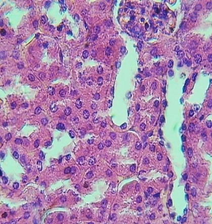
Figure 1:.
Section of renal cortex (control) shows: normal lining cells with and normal glomerulus. H and E stain. 400x.
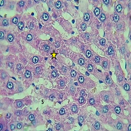
Figure 2:.
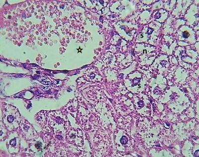
Section of hepatic lobule (5) shows normal hepatocytes (Asterisk), normal sinusoids (S) and kupffer cells. H and E. 400x.
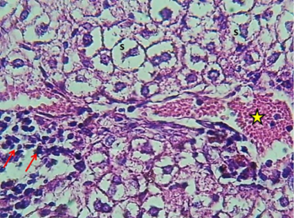
Figure 3:.
Hepatic lobule section (diabetic) showing mild congestion with dilation of portal vein (asterisk) with peri portal aggregation of MNCs (Arrow) and marked cellular swelling with nuclear pyknosis of hepatocytes (S). H and E stain. 400x.
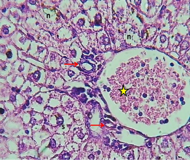
Figure 4:.
section of hepatic lobule (diabetic) showing mild congestion of central vein (Asterisk) with marked peri central histogenesis of newly formed hepatocytes (Arrow) and marked cellular swelling with nuclear pyknosis of hepatocytes (s). and necrosis (n) H and E stain. stain. 400x.
Figure 5:.
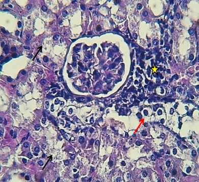
Section of renal cortex (diabetic) showing mild cellular swelling of lining cells (Red arrow) with necrosis (Black arrow) of the lining cells of renal tubules, with little peri glomerular aggregation of MNCs (Asterisk). H and E stain. 400x.
Figure 6:.
Central vein section (diabetic) showing dilation central vein with congestion with little cellular swelling (s) and necrosis (n) of hepatocytes. H and E stain. 400x.
Figure 7:.
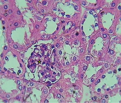
Renal cortex section (diabetic) showing little figures of vascular degeneration of lining cells of renal tubules (v) with normal glomerulus (Asterisk). H and E stain. 400x.
Figure 8:.
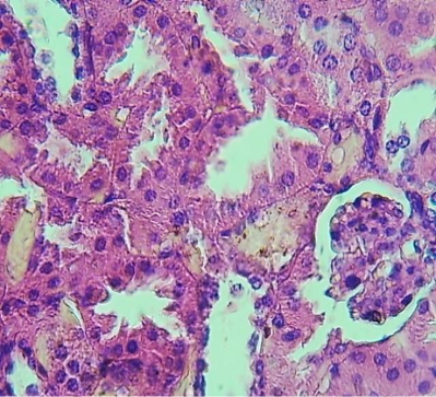
Renal cortex section (diabetic treated with extract) showing normal lining cells of renal tubules with normal cytoarchitecture of glomerular. H and E stain. 400x.
Figure 9:.
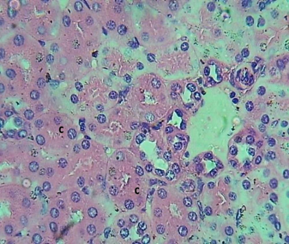
Renal cortex section (diabetic treated with extract) showing mild nephrosis little figures of cloudy swelling of lining cells of renal tubules (C). H and E stain. 400x.
References
Abdlkarem HA, Zainulabdeen JA (2024). A comparative study of vitamin D level and lactate dehydrogenase activity in relation to oxidative stress in women with osteoporosis. J. Fac. Med. Baghdad, 66: 110-115. https://doi.org/10.32007/jfacmedbagdad.6612255[CrossRef]
Abdul-Hussein TA, Fadhil HY (2023). Impact of inflammatory markers, dread diseases and cycle threshold (Ct) Values in COVID-19 progression. Rev. Bionat., 8(1): 33. https://doi.org/10.21931/RB/2023.08.01.33[CrossRef]
Al-Timimi ZK (2019). Histological effect of aspirin on the stomach of male albino Swiss mice (Mus musculus). Iraqi J. Sci., 61(6): 1275-1281. https://doi.org/10.24996/ijs.2020.61.6.4[CrossRef]
Al-Muhammadim QN (2016). Physiological study to investigate the activity of an aqueous extract of Cinnamomum cassiabark on the blood glucose levels in healthy and diabetic rats induced by streptozotocin (stz). Baghdad Sci. J., 13(4): 0681. https://doi.org/10.21123/bsj.2016.13.4.0681[CrossRef]
Alrefaei GI, Almohaimeed HM, Algaidi SA, Almuhayawi MS, AbdAllah FM, Al-Abbas NS, Shaer NA, Mohammedsaleh ZM, Saleh FM, Ayuob N (2023). Cinnamon and ginger extracts attenuate diabetes-induced inflammatory testicular injury in rats and modulating SIRT1 expression. J. Men’s Health, 19(8): 22-32.
Al-Timimi ZK, Taher TA (2020). Hepatotoxic impact of erythromycin succinate after orally repeated exposure in male albino Swiss mice (Mus musculus). Iraqi J. Agric. Sci., 51(5): 1375-1380. https://doi.org/10.36103/ijas.v51i5.1147[CrossRef]
Azemi ME, Namjoyan, F., Khodayar, M. J., Ahmadpour, F., Padok, A. D., Panahi, M. (2012). The antioxidant capacity and anti-diabetic effect of boswellia serrata triana and planch aqueous extract in fertile female diabetic rats and the possible effects on reproduction and histological changes in the liver and kidneys. Jundishapur J. Nat. Pharm. Prod., 7(4): 168-175. https://doi.org/10.17795/jjnpp-6755[CrossRef]
Bacha F, Lee S, Gungor N, Arslanian SA (2010). From pre-diabetes to type 2 diabetes in obese youth: pathophysiological characteristics along the spectrum of glucose dysregulation. Diabetes Care, 33(10): 2225-2231. https://doi.org/10.2337/dc10-0004[CrossRef]
Chaudhary N, Tyagi N (2018). Medicinal plants used for diabetes mellitus: An overview. Int. J. Res. Dev. Pharm. L. Sci., 7(4): 3022-3029. https://doi.org/10.21276/IJRDPL.2278-0238.2018.7(4).3022-3029[CrossRef]
Deepti B, Sowjanya K, Lidiya B, Bhargavi RS, Babu PS (2017). A modern review on Diabetes mellitus: An inhibitory metabolic disorder. J. in silico in vitro Pharmacol., 3: 1-14.
Gwarzo MY, Ahmadu JH, Ahmad MB, Dikko AUA (2014). Serum glucose and malondialdehyde levels in alloxan induced diabetic rats supplemented with methanolic extract of tacazzea apiculata. Int. J. Biomed. Sci., 10(4): 236-242. https://doi.org/10.59566/IJBS.2014.10236[CrossRef]
Jameel MAR, Baker SZ, Nawar MH (2023). Control of the Khapra Beetle Trogoderma granarium (Everts.) (Coleoptera: Dermastidae) using nanoparticles of neem leaf extract. In: IOP conference series: Earth and environmental science. IOP Publishing. 1225(1): 012087. https://doi.org/10.1088/1755-1315/1225/1/012087[CrossRef]
Jimoh A, Tank Y, Mohammed A (2013). Anti-diabetic effect of methanolic leaf extract of Cissus cornifolia on alloxan-induce hyperglycemia in Wistar rats. Ann. Biol. Res., 4(3): 46-54.
Kareem Z, Galeel S (2022). Physiological and biochemical effects study on aqueous extract of thymus vulgaris in alloxan induced diabetic male albino rats. Biochem. Cell. Arch., 22: 779-783.
Kavitha JV, Rosario, J. F., Chandran, J., Anbu, P. (2007). Hypoglycemic and other related effects of Boswellia glabra in alloxan-induced diabetic rats. Indian J. Physiol. Pharmacol., 51(1): 29-39.
Kirkham S, Akilen R, Sharma S, Tsiami A (2009). The potential of cinnamon to reduce blood glucose levels in patients with type 2 diabetes and insulin resistance. Diabetes Obes. Metab., 11(12): 1100-1113. https://doi.org/10.1111/j.1463-1326.2009.01094.x[CrossRef]
Lee LF, Xu B, Michie SA, McDevitt HO (2005). The role of TNF-α in the pathogenesis of type 1 diabetes in the nonobese diabetic mouse: Analysis of dendritic cell maturation. Proc. Natl. Acad. Sci., 102(44): 15995-16000. https://doi.org/10.1073/pnas.0508122102[CrossRef]
Mang B, Wolters M, Schmitt B, Kelb K, Lichtinghagen R, Stichtenoth DO, Hahn A (2006). Effects of a cinnamon extract on plasma glucose, HbA, and serum lipids in diabetes mellitus type 2. Eur. J. Clin. Invest., 36(5): 340-344. https://doi.org/10.1111/j.1365-2362.2006.01629.x[CrossRef]
Mothana RA (2011). Anti-inflammatory, antinociceptive and antioxidant activities of the endemic Soqotraen Boswellia elongata Balf. f. and Jatropha unicostata Balf. f. in different experimental models. Food Chem. Toxicol., 49(10): 2594-2599. https://doi.org/10.1016/j.fct.2011.06.079[CrossRef]
Mothana RAA, Gruenert, R., Lindequist, U., Bednarski, P.J. (2007). Study of the anticancer potential of Yemeni plants used in folk medicine. Pharmazie, 62(4): 305-307.
Nawar MH, Yaaqoob LA, Hatem MW, Mohammed AK (2024). The effect of nanoparticles preparation from extract of Dodonaea Viscosa L. leaves on the biological performance of the Great Waxworm. In: AIP Conf. Proc. AIP Publishing. 2922(1). https://doi.org/10.1063/5.0183841[CrossRef]
Obaid RM, Yaseen FT, Mukhlif MY (2020). Blood cells depletion after chemotherapy in Iraqi women with breast cancer. Indian J. Forensic Med. Toxicol.,14(4): 3379-3382.
Obaid RM, Yaseen FT, Salim AK (2020). Correlation between vitamin D3 (cholecalciferol) and thyroid diseases in Iraqi patients. Ann. Trop. Med. Publ. Health., 23(S16): SP231603. https://doi.org/10.36295/ASRO.2020.231603[CrossRef]
Obaid RM, Yaseen FT, Kadhim NK, Salim DH, Sabar ZT, Abd DS, Hasan AF (2025). Changes in the level of lipid profile in diabetes mellitus in samples patients. J. Biosci. Appl. Res., 11: 331-336. https://doi.org/10.21608/jbaar.2025.350090.1134[CrossRef]
Obaid RM (2021). A prevalence of menstrual dysfunction and hirsutism in women with Polycystic Ovary Syndrome (PCOS). Biochem. Cell. Arch. 21: 2653-2656. https://connectjournals.com/03896.2021.21.2653.
Okigbo R, Anuagasi C, Amadi J (2009). Advances in selected medicinal and aromaticplants indigenous to Africa. J. Med. Plants Res., 2: 086-095.
Oudah HK, Jawad MM, Abed EH (2017). Role of enzymes catalase, peroxidase and amino acid (proline) in Raphanus sativus and Lepidium sativus in exposure levels different water pollution of ion lead. Iraqi J. Sci., 58(2A): 592-599.
Pari L, Saravanan R (2004). Antidiabetic effect of diasulin an herbal drug on blood glucose, plasma insulin and hepatic enzymes of glucose metabolism in hyperglycaemic rats. Diabetes Obesity Metab., 6: 286-292. https://doi.org/10.1111/j.1462-8902.2004.0349.x[CrossRef]
Raheem BM, Obaid RM, Ali BR, Al-Fahham AA (2024). The role of TNF-α and IL-6 SNP in polycystic ovary syndrome susceptibility. Pol. Merkur. Lek., 52(3): 286-291. https://doi.org/10.36740/Merkur202403104[CrossRef]
Ramanathan M, Jaiswal AK, Bhattacharya (1999). Superoxide dismutase, catalase and glutathione peroxidase activities in the brain of streptozotocin induced diabetic rats. Indian J. Exp. Biol., 37: 182-183.
Salah MM, Al-Chalabi, Duha MM, Ahmed AJ, Khalid SA-A (2020). Benefit effect of ethanolic extract of Bay leaves (Laura nobilis) on blood sugar level in adult diabetic rats induced by alloxan monohydrate. J. Ann. Trop. Med. Publ. Health, 23(1). https://doi.org/10.36295/ASRO.2020.231608[CrossRef]
Salih LA, Nawar MH, Muuhsen SM (2021). Analytical study of the royal-jelly effect on sperms morphological characteristics and some liver function parameters and their correlation with some antioxidants in male albino rats treated with pregabalin drug. J. Phys. Conf. Ser., 2114(1): 012056. https://doi.org/10.1088/1742-6596/2114/1/012056[CrossRef]
Salih ND, Kumar GH, Noah RM, Muslih RK (2014). The effect of streptozotocin induced diabetes mellitus on liver activity in mice. Glob. J. Adv. Pure Appl. Sci., 4: 66-74. Available from: http://www.world-education-center.org/index.php/paas
Shehata AM, Quintanilla-Fend, L., Bettio, S., Singh, C. B., Ammon, H. P. T. (2011). Prevention of multiple low-dose streptozotocin (MLD-STZ) diabetes in mice by an extract from gum resin of Boswellia serrata (BE). Phytomedicine, 18(12): 1037-1044. https://doi.org/10.1016/j.phymed.2011.06.035[CrossRef]
Sun H, Saeedi P, Karuranga S, Pinkepank M, Ogurtsova K, Duncan BB, Magliano, D. J. (2022). IDF diabetes atlas: Global, regional and country-level diabetes prevalence estimates for 2021 and projections for 2045. Diabetes Res. Clin. Pract., 183: 109-119. https://doi.org/10.2337/db22-er08a[CrossRef]
Tripathi V, Verma JJ (2014). Current updates of Indian antidiabetic medicinal plants. Int. Pharm. Chem., 4: 114-118.
Wang TJ, Larson MG, Vasan RS, Cheng S, Rhee EP, McCabe E (2011). Metabolite profiles and the risk of developing diabetes. Nat. Med., 17(4): 448-453. https://doi.org/10.1038/nm.2307[CrossRef]