Place of Work
The study was conducted at the High Security Animal Disease Laboratory (HSADL), IVRI, Bhopal, and Department of Veterinary Microbiology, College of Veterinary Science and Animal Husbandry, Mhow, Madhya Pradesh, India.
Virus Isolate
The BVDV isolate (Ind S–17555), initially isolated from a sheep and available at HSADL, IVRI, Bhopal, was used in this study to investigate the physio–chemical properties of BVDV.
Cell Line
Sheep Fetal Thymus Riems (SFTR) cell line obtained from Riems, Germany and available at HSADL, IVRI, Bhopal, was used in this study. The cells were grown in EMEM medium containing 10% horse serum and were free of any adventitious pestivirus contamination.
Immunoconjugates
Anti–mouse IgG–peroxidase conjugate, raised in rabbits (Sigma, cat.no.A–9044) and anti–bovine IgG (whole molecule) peroxidase conjugate, raised in rabbits (Sigma, cat. No.A–7414) were used in the imunoperoxidase monolayer assay (IPMA).
Effect of Temperature
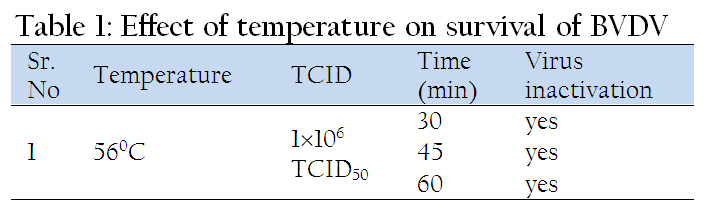
In this study 1x10⁶ TCID50 dilution of BVDV was used for determining the effect of temperature on its viability. One ml of virus suspension in EMEM was treated at 56oC for 30, 45, and 60 min, respectively in circulating water bath. Then preformed monolayer of SFTR cells in 6 well tissue culture plate was infected with 300 µl of the treated inoculums. The plate was incubated at 37oC for 1h for adsorption of virus. The inoculum was removed, monolayer was rinsed slowly with 500µl EMEM, and 2 ml EMEM supplemented with 2% horse serum (infection medium) was added to the wells. The plate was incubated at 37oC in a CO2 incubator for three to four days.
Immunoperoxidase Momnolayer Assay (IPMA)
After completion of incubation period of three to four days, the plate was tested by IPMA for detection of live/survived virus as per the method described by Wood et al. (2004) with certain modifications. The medium was removed and plate was rinsed with 1X PBS. The cells were then heat fixed by keeping the plate at 80oC for 1h in hot air oven and then allowed to cool to room temperature for 20 min. Cells were rehydrated by adding 300 µl of PBS (1X). Then 50 µl of 1:120 diluted WB112 Mab (specific for NS3 region) was added to each well and incubated at 4oC. After overnight incubation the wells were washed thrice (each of 5 min duration) with 1x PBST (0.05% Tween–20). Then 50µl of HRPO conjugated anti–mouse antibodies (Sigma), diluted to 1:300, was added to each well and incubated for 1 h at 37oC. Again four washings were given with 1X PBST (0.05% Tween–20). Stock chromogen mixture was prepared by dissolving 100mg of AEC (3 amino–9–ethylcarbazole) in 15 ml DMF (N’N’ dimethyl formamide). From this stock, AEC working chromogen substrate was prepared by adding 300 µl of stock chromogen in 5 ml of acetate buffer (pH 4) and 5 µl of hydrogen peroxide (H2O2). 50 µl of this working chromogen substrate was dispensed in each well and plate was incubated for 20 min at room temperature in dark. Rinsing the plate under tap running water stopped the reaction. Development of red color in the cell cytoplasm was considered positive for infectivity / presence of BVD virus, while no color in the cell cytoplasm was considered negative for infectivity or / absence of BVD virus as shown in Figure 1.
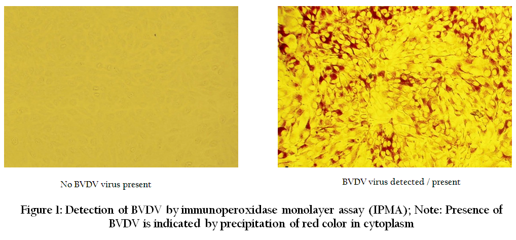
Figure 1: Detection of BVDV by immunoperoxidase monolayer assay (IPMA); Note: Presence of BVDV is indicated by precipitation of red color in cytoplasm
Effect of Dyes
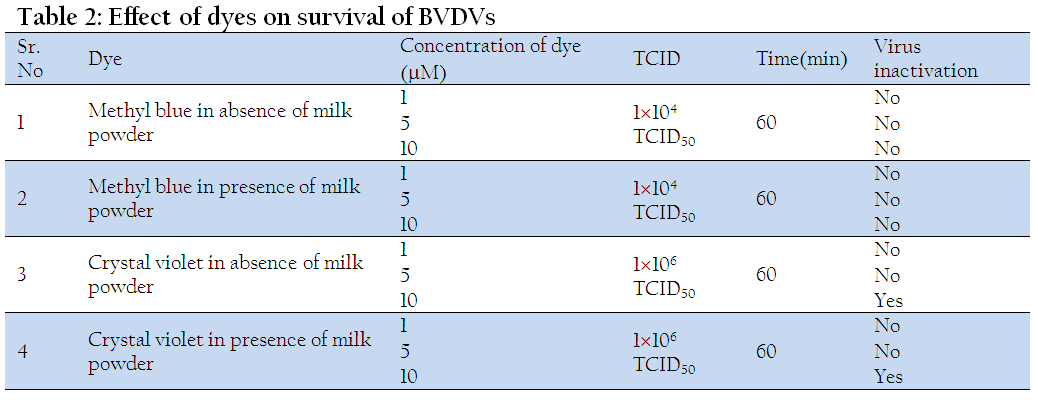
The effect of 1, 5 and 10µM concentrations of methyl blue and crystal violet dyes on BVDV was studied in the presence and absence of organic matter (milk powder). The BVDV was diluted (1104 TCID50 for methyl blue and 1x10⁶ TCID50 for crystal violet) in EMEM. Different concentrations (1, 5 and 10µM) of dye were prepared in 1.5 ml eppendorf tubes by adding 1, 5, 10µl of each of dye (from stock 1mM solution) in corresponding tube and final volume was made up to 1000µl by adding the diluted virus. The material was then kept under artificial light (100 lux) at room temperature for 1h. The preformed monolayer of SFTR cells (3×10⁵ cells/ml) in 6 well tissue culture plate was infected with 300 µl of the treated inoculums and remaining procedure was same as described earlier.
For studying the effect of dyes on BVDV in presence of milk powder, the virus was diluted in EMEM supplemented with 1% milk powder and all other procedures followed were same as described above.
Effect of Alcohols
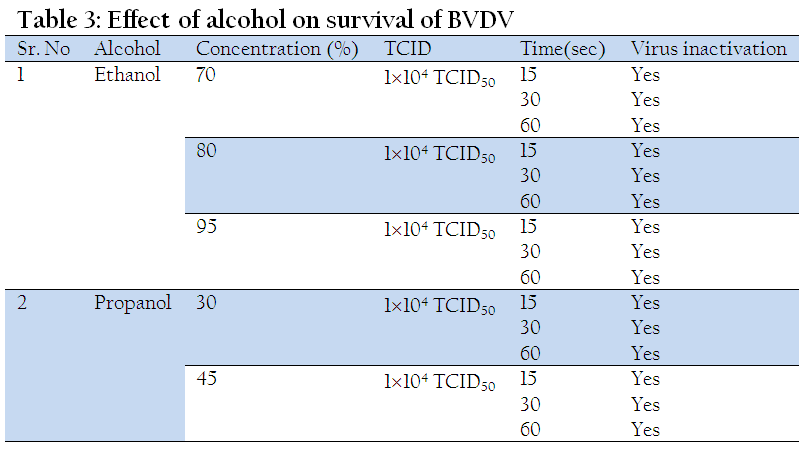
For studying the effect of alcohols treatment was given as 8 parts alcohol +1 part virus (1x10⁴ TCID50 of BVDV) +1 part medium.
Effect of Ethanol
To study the effect of 70 % ethanol, 80 µl of 87.5% ethanol, 10 µl diluted virus (1x10⁴ TCID50 ), and 10µl EMEM were dispensed in triplicate and mixed properly in an eppendorf tube, while for 80% ethanol 80 µl of 100% ethanol was mixed with identical volumes of diluted virus and EMEM. For 95% ethanol, 90 µl of 100% ethanol and 10 µl diluted virus (1x10⁴ TCID50 ) was dispensed in triplicate and mixed properly. Tubes were incubated at 20oC in a circulating water bath for 15, 30 and 60 sec, respectively and then ice–cold 900 µl EMEM was added in each tube. Subsequently preformed monolayer of SFTR cells in six well cell culture plates was infected using 300 µl of inoculum and remaining procedure was same as described earlier.
Effect of Different pH
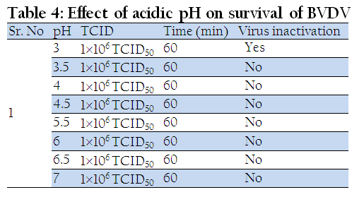
One ml of diluted virus (1x10⁶ TCID50 ) in EMEM was taken in each 2 ml eppendorf tubes and then pH of the medium was adjusted to 3, 3.5, 4, 4.5, 5, 5.5, 6, 6.5, and 7 with glacial acetic acid. The material was then kept for 1 h at room temperature and then pH of the inocula was adjusted to 7 by using 0.1M NaOH. Preformed monolayer of SFTR cells in six well plates was infected with 300 µl of treated inoculums and then remaining procedure was followed same as that described earlier.
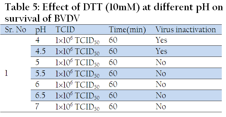
Effect of DTT at Different pH
DTT is a disulphide bond reducing agent. 450 µl of diluted virus (1x10⁶ TCID50 ) in EMEM and 50 µl DTT (stock 100mM) were dispensed in seven 2ml eppendorf tubes to make final concentration of 10mM DTT in each tube. The pH of the medium was adjusted to 4, 4.5, 5, 5.5, 6, 6.5, and 7 with HCl and kept for 1 h at room temperature. After completion of incubation period the pH of the inocula was adjusted to 7 by using 0.1M NaOH. Preformed monolayer of SFTR cells in six well plates was infected with 300 µl of treated inoculums and then remaining procedure was followed same as described earlier.
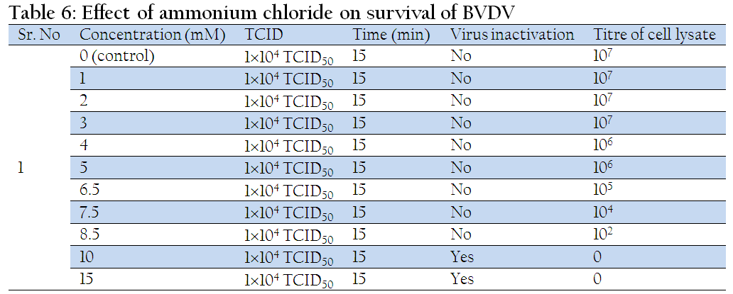
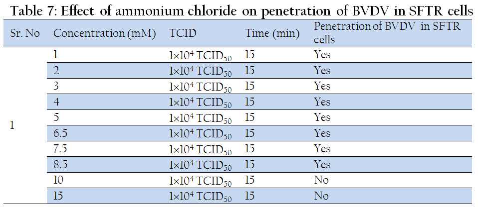
Effect of Ammonium Chloride
Different concentrations of ammonium chloride – 1, 2, 3, 4, 5, 6.5, 7.5, 8.5 and 10mM were prepared in 2 ml eppendorf tubes by adding 40, 80, 120, 160, 200µl, 260, 300, 340, and 400µl of ammonium chloride solution, respectively from 50mM stock and final volume was made up to 2000 µl by adding EMEM. The preformed monolayer of SFTR cells in six well tissue culture plates was inoculated with 500µl respective concentration of ammonium chloride and kept at 37oC for 15 min. The media was removed from wells, cells was infected with 300µl of diluted virus (1x10⁴ TCID50 ) and incubated at 37oC for 1 h for adsorption of virus. Then 1.5 ml EMEM containing respective amount of ammonium chloride concentration with 2% horse serum was added in the respective wells. The plate was incubated at 37oC in 5% CO2 incubator for 3 days and rest procedure was followed same as described previously. The virus titration of the cell lysate was carried out by IPMA as per procedure of Wood et al. (2004).
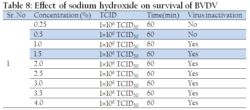
Effect of Sodium Hydroxide
Different concentrations of sodium hydroxide – 0.25, 0.5, 1.0, 1.5, 2.0, 2.5, 3.0, 3.5 and 4.0 percent were prepared by adding 6.25, 12.5, 25, 37.5, 50, 62.5, 75, 87.5 and 100µl of NaOH (from stock 10M NaOH), respectively and final volume was made up to 1000 µl by adding the diluted virus (1x10⁶ TCID50 ). The pH obtained was 8, 9, and 12 for 0.25, 0.5 and 1.0 percent NaOH, respectively, and above 14 for other concentrations. The treated inoculums were kept at room temperature for 1 h and then pH of the inocula was adjusted to 7 by using 0.1 M HCl. Monolayer of SFTR cell in 6 well tissue culture plate was infected with 300 µl of inoculums from the treated material and remaining procedure followed was same as described earlier.
Effect of Trypsin
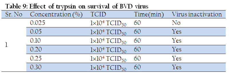
Different concentrations of trypsin such as, 0.025, 0.05, 0.10, 0.20, 0.25 and 0.3 percent were made by adding 5, 10, 20, 40, 50, and 60µl trypsin (from 5% trypsin stock) respectively, and final volume was made up to 1000 µl by adding diluted BVDV (1x10⁴ TCID50 ). The inuculum was incubated at 37oC for 1 h in a circulating water bath and then horse serum was added to neutralize trypsin. Preformed monolayer of SFTR cells in six well cell culture plate was infected using 300 µl of inoculums from the treated material and remaining procedure followed was same as described earlier.
Effect of Chloroform
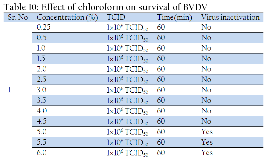
In 1.5 ml eppendorf tube different concentrations of chloroform such as 0.25, 0.5, 1, 1.5, 2, 2.5, 3, 3.5, 4, 4.5, 5, 5.5 and 6 percent were made by adding 2.5, 5, 10, 15, 20, 25, 30, 35, 40, 45, 50, 55 and 60µl (from stock of 100% chloroform solution), respectively and final volume was made up to 1000 µl by adding diluted virus (1x10⁶ TCID50 ) in the respective tubes. Preformed monolayer of SFTR cells in six well cell culture plates was infected using 300 µl of inoculum from the treated material and remaining procedure followed was same as described earlier.