
Chest CT imaging can be helpful in early diagnosis of COVID-19 instead of relying on real time polymerase chain reaction (RT-PCR) that can give false negative result. Nasopharyngeal samples from a 22 years old man were detected as negative for COVID-19 for consecutive three RT-PCR tests. Complete blood count (CBC), D-dimer, serum ferritin, eosinophil sedimentation rate (ESR) test, tuberculosis test, real time PCR and high-resolution computed tomography (HRCT) were done to rule out the cause of flu like symptoms. HRCT reveals a haze area in the right perihilar region adjacent to medial part of horizontal fissure on the 3rd day of manifestation of symptoms. Radiological studies showed early consolidation of COVID-19 whereas RT-PCR showed negative results. Chest CT imaging is a highly sensitive technique that has also been used in detection of corona virus. This case study emphasizes the importance of HRCT for early and confirm diagnosis of COVID-19 whereas RT-PCR results can vary. This process may show negative results and is time consuming.
Short Communication · Volume 55, Issue 1, 13 November 2022 · Open Access
Patient with Suspected COVID-19 Infection Tests Negative in Nasopharyngeal Swabs but is Confirmed by Laboratory Tests and Chest CT Scan: A Case Report
· ·
Affiliations & Notes ⌄
- 1Division of Molecular Virology and infectious diseases, Centre of excellence in Molecular Biology, University of the Punjab, Lahore, Pakistan * Corresponding author: samiaraza@live.com
Article Info ⌄
- Journal:
- Pakistan Journal of Zoology
- Published:
- 13 November 2022

From the article
- HRCT showed a ~24*18 mm ill-defined haze area in right perihilar region adjacent to medial part of horizontal fissure, which was likely early consolidation of COVID-19.
- RT-PCR showed consistent negative results whereas HRCT showed the true prognosis of the disease.
- Blood tests showed raised levels of serum ferritin, D-Dimer and ESR (inflammatory factors) that indicated the presence of SARS-CoV-2.
- HRCT is a powerful tool that can diagnose COVID-19 patients with negative RT-PCR.
Selected from the article's own text.
Abstract
Materials and methods
A 22 years old male lab technician was constantly vulnerable to COVID-19 patients. But then despite of observing all the SOPs, he reported a fever (39oC) with coarse breath sounds during auscultation accompanied by severe cough, body aches, diarrhea, absolute loss of taste and smell and shortness of breath. Oxygen saturation was below 80% and pulse rate was 82.
COVID-19 was diagnosed by taking the blood sample of the patient to find the levels of D-Dimer, ferritin, ESR, TLC, neutrophils, lymphocyte, eosinophil, monocyte, thrombocyte. Nasopharyngeal swab sample was taken to test the presence of viral nucleic acid in the patient. RT-PCR was done by SARS-Cov-2 Real-TM-Sacace Biotechnologies. Plain HRCT was done to explore the physiology of the lungs. Abnormal levels of D-dimer, ferritin, ESR and atypical physiology of lungs confirmed COVID-19 infection. Antibody level was detected by Elecsys anti-SARS-CoV-2 S.
On the basis of the diagnostic assessment the concerned doctors followed a regimen of recognized medicines using antiviral infusions through intravenous routes. On the 16th day blood tests, RT-PCR and HRCT were held again. The patient showed normal reports. Further, it was confirmed by SARS-CoV-2 antibody test on the 25th day of infection. A reasonable account of antibodies was found in the serum.
Results
Laboratory test reports showed total leucocyte count (TLC) 9800 cells/mm3 (normal range: 4,300 and 10,800 cells/ mm3), neutrophils 67% (normal range: 40% to 60%), lymphocytes 23% (normal range: 18-45% of white blood cells), monocytes 7% (normal range: 1-10% of body’s white blood cells), eosinophils 2% (normal range: 0.0-6.0 % of blood), thrombocyte was 0.25% (normal range: 0.23-0.24%), D-dimer was 15 mg/L (normal range: < 0.50 mg/L), whereas serum ferritin 313.2 µg/L (normal range: 30-220 µg/L) and eosinophil sedimentation rate (ESR) was 61 mm/hr( normal range: 0-15 mm/hr). The RT-PCR showed negative results with nasopharyngeal swabs for consecutive three time during early stage on days 1 (9th June, 2021), 3 (11th June, 2021), and 7 (15th June, 2021) of the infection.
HRCT showed a ~24*18 mm ill-defined haze area in right perihilar region adjacent to medial part of horizontal fissure, which was likely early consolidation of COVID-19. Minimal apical interlobular septal thickening was seen as in Figure 1.
Discussion
The uniqueness of the study case is the major strength of this work. In earlier studies no such case had been reported. Here we see a case which qualifies that there is no final word in scientific investigation. RT-PCR showed consistent negative results whereas HRCT showed the true prognosis of the disease. Thus, the objective of providing a quick and sure relief for the patient was ensured. Sample collection, transport, storage and processing of the RT-PCR sample could inhibit successful detection of the virus but in the present case study repeated tests confirmed the integrity of the results. Sometimes unknown factors could hinder the process of diagnosis of disease that may include virus variant formation due to high spread rate among the population.
Looking back at the patient’s history, it was found that he got infected with coronavirus on the 9th of June, 2021 and suffered from COVID-19 disease symptoms. He was prescribed with RT-PCR for COVID-19 and blood tests. His nasopharyngeal sample was taken but RT-PCR was negative. Blood tests showed raised levels of serum ferritin, D-Dimer and ESR (inflammatory factors) that indicated the presence of SARS-CoV-2. Therefore, on the 10th June, 2021 HRCT was done that ill-defined area in right perihilar region that indicated the beginning of COVID-19 consolidation. RT-PCR was repeated on the 3rd and 7th day of the start of infection but it remained negative.
Globally recognized authentic test is RT-PCR and it has been widely used for the detection of COVID-19 but in the present case the results were continuously negative that were against the obvious symptoms. Thus, it generated the need to perform other tests. Keeping in view the raised D-dimer value, ESR and serum ferritin levels, high resolution computed tomography (HRCT) of his chest was done. Increased values of ferritin in serum, d-dimer and ESR values are indicators of corona virus infection ; . HRCT initiates the screening and diagnoses COVID-19 . In this case, HRCT confirmed the consolidation of COVID-19. Now it was a confirmed case of COVID-19. The patient was immediately isolated to prevent the spread to the community. It is stated that during early days of infection RT-PCR showed negative results due to low viral load but after a few more days it indicated the presence of pathogenic virus . In the light of the above claim consecutive RT-PCR tests were conducted and were found negative. The patient’s symptoms resolved after 14 days of infection. Even then RT-PCR did not show any positive result. To diagnose other potential pathogens causing these physiological changes in the lungs, a tuberculosis test was also performed but also found negative. Follow-up test of level of SARS-CoV-2 antibodies confirmed the infection of COVID-19.
Hence, this study shows that HRCT is more sensitive in detecting COVID-19 than RT-PCR.
COVID-19 disease is diagnosed on the basis of epidemiological features, clinical symptoms, chest CT scan and laboratory findings. Negative tuberculosis test (Arslan-Gulen et al., 2021), negative COVID-19 RT-PCR and normal range of TLC cannot rule out the presence of COVID-19 infection whereas chest CT scan can provide clear picture even at early stages of infection ; ; ; . Therefore, chest CT/HRCT can be considered more reliable than RT-PCR for quick and accurate diagnosis . HRCT is a powerful tool that can diagnose COVID-19 patients with negative RT-PCR.
This case study opens new horizons to find more factors that can indicate the COVID-19 infection. Further studies in this regard will help in deciphering the pathophysiology of SARS-CoV-2. This study attracts the attention towards the reliable and early detection method instead of relying solely on RT-PCR. Real time polymerase chain reaction (RT-PCR) is considered as the highly specific and moderately sensitive assay for the detection of virus . Negative RT-PCR result does not rule out the presence of SARS-CoV-2 infection. Therefore, in the presence of symptoms HRCT can be done to confirm the beginning of consolidation in lungs.
Informed consent
Written consent from the patient was obtained for conducting research.
Funding
This research did not receive any specific grant from funding agencies in the public, commercial, or not-for-profit sectors.
Statement of conflict of interest
The authors have declared no conflict of interest.
Figures
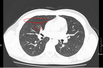
HRCT of second day of infection. It shows haze area in right perihilar region and minimal apical interlobular septal thickening.
References
- Ai, T., Yang, Z., Hou, H., Zhan, C., Chen, C., Lv, W., Tao, Q., Sun, Z., and Xia, L., 2020. Radiology, 296: E32-40. https://doi.org/10.1148/radiol.2020200642[CrossRef]
- Arslan-Gulen, T., Bayraktar, M., Yaksi, N. and Kayabas, U., 2021. J. Med. Virol., https://doi.org/10.1002/jmv.27414[CrossRef]
- Baines, D.K., 2020. Hosts Viruses, 7: 7-9. http://dx.doi.org/10.17582/journal.hv/2020/7.1.7.9[Full text][CrossRef]
- Brogna, B., Bignardi, E., Brogna, C., Alberigo, M., Grappone, M., Megliola, A., Salvatore, P., Fontanella, G., Mazza, E.M., and Musto, L., 2021. Radiography, 27: 743-747. https://doi.org/10.1016/j.radi.2020.09.012[CrossRef]
- Feng, H., Liu, Y., Lv, M., and Zhong, J., 2020. Japan J. Radiol., 38: 409-410. https://doi.org/10.1007/s11604-020-00967-9[CrossRef]
- Gupta-Wright, A., Macleod, C.K., Barrett, J., Filson, S.A., Corrah, T., Parris, V., Sandhu, G., Harris, M., Tennant, R., Vaid, N., and Takata, J., 2021. Br. Med. J. Open, 11: e047110. https://doi.org/10.1136/bmjopen-2020-047110[CrossRef]
- Hanif, N., Rubi, G., Irshad, N., Ameer, S., Habib, U., and Zaidi, S.R., 2021. J. Coll. Phys. Surg. Pak., 30: S1-S6. https://doi.org/10.29271/jcpsp.2021.Supp1.S1[CrossRef]
- Hassan, S.A., Sheikh, F.N., Jamal, S., Ezeh, J.K., and Akhtar, A., 2020. Cureus, 12: e7355. https://doi.org/10.7759/cureus.7355[CrossRef]
- Hui, D.S., Azhar, E.I., Madani, T.A., Ntoumi, F., Kock, R., Dar, O., Ippolito, G., Mchugh, T.D., Memish, Z.A., Drosten, C., Zumla, A., and Petersen, E., 2020. Int. J. Infect. Dis., 91: 264-266. https://doi.org/10.1016/j.ijid.2020.01.009[CrossRef]
- Hussein, A.M., Taha, Z.B., Malek, A.G., Rasul, K.A., Hazim, D.Q., Ahmed, R.J., and Mohmed, U.B., 2021. Materialstoday Proc., https://doi.org/10.1016/j.matpr.2021.04.009[CrossRef]
- Iwamura, A.P., Tavares da Silva, M.R., Hümmelgen, A.L., Soeiro Pereira, P.V., Falcai, A., Grumach, A.S., Goudouris, E., Neto, A.C., and Prando, C., 2021. Rev. Med. Virol., 31: e2199. https://doi.org/10.1002/rmv.2199[CrossRef]
- Kanji, J.N., Zelyas, N., MacDonald, C., Pabbaraju, K., Khan, M.N., Prasad, A., Hu, J., Diggle, M., Berenger, B.M. and Tipples, G., 2021. Virol. J., 18: 1-6. https://doi.org/10.1186/s12985-021-01489-0[CrossRef]
- Kanne, J.P., 2020. Radiology, 295: 1. https://doi.org/10.1148/radiol.2020200241[CrossRef]
- Lu, H., Stratton, C.W., and Tang, Y.W., 2020. J. Med. Virol., 92: 401–402. https://doi.org/10.1002/jmv.25678[CrossRef]
- Seibert, F.S., Toma, D., Bauer, F., Paniskaki, K., Anft, M., Rohn, B.J., Wang, S., Racovitan, D., Babel, N., and Westhoff, T.H., 2020. J. Med. Case Rep., 14: 1-4. https://doi.org/10.1186/s13256-020-02551-1[CrossRef]
- Tahan, S., Parikh, B.A., Droit, L., Wallace, M.A., Burnham, C.A., and Wang, D., 2021. J. clin.Micrbiol., 59. https://doi.org/10.1128/JCM.00075-21[CrossRef]
- Watson, J., Whiting, P.F., and Brush, J.E., 2020. Br. Med. J., 369:m1808. https://doi.org/10.1136/bmj.m1808[CrossRef]
- Xie, X., Zhong, Z., Zhao, W., Zheng, C., Wang, F., and Liu, J., 2020. Radiology, 296: E41-E45. https://doi.org/10.1148/radiol.2020200343[CrossRef]
- Yang, Y., Peng, F., Wang, R., Guan, K., Jiang, T., Xu, G., Sun, J., and Chang, C., 2020. J. Autoimmun., 109: 102434. https://doi.org/10.1016/j.jaut.2020.102434[CrossRef]
- Zeng, F., Huang, Y., Guo, Y., Yin, M., Chen, X., Xiao, L., and Deng, G., 2020. Int. J. Infect. Dis., 96: 467-474. https://doi.org/10.1016/j.ijid.2020.05.055[CrossRef]
- Zhou, P., Yang, X.L., Wang, X.G., Hu, B., Zhang, L., Zhang, W., Si, H.R., Zhu, Y., Li, B., Huang, C.L. and Chen, H.D., 2020. Nature, 579: 270-273. https://doi.org/10.1038/s41586-020-2012-7[CrossRef]
Services
Article usage statistics
Loading…