Pasteurella Multocida: Composition, Genetics and Biosynthesis of Capsular Polysaccharides (CPS) of Serotypes A, B, D, E and F
· · · · ·
Affiliations & Notes ⌄
1Health Biotechnology Department, National Institute for Biotechnology and Genetic Engineering, Faisalabad, Pakistan
2Chemistry Department, University of Agriculture, Faisalabad, Pakistan. *Correspondence | Waqar Siddique, Health Biotechnology Department, National Institute for Biotechnology and Genetic Engineering, Faisalabad, Pakistan
P. multocida has many capsular serotypes i.e. A, B, D, E, F whose capsules are made up of hyaluronic acid, mannosaminuronic acid, heparosan, mannosaminuronic acid, and chondroitin respectively.
R1 of capsular biosynthetic gene locus encodes proteins to make ATP binding cassette transport system while R2 and R3 are responsible for the polymerization of polysaccharides and lipidation of polysaccharides respectively.
CPS is synthesized by the initiation, extension and export of GAG units which is regulated by many genes at transcriptional and translational level.
Selected from the article's own text.
Abstract
Pasteurella multocida is a gram negative bacterium and gaining importance due to its role in economic losses causing different diseases such as bovine hemorrhagic septicemia, swine atrophic rhinitis, avian fowl cholera etc. Capsular Polysaccharide (CPS) is one of the most important virulence factors that is currently highly being studied to use it as a vaccine candidate individually or as a conjugated vaccine with the immunogenic proteins. This review collects the latest data regarding the chemical composition, genetics and biosynthesis of CPS of all capsular serotypes (A, B, D, E and F) of P. multocida. These serotypes are primarily made up of hyaluronic acid, mannosaminuronic acid, heparosan, mannosaminuronic acid, and chondroitin respectively. The genes involved in CPS biosynthesis are divided into three regions (R1, 2, 3). Regions 1 encodes proteins to make ATP binding cassette transport system, Region 2 consists of the genes that encode for the synthases for the polymerization of serotype specific polysaccharides, Region 3 encodes proteins responsible for lipidation as well as surface attachment of polysaccharides. CPS is synthesized in majorly three steps; initiation of GAG (glycosaminoglycan) synthesis, extension of GAG disaccharide units and export of GAG. There is a further need to study the genetics and biosynthesis of CPS to use it as a successful vaccine candidate.
Pasteurellaceae is a family, initially proposed by Pohl in 1979 , which represents the commensals or vertebral parasites, unable to survive well outside the natural host. Through the application of advance genetic based taxonomy and identification methods, the family of Pasteurellaceae is now consisting of 28 genera since 1995, including the genus, Gallibacterium. Recently, Actinobacillus, Pasteurella and Haemophilus have also been added in this family as new genera ; ; . There are so many devastating and common pathogens that include Actinobacillus pleuropneumoniae, Pasteurella multocida, Avibacterium paragallinarum and Mannheimia haemolytica. Pasteurella genus contains coccobacillary to rod shaped bacteria whose width ranges from 0.3–1.0 μm while their length ranges from 1.0–2.0 μm ; . They can be grown under aerobic, anaerobic and even under the microaerophilic condition at temperature ranging from 37 ℃ to 41 ℃. This genus is currently containing 12 species .
It is a coccobacillus, non-motile and penicillin sensitive gram-negative bacterium ; . It was first identified in a bird infected by cholera in 1878 and successfully isolated in 1880 by a French microbiologist named Louis Pasteur. This bacterium was named after the name of the scientist . There are four subspecies of P. multocida which include the type strain: Multocida, septica, gallicida and tigris . P. multocida strains are classified into serogroups or serotypes (A, B, D, E and F) on the basis of capsular antigens. This system was developed by Carter in 1955 and launched for the classification in 1963 based on haemagglutination ; . They are also categorized into 1–16 serotypes which are based primarily on lipopolysaccharide (LPS) antigens ; .
P. multocida is a major pathogen among the members of Pasteurella, responsible for many animal diseases such as bovine hemorrhagic septicemia, swine atrophic rhinitis, and avian fowl cholera and severe economic losses in poultry and cattle industry ; ; . The processes by which these bacteria infect the mucosa, elude the innate immunity, and cause the systemic illnesses are being explained slowly. Capsule and lipopolysaccharide have been identified as the key virulence factors so far . The capsule appears to have a role in bacterial phagocytosis evasion and complement resistance, whereas full lipopolysaccharide is required for bacterial survival in the host . P. multocida toxin (PMT), putative surface adhesions, and iron acquisition proteins are among the additional virulence components discovered by both directed and random mutagenesis . Many critical virulence factors, including those necessary for adhesion and penetration of host cells, as well as for survival in a poorly nutritious and hostile environment, are likely to remain unknown . These diseases are associated with the particular serotype/es.
Hemorrhagic Septicemia (HS) majorly affects the livestock animals such as cattle, buffalo, camel and pigs (De Alwis, 1999; predominantly infected either by serotype B:2 (Asian and European serotype) or serotype E:2 (African serotype) of P. multocida ; . The observed clinical signs in affected animals are the elevation of temperature ranging from 102 to 107oF, chest and neck swelling, salivation, inappetence and protrusion of tongue. Sometimes, development of tympany, eye discharges and oedema have also been observed ; ; .
Progressive Atrophic Rhinitis (AR) is particularly caused by specific toxins produced by P. multocida serotype A and/or D i.e. PMT ; ; . Major clinical symptoms include: nasal crusting, mucosal atrophy, paradoxical feeling of nasal congestion, decreased growth, facial distortion, enlarged nasal space and fetor while epistaxis, sinusitis and anosmia are also accompanied with these symptoms .
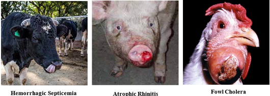
Fowl Cholera or FC, also known as avian cholera, is a serious septicemic contagious disease of poultry and wild birds that can either be present in acute or chronic form and its etiological agents are P. multocida strains (especially serovar A:1, A:3, A:4 and serovar D to a lesser extent) ; . Generally, acute FC is caused by the serotype A whose symptoms include anorexia, fever, ruffled feathers, diarrhoea, oral mucous discharge and increase in breathing rate ; . Apparent condition of the animals suffering from HS, AR and FC has been shown in the Figure 1.
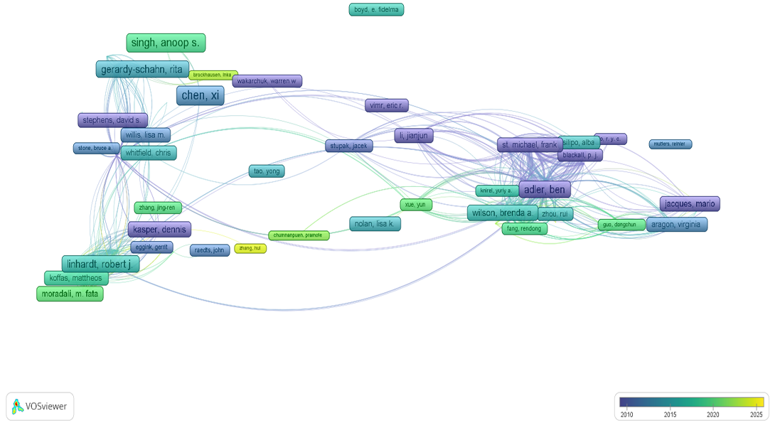
In Figure 2, there is a visualization map of authors from 2010 to 2025 and this map was made by VOS viewer (version 1.6.20). Data was extracted using the Dimensions database using the terms, “Pasteurella multocida AND Capsular polysaccharides AND composition AND genetics AND biosynthesis”. The map shows the clusters of authors who frequently contribute and collaborate in the composition, genetics, and biosynthesis of CPS of P. multocida. Authors in yellow indicate recent active involvement in the research field.
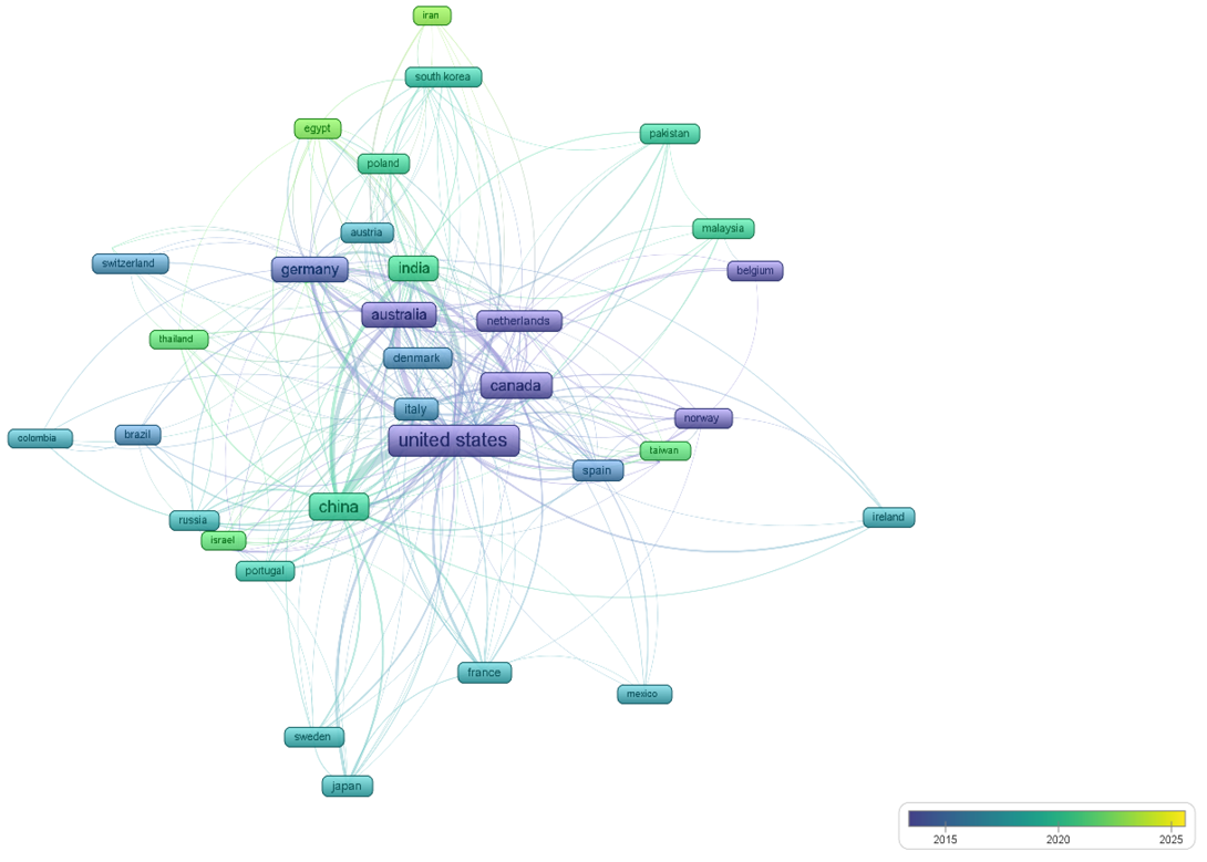
In Figure 3, there is a visualization map of countries that have contributed so far. This map is made by VOSviewer. Data was extracted using the Dimensions database using the terms, “Pasteurella multocida AND Capsular polysaccharides AND composition AND genetics AND biosynthesis”. The map shows the clusters of countries who frequently collaborate in the composition, genetics, and biosynthesis of CPS of P. multocida. Countries in yellow indicate recent active involvement in this area of research. United States stands out as a central node with numerous connections, indicating its significant contribution. Moreover, Canada, Australia, Germany, India, and China are also leading in this research field.
Virulence factors of P. multocida
Virulence is the measure of the pathogenicity of an organism. Virulence factors or pathogenicity factors are the molecules, cellular structures or regulatory systems which enable the microorganisms for colonization, immune-evasion, immunosuppression, getting nutrients and entry/exit causing the virulence in host cell ultimately . There are various virulence factors in P. multocida, involved in its pathogenicity including CPS, LPS, toxins, fimbriae, adhesins, iron regulating and iron acquisition proteins, hyaluronidase and several outer membrane proteins ; ; ; ; . Studies have shown that capsule of P. multocida is very important in the development of vaccines. A lot of research is being carried out to develop the vaccines to prevent the deaths of livestock especially in the developing countries. Due to the importance of capsular polysaccharides (CPS) and their diversity in different serotypes, they are being studied well .
Capsule
Bacterial capsule is a large polysaccharide structure lying outside the cell and can be a cause of many diseases ; . It is not confined to gram negative bacteria but also present in gram positive bacteria. In contrast to slime, capsule is thicker, tightly bound to the cell wall, well organized, difficult to be washed off and consists only of polysaccharides . Capsule and slime are sometimes collectively called as glycocalyx . Monosaccharides are joined together through glycosidic linkages to make capsular polysaccharide . Bacteria containing capsule are known as polysaccharide encapsulated bacteria and some of these bacteria make smooth or rough colonies on agar media . P. multocida also contains capsule whose chemical composition varies in its different serotypes (A, B, D, E and F) .
Capsule is an important virulence factor which means that it helps bacteria to cause disease by protecting them from phagocytosis by macrophages . As it contains water so it also protects from desiccation ; . Capsule is also important for the structural integrity and controlling the flow of nutrients. It may also act as a signal for the host to produce the needed nutrients. Capsule also helps in the adherence of bacterial cells with each other and to non-living surfaces as well.
Potential for CPS based vaccine development
CPS itself and its conjugate with other proteins have a strong potential for vaccine development. It has been observed that although CPS is essential for virulence, the antibody responses elicited against CPS are not necessarily the primary mediators of protective immunity . Instead, other surface-exposed proteins, such as outer membrane proteins (OMPs) and lipoproteins, may serve as better vaccine targets . Indeed, several recombinant protein vaccines targeting OMPs (e.g., PlpE and OmpH) have shown promising results in preclinical studies ; .
The challenge in developing an effective CPS-based vaccine for P. multocida lies in the intrinsic T cell-independent nature of polysaccharide antigens. Such antigens typically generate a short-lived immune response with poor immunological memory. To overcome this limitation, conjugate vaccines (chemically linked CPS to a protein carrier) have been developed for other pathogens, significantly improving immunogenicity. However, for P. multocida, the high variability in capsule composition across serogroups (A, B, D, E, and F) poses an additional hurdle for designing a broadly protective CPS-based vaccine . This serogroup heterogeneity means that a vaccine targeting the capsule of one serogroup might not protect against another. Recent work has identified novel surface lipoproteins such as PmSLP, which are highly conserved among bovine isolates and can elicit robust and long-lasting immune responses in both mice and cattle .
Structure and composition of capsule
Although, most of the P. multocida strains produce the capsule made up of polysaccharides, different strains with different chemical composition of capsule have been identified .
Serotype A
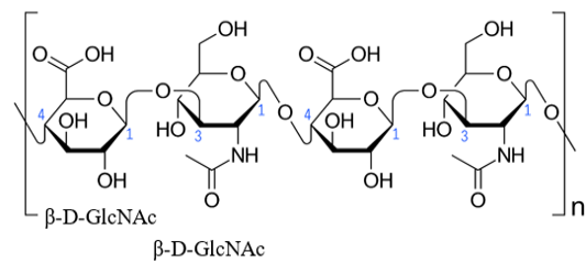
Serotype A has capsule primarily made up of hyaluronic acid (HA) or hyaluronan which is a linear polysaccharide consisting of β-D-glucuronic acid (β-D-GlcA) and β-D-N-acetylglucosamine (β-D-GlcNAc) repeating units joined together with the help of alternating β-1,4 and β-1,3 glycosidic linkages ; ; . Its chemical structure has been shown in the Figure 4.
Serotypes B and E
Previous studies: The exact chemical composition of capsular polysaccharides of serotype B and E had not been determined till 2021 . According to the previous studies it was shown that the capsule of serotype B consists of mannose (Man), arabinose (Ara) and galactose (Gla) with the ratio of 2.0:0.5:0.8. The molecular weight was found to be 90 kDa in ziwittergent buffer by gel filtration and 900 kDa in Phosphate Buffer Saline (PBS) showing that it exists in decamer form in its native state . On the other hand, chemical composition of capsule of serotype E remained totally unclear till 2021.
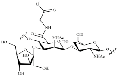
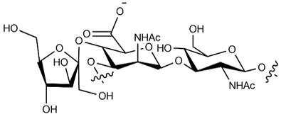
Latest studies: Recently, the capsular polysaccharides of serotypes B and E have been determined and compared with respect to their structures, genetics and serology. Full nuclear magnetic resonance (NMR) spectroscopy revealed that there is a high similarity between CPS repeat units of both serotypes with minor differences. In serotype B, mannosaminuronic acid (ManNAcA) is substituted by fructose (Fruc) at the position 3 and by GlcNAc at position 4. In serotype E, linkages are reversed. At position 3, ManNAcA is substituted by GlcNAc and by fructose at position 4. The presence of glycine in the capsular polysaccharide of serotype B is a unique characteristic ; . Chemical composition of capsule of serotype B and E has been shown in the Figures 5, 6.
Serotype D
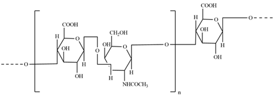
Serotype D has capsule made up of heparosan which is a linear polysaccharide consisting of β-D-glucuronic acid (β-D-GlcA) and α-D-N-acetylglucosamine (α-D-GlcNAc) repeating units joined together with the help of 1,4 glycosidic linkages as shown in the Figure 7.
Serotype F
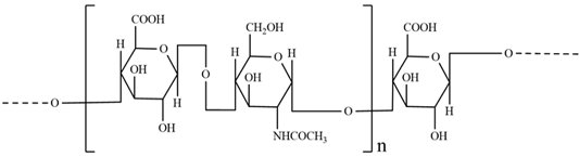
Serotype F has capsule made up of chondroitin which is a linear polysaccharide consisting of β-D-glucuronic acid (β-D-GlcA) and β-D-N-acetylgalactosamine (β-D-GalNAc) repeating units joined together with the help of 1,3 glycosidic linkages with alternating units linked by 1,4 glycosidic linkages as shown in the Figure 8.
Genetics of CPS
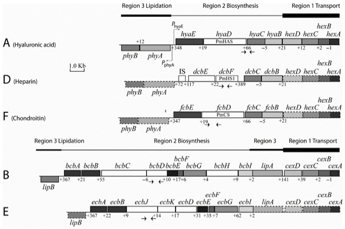
Genetic sequences of all genes of all five serotypes have been determined that are involved in the capsular biosynthesis and by comparing their genetic organization with other gram-negative bacteria it has been shown that there is similarity of these sequences with group II capsule biosynthetic operons . The genes can be grouped into three regions (R1-3) .
The first group (Region 1) produces a set of proteins that function as a delivery system, transporting essential materials to the cell’s surface. Regions 1 is highly conserved among all the serotypes of P. multocida and encode proteins to make ATP binding cassette transport system (a protein complex) . This transport system plays role in the export of polysaccharides to the surface of bacteria. Role of hex in serotype A:1 and role of cexA in serotype B:2 in the transport of polysaccharides to the surface have been determined and confirmed by mutagenesis .
The second group (Region 2) contains genes that act like construction workers, piecing together the sugar building blocks to form the capsule. Region 2 consists of the genes that encode for the synthases for the polymerization of serotype specific polysaccharides. Synthase PmHAS is responsible for the polymerization of hyaluronic acid, synthase PmHS1 is responsible for the polymerization of heparin and synthase PmCS is responsible for the polymerization of chondroitin in serotypes A, D and F respectively ; . Roles of these synthases have been confirmed by mutagenesis and/or functional analysis. These synthases as bifunctional transferases can add both glucuronic acid and N-acetyl glucosamine or N-acetyl galactosamine in growing polysaccharide molecule. Mutagenesis has also confirmed the role of bcbH encoded protein in type B polysaccharide synthesis .
The third group (Region 3) makes sure the capsule is properly attached to the bacterial cell, much like an anchoring system. This coordinated process is crucial for the bacterium’s ability to protect itself and cause disease. Region 3 is also highly conserved though all the serotypes of P. multocida consisting of the genes which encode proteins responsible for lipidation as well as surface attachment of polysaccharides . R3 has two genes; phyAB and lipAB. These genes encode proteins that are responsible for the substitution of phospholipids for the anchorage of CPS . However, in serotypes B and E, loci lipA is located adjacent to Region 1 for the diagrammatical representation of the synthesis of capsular polysaccharide . Genetic organization of capsule biosynthetic loci of all serotypes of P. multocida has been shown in the Figure 9.
Later on, outside the capsule biosynthetic region, a cryptic heparin synthase gene was discovered in serotype D which encodes heparin synthase. Homologous genes were also discovered in other A, D and F serotypes. It was proposed without any experimental investigation that these synthases may give variations in capsule composition .
The development of the typing of P. multocida with the help of multiplex PCR based on the gene sequences present in region 2 was facilitated by the clarification on the genetic basis for capsule biosynthesis . As this PCR-based typing is much easier and reliable so this has replaced the conventional serotyping and now is being used as a gold standard throughout the world . Functions of ORFs of different serotypes has been summarized in the Table 1.
Table 1: Function of ORFs of P. multocida.
Serotype
ORFs
Function of encoded proteins
Serotype A, D, F
hexA (R1)
ATP dependent or binding protein
hexB (R1)
Proteins present in inner membrane
hexC (R1)
Proteins present in inner membrane and periplasmic domain
hexD (R1)
Proteins present in outer membrane (lipoprotein)
hyaB/dcbB/fcbB (R2)
Glycosyltransferases
hyaC/dcbC/fcbC (R2)
Changes the UDP-glucose to UDP-glucuronic acid as UDP glucose dehydrogenase ;
hyaD/dcbF/fcbD (R2)
Extension of polymer as PmHAS/PmHS/PmCS ;
hyaE/dcbE/fcbE (R2)
Initiation of transfer of enzymes ;
phyA/phyB (R3)
Glycosyltransferase activity ,
Serotype B, E
cexA (R1)
ATP dependent or binding protein
cexB (R1)
Proteins present in inner membrane
cexC (R1)
Proteins present in inner membrane and periplasmic domain
cexD (R1)
Proteins present in outer membrane (lipoprotein)
bcbA/ecbA (R2)
UDP N acetylglucosamine 2 epimerase
bcbB/ecbB (R2)
UDP-N-acetylminnosaminuronic acid dehydrogenase
bcbC/ecbK (R2)
Extension of polymer as a putative glycosyltransferase
bcbDEFGI/ecbDEFGI (R2)
Unknown
bcbH/ecbJ (R2)
Unknown
lipA/lipB (R3)
Lipidation
Table 2: Summary of CPS composition, genetics and virulence in serotype A, B, D, E, and F.
Serotype
Structure (capsule composition)
Genetics (key genes/regions)
Virulence (associated diseases)
A
Hyaluronic acid (HA): Alternating β-1,4 and β-1,3 linkages of β-D-glucuronic acid (GlcA) and β-D-N-acetylglucosamine (GlcNAc).
R1: Contains conserved hexA-D genes; encoding components of the ABC transport system.
Heparosan: β-1,4 linkages of β-D-GlcA and α-D-GlcNAc.
R1: Utilizes the conserved hexA–D gene cluster as in serotypes A and F. R2: Contains PmHS1; Heparin polymerization R3: Contains phyAB, lipAB genes for lipidation and surface attachment of CPS.
Atrophic Rhinitis (AR; with PMT toxin), Fowl Cholera (less common).
E
ManNAcA substituted with GlcNAc (position 3) and fructose (position 4); structurally similar to B.
R1: Also contains the cexA–D genes. R2: Possesses a bcb/ecb gene cluster for the altered linkage pattern. R3: Contains phyAB, lipAB genes for lipidation and surface attachment of CPS.
Chondroitin: Alternating β-1,3 and β-1,4 linkages of β-D-GlcA and β-D-N-acetylgalactosamine (GalNAc).
R1: Shares the conserved hexA–D gene cluster with serotypes A and D. R2: Contains PmCS gene for the chondroitin synthesis. R3: Contains phyAB, lipAB genes for lipidation and surface attachment of CPS.
Less clearly defined; associated with general infections (e.g., avian or livestock diseases).
Genetic organization is divided into three distinct regions: Region 1, which is highly conserved and encodes components of the ATP-binding cassette (ABC) transport system responsible for polysaccharide export; Region 2, which contains the genes encoding serotype-specific synthases that catalyze the polymerization of sugar units into the growing capsule; and Region 3, which comprises genes involved in the lipidation and surface attachment of the capsule.
Summary of CPS composition, Genetics and Virulence in Serotype A, B, D, E, and F has been shown in Table 2 for a holistic understanding.
Mechanism of biosynthesis of capsule
The capsular glycosaminoglycan (GAG) of P. multocida is produced in the inner membrane of bacteria and then transported to outside through the ATP binding cassette (ABC) transporter which belongs to the translocases and is present from prokaryotes to humans . Synthesized glycolipids attach to the cell wall by making hydrogen bonds. The biosynthesis of CPS involves initiation of GAG synthesis, extension of GAG disaccharide units and export of GAG .
In addition to the CPS based vaccine development, we also need to extensively study the CPS biosynthesis and the enzymes involved in this biosynthesis to find potential drug targets. Small molecules blocking PmHAS (serotype A), PmHS (serotype D), or PmCS (serotype F) could prevent capsule assembly. Compounds targeting HexA/CexA (ATP-binding proteins in R1) could halt CPS export, rendering bacteria susceptible to host defenses.
Initiation
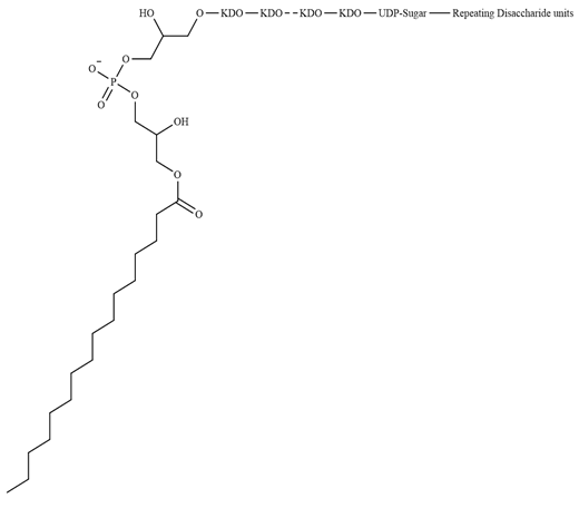
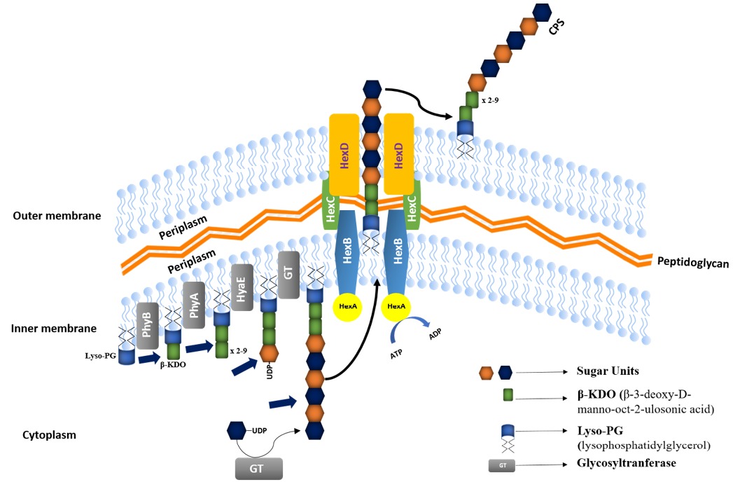
In the initiation, GAG glycolipid terminal is synthesized where three steps are involved. (I) PhyB (glycosyltransferase) similar to E. coli protein KpsS, transports the first eight carbon sugar, β-3-deoxy-D-manno-oct-2-ulosonic acid (β-KDO) to lysophosphatidylglycerol (lyso-PG) receptor ; , b; . (II) PhyA (glycosyltransferase) similar to E. coli protein KpsC, adds five to nine β-KDOs to synthesize poly-β-KDO linker chain , b). (III) HyaE transports and adds UDP sugar molecules to poly-β-KDO linker chain and this is the first residue of glycolipid terminal . So, the bacterium starts by building a “starter anchor” made of sugars and fats (a glycolipid). This anchor acts like a foundation for the capsule. Structure of glycolipid has been shown in the Figure 10.
Extension
The extension or polymerization of GAG disaccharide units is essentially similar in serotypes A, D and F ; ; . PmHAS in serotype A, PmHS in serotype D and PmCS in serotype F act as bifuntional glycosyltransferase which catalyze the transfer of both β-D-acetylglucosamine (β-D-NGlcNAc) and β-D-N-acetylgalactosamine (β-D-GlcUA) using 2 transferase active sites. These proteins add the activated sugar molecules to the glycolipid terminal, release UDP and form the polysaccharide of the relevant serotype ; .
Here, n is the degree of polymerization which is 20-100 and 103-104 in serotypes D and F, respectively.
In simple words, enzymes add sugar molecules to the anchor. These sugars are carried into place by UDP molecules acting as a “delivery truck”. The enzymes link them into long chains.
Export
The export of GAG outside the cell surface mainly depends upon the ABC transport system a). The encoded protein products of first four genes (Region 1) together make an ATP transport system. HexA encoded by hexA, as an ATP binding protein delivers the energy for the GAG transportation. HexB encoded by hexB, is the protein of inner membrane. HexC encoded by hexC, is the protein of inner membrane that has a periplasmic domain. HexD encoded by hexD, is
the protein of outer membrane . GAG chain passes through the inner membrane, periplasmic space and outer membrane via a channel made by HexB/C/D with the help of ATP binding domain and ultimately makes the covalent bond with phospholipid at the cell surface . Mechanism of biosynthesis of capsule has been shown in the Figure 11.
The process is depicted in three main stages. Initiation: A glycolipid precursor is synthesized by the sequential addition of β-KDO units to a lipid receptor, laying the foundation for capsule assembly. Extension: Bifunctional glycosyltransferases (such as PmHAS, PmHS, or PmCS, depending on the serotype) add sugar units, forming a polysaccharide chain by polymerizing glycosaminoglycan (GAG) disaccharide units. Export: The fully assembled capsule is translocated to the bacterial surface through the action of the ABC transporter system, which ensures its proper attachment.
Expression and regulation of biosynthetic loci of P. multocida
Regulatory sequences: Expression of capsular biosynthetic loci depends upon the operon. Contrary to R3, R1 and R2 genes make a single transcriptional unit because they undergo the transcription in the same direction. The promoter of both R1 and R2 exists in between R2 and R3 , 2010). In serotype A, R2 and R3 promoter is present between phyA and hyaE that is recognized by a transcription factor, σ70. -35 box and -10 box in the promoter are separated from each other by 17bp. R1 and R2 transcription initiation site is present at the 37bp upstream of start codon of hyaE , while ribosome binding site (RBS) is at 8bp upstream to stop codon of hexA and 4bp downstream of stop codon of hexA. Regulatory sequences of R3 are not well known yet however this is supposed to be not involve σ70 ; .
Transcriptional regulation: There is a factor for inversion stimulation (Fis) protein, which performs the transcriptional regulation and regulates the expression of capsule ; . This nucleoid associated protein is composed of 99 amino acids and has two of its biding sites in promotor region. From 73 to 94 amino acids, there is a DNA binding motif that can bind with promoter region and positively regulate the GAG genes ; . If mutation occurs in Fis gene, it causes production of acapsular strains even in the presence of intact nucleotide sequences. Fis gene is not always present in the capsular gene cluster in all serotypes. This gene also regulates the transcription of some other virulence factors for example LPS, Pasteurella Lipoprotein E (plpE) etc. .
Translational regulation
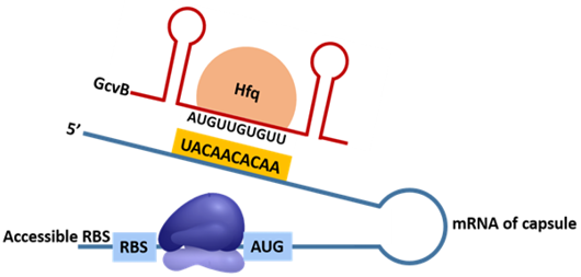
There is a protein named as host factor for Qβ (Hfq) which is an RNA chaperone protein encoded by hfq gene and regulates the translation of capsule biosynthesis loci ; . This protein together with GcvB (a small non-coding RNA molecule) as an Hfq-GcvB complex, targets the mRNA transcribed by capsular biosynthesis gene and positively regulates the gene expression . Hfq-GcvB complex, binds with the target mRNA via specific seed sequence, opens the secondary structure and exposes the RBS and AUG (start codon) ; . Ribosome then binds with RBS and AUG, activates the inhibited mRNA and initiates the translation. The seed sequence which is present on mRNA is 5’-AUGUUGUGUU-3′ while the complementary sequence present on GcvB is 5’-AACACAACAU-3′ . Mechanism of regulation of biosynthetic loci of P. multocida has been shown in the Figure 12.
The figure highlights both transcriptional and translational control mechanisms that modulate the expression of capsule biosynthetic genes. The binding of general transcription factors such as σ70 and regulatory proteins like Fis to specific promoter regions initiates and modulates the transcription of the gene clusters. The interaction of the RNA chaperone Hfq with the small RNA GcvB facilitates the exposure of ribosome binding sites on the mRNA, enhancing translation.
Conclusion
P. multocida has many capsular serotypes i.e. A, B, D, E, F whose capsules are made up of hyaluronic acid, mannosaminuronic acid, heparosan, mannosaminuronic acid, and chondroitin respectively. R1 of capsular biosynthetic gene locus encodes proteins to make ATP binding cassette transport system while R2 and R3 are responsible for the polymerization of polysaccharides and lipidation of polysaccharides respectively. CPS is synthesized by the initiation, extension and export of GAG units which is regulated by many genes at transcriptional and translational level.
Novelty Statement
This review article has gathered all the recent informations and studies about the composition, genetics, and biosynthesis of CPS of all serotypes of P. multocida which we do not find in any other article so that we can find the solutions of related diseases in animals.
Author’s Contribution
Waqar Siddique wrote this article under the guidance of Sadia Mahboob and Waqar Rauf. Sobia Jabeen and Zubera Naseem helped in drawing the chemical structures of CPS using ChemDraw. Fiza Shafaqat helped in reviewing the article and mentioned the areas of improvement.
Conflict of interest
The authors have declared no conflict of interest.
Figures
Figure 1:.
Apparent condition of animals with HS, AR and FC.
Figure 2:.
Visualization map of Authors contributing to the composition, genetics, and biosynthesis of CPS of P. multocida.
Figure 3:.
Visualization map of Countries contributing to the composition, genetics, and biosynthesis of CPS of P. multocida.
Figure 4:.
Chemical structure of heparin of serotype A.
Figure 5:.
Chemical structure of capsule of serotype B.
Figure 6:.
Chemical structure of capsule of serotype E.
Figure 7:.
Chemical structure of capsule of serotype D.
Figure 8:.
Chemical structure of capsule of serotype F.
Figure 9:.
Genetic organization of capsule biosynthetic loci.
Figure 10:.
Glycolipid structure of the GAG terminal of P. multocida.
Figure 11:.
Mechanism of biosynthesis of capsule.
Figure 12:.
Mechanism of regulation of biosynthetic loci of P. multocida.
References
Almoheer R, Abd-Wahid ME, Zakaria HA, Jonet MAB, Al-Shaibani MM, Al-Gheethi A, Addis SNK (2022). Spatial, temporal, and demographic patterns in the prevalence of hemorrhagic septicemia in 41 countries in 2005–2019: A systematic analysis with special focus on the potential development of a new-generation vaccine. Vaccines, 10(2): 315. https://doi.org/10.3390/vaccines10020315[CrossRef]
Argüello-García R, Ortega-Pierres G (2021). Giardia duodenalis virulence to be, or not to be. Current tropical medicine reports. pp. 1-11. https://doi.org/10.1007/s40475-021-00248-z[CrossRef]
Arumugam N, Ajam N, Blackall P, Asiah N, Ramlan M, Maria J, Yuslan S, Thong KL (2011). Capsular serotyping of Pasteurella multocida from various animal hosts-a comparison of phenotypic and genotypic methods. Trop. Biomed., 28(1): 55-63.
Asghar F, Babar M T, Ali A, Ali M (2023). Hemorrhagic septicemia.
Bagchi A (2015). Structural characterization of Fis. A transcriptional regulator from pathogenic Pasteurella multocida essential for expression of virulence factors. Gene, 554(2): 249-253. https://doi.org/10.1016/j.gene.2014.10.044[CrossRef]
Baselga R, Albizu I, Amorena B (1994). Staphylococcus aureus capsule and slime as virulence factors in ruminant mastitis. A review. Vet. Microbiol., 39(3-4): 195-204. https://doi.org/10.1016/0378-1135(94)90157-0[CrossRef]
Boyce JD, Adler B (2001). Acapsular Pasteurella multocida B: 2 Can stimulate protective immunity against pasteurellosis. Infect. Immun., 69(3): 1943-1946. https://doi.org/10.1128/IAI.69.3.1943-1946.2001[CrossRef]
Boyce JD, Chung JY, Adler B (2000). Genetic organisation of the capsule biosynthetic locus of Pasteurella multocida M1404 (B:2). Vet. Microbiol., 72(1-2): 121-134. https://doi.org/10.1016/S0378-1135(99)00193-5[CrossRef]
Boyce JD, Chung JY, Adler B (2000). Pasteurella multocida capsule: Composition, function and genetics. J. Biotechnol., 83(1-2): 153-160. https://doi.org/10.1016/S0168-1656(00)00309-6[CrossRef]
Boyce JD, Harper M, Wilkie I, Adler B (2010). Pasteurella. Pathogenesis of bacterial infections in animals: John Wiley and Sons, pp. 325-346. https://doi.org/10.1002/9780470958209.ch17[CrossRef]
Boyce JD, Lo RY, Wilkie I, Adler B (2004). Pasteurella and mannheimia. Pathogenesis of bacterial infections in animals. pp. 273-285. https://doi.org/10.1002/9780470344903.ch20[CrossRef]
Cao X, Gu L, Gao Z, Fan W, Zhang Q, Sheng J, Zhang Y, Sun Y (2024). Pathogenicity and genomic characteristics analysis of pasteurella multocida serotype a isolated from argali hybrid sheep. Microorganisms, 12(6): 1072. https://doi.org/10.3390/microorganisms12061072[CrossRef]
Carmichael GG (1975). Isolation of bacterial and phage proteins by homopolymer RNA-cellulose chromatography. J. Biol. Chem., 250(15): 6160-6167. https://doi.org/10.1016/S0021-9258(19)41172-1[CrossRef]
Carter G (1955). Studies on Pasteuvella multocida. I. A hemagglutination test for the identification of serological types.
Casadevall A, Pirofski LA (2009). Virulence factors and their mechanisms of action: The view from a damage–response framework. J. Water Health, 7(S1): S2-S18. https://doi.org/10.2166/wh.2009.036[CrossRef]
Chong CWH, Espinosa-Gongora C, Blackall PJ, Sinclair HA, Bojesen AM, Omaleki L, Turni C (2021). Molecular identification of members of the family Pasteurellaceae from the oral cavity of koalas (Phascolarctos cinereus) and their relationship with isolates from koala bite wounds in humans. J. Zoo Wildl. Med., 51(4): 771-779. https://doi.org/10.1638/2020-0041[CrossRef]
Christensen H, Sajid SM, Bisgaard M, Magistrali CF, Massacci FR, Liman M, Menke T, Bischoff H, Olsen JE (2022). Prediction of Pasteurella multocida serotypes based on whole genomic sequences. Vet. Microbiol., 271:109492. https://doi.org/10.1016/j.vetmic.2022.109492[CrossRef]
Chung JY, Zhang Y, Adler B (1998). The capsule biosynthetic locus of Pasteurella multocida A:1. FEMS Microbiol. Lett., 166(2): 289-296. https://doi.org/10.1111/j.1574-6968.1998.tb13903.x[CrossRef]
Dabo SM, Taylor JD, Confer AW (2007). Pasteurella multocida and bovine respiratory disease. Anim. Health Res. Rev., 8(2): 129-150. https://doi.org/10.1017/S1466252307001399[CrossRef]
Daffe M, Etienne G (1999). The capsule of Mycobacterium tuberculosis and its implications for pathogenicity. Tuber Lung Dis., 79(3): 153-169. https://doi.org/10.1054/tuld.1998.0200[CrossRef]
De-Alwis M (1999). Haemorrhagic septicaemia. Australian Center for International Agricultural Research, Canberra, Australia.
DeAngelis PL (1996). Enzymological characterization of the Pasteurella multocida hyaluronic acid synthase. Biochemistry, 35(30): 9768-9771. https://doi.org/10.1021/bi960154k[CrossRef]
DeAngelis PL (1999). Molecular directionality of polysaccharide polymerization by the Pasteurella multocida hyaluronan synthase. J. Biol. Chem., 274(37): 26557-26562. https://doi.org/10.1074/jbc.274.37.26557[CrossRef]
DeAngelis PL, Gunay NS, Toida T, Mao WJ, Linhardt RJ (2002). Identification of the capsular polysaccharides of type D and F Pasteurella multocida as unmodified heparin and chondroitin, respectively. Carbohydrate Res., 337(17): 1547-1552. https://doi.org/10.1016/S0008-6215(02)00219-7[CrossRef]
DeAngelis PL, Jing W, Drake RR, Achyuthan AM (1998). Identification and molecular cloning of a unique hyaluronan synthase from Pasteurella multocida. J. Biol. Chem., 273(14): 8454-8458. https://doi.org/10.1074/jbc.273.14.8454[CrossRef]
DeAngelis PL, Padgett-McCue AJ (2000). Identification and molecular cloning of a chondroitin synthase from Pasteurella multocida type F. J. Biol. Chem., 275(31): 24124-24129. https://doi.org/10.1074/jbc.M003385200[CrossRef]
DeAngelis PL, White CL (2004). Identification of a Distinct, cryptic hparosan snthase from Pasteurella multocida types, D, and F. J. Bacteriol., 186(24): 8529-8532. https://doi.org/10.1128/JB.186.24.8529-8532.2004[CrossRef]
Dorman CJ, Deighan P (2003). Regulation of gene expression by histone-like proteins in bacteria. Curr. Opin. Genet. Dev., 13(2): 179-184. https://doi.org/10.1016/S0959-437X(03)00025-X[CrossRef]
Eid S, Marouf S, Hefny HY, Al-Atfeehy NM (2019). Pasteurellaceae members with similar morphological patterns associated with respiratory manifestations in ducks. Vet. World, 12(12): 2061-2069. https://doi.org/10.14202/vetworld.2019.2061-2069[CrossRef]
Fegan JE, Waeckerlin RC, Tesfaw L, Islam EA, Deresse G, Dufera D, Assefa E, Woldemedhin W, Legesse A, Akalu M (2024). Developing a PmSLP3-based vaccine formulation that provides robust long-lasting protection against hemorrhagic septicemia–causing serogroup B and E strains of Pasteurella multocida in cattle. Front. Immunol., 15(1392681). https://doi.org/10.3389/fimmu.2024.1392681[CrossRef]
Fenwick BW, Rycroft AN (2022). Pasteurellaceae: Actinobacillus. Vet. Microbiol., pp. 118-128. https://doi.org/10.1002/9781119650836.ch11[CrossRef]
Foster G, Ross H, Malnick H, Willems A, Hutson R, Reid R, Collins M (2000). Phocoenobacter uteri gen. nov., sp. nov., a new member of the family Pasteurellaceae Pohl (1979) 1981 isolated from a harbour porpoise (Phocoena phocoena). Int. J. Syst. Evol. Microbiol., 50(1): 135-139. https://doi.org/10.1099/00207713-50-1-135[CrossRef]
Fröhlich KS, Vogel J (2009). Activation of gene expression by small RNA. Curr. Opin. Microbiol., 12(6): 674-682. https://doi.org/10.1016/j.mib.2009.09.009[CrossRef]
Guan L, Xue Y, Ding W, Zhao Z (2019). Biosynthesis and regulation mechanisms of the Pasteurella multocida capsule. Res. Vet. Sci., 127: 82-90. https://doi.org/10.1016/j.rvsc.2019.10.011[CrossRef]
Guan LJ, Yang JQ, Xu QY, Feng YF, Zhang XC, Tang B, Zhao ZQ (2023). Immunogenicity and efficacy of serogroup A and D bacterins against Pasteurella multocida in mice. Front. Vet. Sci., 10: 1132536. https://doi.org/10.3389/fvets.2023.1132536[CrossRef]
Gulliver EL, Wright A, Lucas DD, Megroz M, Kleifeld O, Schittenhelm RB, Powell DR, Seemann T, Bulitta JB, Harper M, Boyce JD (2018). Determination of the small RNA GcvB regulon in the Gram-negative bacterial pathogen Pasteurella multocida and identification of the GcvB seed binding region. RNA. 24(5): 704-720. https://doi.org/10.1261/rna.063248.117[CrossRef]
Hara Y, Nathan S (2022). Outer membrane proteins as potential candidate vaccine targets. Post-genomic Approaches in Drug and Vaccine Development: River Publishers, pp. 277-322. https://doi.org/10.1201/9781003339090-11[CrossRef]
Harper M, Boyce JD, Adler B (2006). Pasteurella multocida pathogenesis: 125 years after Pasteur. FEMS Microbiol. Lett., 265(1): 1-10. https://doi.org/10.1111/j.1574-6968.2006.00442.x[CrossRef]
Harper M, Boyce JD, Adler B (2012). The key surface components of Pasteurella multocida: Capsule and lipopolysaccharide. Curr. Top. Microbiol. Immunol., 361: 39-51. https://doi.org/10.1007/82_2012_202[CrossRef]
Hashimi T, Joyce D, Nasir SM, Masarudin MJ, Salleh A, Othman S (2023). Characterisation of the putative antigenic genes of the outer membrane proteins of Pasteurella multocida B: 2 Strain PMTB2. 1 through in silico Analysis. Pertanika J. Trop. Agric. Sci., 46(1): 277-313. https://doi.org/10.47836/pjtas.46.1.16[CrossRef]
Hassan AK, Mustafa AA (1985). Isolation of Pasteurella multocida type B from an outbreak of haemorrhagic septicaemia in camels in the Sudan. Rev. Elev. Med. Vet. Pays. Trop., 38(1): 31-33.
Jiménez-Guerra G (2024). Six years of Pasteurella spp. in the Pitiusas. https://doi.org/10.20944/preprints202305.0941.v1[CrossRef]
Jogi J, Nayak A, Rai A, Dubey A (2023). Immunoprotection in mice immunized with native OmpH, recombinant OmpH and HS alum precipitated vaccine of Pasteurella multocida P52 against P. multocida challenge.
Kadioglu A, Weiser JN, Paton JC, Andrew PW (2008). The role of Streptococcus pneumoniae virulence factors in host respiratory colonization and disease. Nat. Rev. Microbiol., 6(4): 288-301. https://doi.org/10.1038/nrmicro1871[CrossRef]
Kane TA, White CL, DeAngelis PL (2006). Functional characterization of PmHS1, a Pasteurella multocida heparosan synthase. J. Biol. Chem., 281(44): 33192-33197. https://doi.org/10.1074/jbc.M606897200[CrossRef]
Khamesipour F, Momtaz H, Azhdary Mamoreh M (2014). Occurrence of virulence factors and antimicrobial resistance in Pasteurella multocida strains isolated from slaughter cattle in Iran. Front. Microbiol., 5: 536. https://doi.org/10.3389/fmicb.2014.00536[CrossRef]
Kubatzky KF (2012). Pasteurella multocida and immune cells. Curr. Top Microbiol. Immunol., 361: 53-72. https://doi.org/10.1007/82_2012_204[CrossRef]
Liva GA, Karatzanis AD, Prokopakis EP (2021). Review of rhinitis: Classification, types, pathophysiology. J. Clin. Med., 10(14): 3183. https://doi.org/10.3390/jcm10143183[CrossRef]
Mégroz M, Kleifeld O, Wright A, Powell D, Harrison P, Adler B, Harper M, Boyce JD (2016). The RNA-binding chaperone Hfq is an important global regulator of gene expression in pasteurella multocida and plays a crucial role in production of a number of virulence factors, including hyaluronic acid capsule. Infect. Immun., 84(5): 1361-1370. https://doi.org/10.1128/IAI.00122-16[CrossRef]
Michael FS, Cairns CM, Fleming P, Vinogradov EV, Boyce JD, Harper M, Cox AD (2021). The capsular polysaccharides of Pasteurella multocida serotypes B and E: Structural, genetic and serological comparisons. Glycobiology, 31(3): 307-314. https://doi.org/10.1093/glycob/cwaa069[CrossRef]
Møller T, Franch T, Højrup P, Keene DR, Bächinger HP, Brennan RG, Valentin-Hansen P (2002). Hfq: A bacterial Sm-like protein that mediates RNA-RNA interaction. Mol. Cell., 9(1): 23-30. https://doi.org/10.1515/HF.2002.081[CrossRef]
Mushtaq A, Singh S, Gazal S, Jadhao A, Kantale R, Basak G, Singh P (2022). Haemorrhagic septicemia: A persistent nuisance in Indian livestock review. Differentiation, 71: 156.
Omaleki L, Blackall PJ, Cuddihy T, White RT, Courtice JM, Turni C, Forde BM, Beatson SA (2022). Phase variation in the glycosyltransferase genes of Pasteurella multocida associated with outbreaks of fowl cholera on free-range layer farms. Microb. Genom., 8(3): 000772. https://doi.org/10.1099/mgen.0.000772[CrossRef]
Orynbayev M, Sultankulova K, Sansyzbay A, Rystayeva R, Shorayeva K, Namet A, Fereidouni S, Ilgekbayeva G, Barakbayev K, Kopeyev S, Kock R (2019). Biological characterization of Pasteurella multocida present in the Saiga population. BMC Microbiol., 19(1): 37. https://doi.org/10.1186/s12866-019-1407-9[CrossRef]
Ovchinnikova OG, Mallette E, Koizumi A, Lowary TL, Kimber MS, Whitfield C (2016). Bacterial beta-Kdo glycosyltransferases represent a new glycosyltransferase family (GT99). Proc. Natl. Acad. Sci. U.S.A., 113(22): E3120-9. https://doi.org/10.1073/pnas.1603146113[CrossRef]
Pasomboon P, Chumnanpuen PTEK (2021). Comparison of hyaluronic acid biosynthetic genes from different strains of Pasteurella multocida. Bioinf. Biol. Insights, 15:11779322211027406. https://doi.org/10.1177/11779322211027406[CrossRef]
Peng Z, Wang X, Zhou R, Chen H, Wilson BA, Wu B (2019). Pasteurella multocida: Genotypes and genomics. Microbiol. Mol. Biol. Rev., 83(4): e00014-19. https://doi.org/10.1128/MMBR.00014-19[CrossRef]
Peterson JW (1996). Bacterial pathogenesis. In: th, Baron S, editors. Med Microbiol. Galveston (TX): University of Texas Medical Branch at Galveston. https://doi.org/10.1128/jb.178.14.4189-4199.1996[CrossRef]
Reckseidler-Zenteno SL (2012). Capsular polysaccharides produced by the bacterial pathogen Burkholderia pseudomallei. The complex world of polysaccharides. pp. 128-152.
Reuben RC, Sarkar SL, Ibnat H, Setu MAA, Roy PC, Jahid IK (2021). Novel multi-strain probiotics reduces Pasteurella multocida induced fowl cholera mortality in broilers. Sci. Rep., 11(1): 8885. https://doi.org/10.1038/s41598-021-88299-0[CrossRef]
Richardson NI, Ravenscroft N, Kuttel MM (2023). Conformational comparisons of Pasteurella multocida types B and E and structurally related capsular polysaccharides. Glycobiology, 33(9): 745-754. https://doi.org/10.1093/glycob/cwad049[CrossRef]
Rimler RB, Rhoades KR (1987). Serogroup F, a new capsule serogroup of Pasteurella multocida. J. Clin. Microbiol., 25(4): 615-618. https://doi.org/10.1128/jcm.25.4.615-618.1987[CrossRef]
Roberts IS (1996). The biochemistry and genetics of capsular polysaccharide production in bacteria. Annu. Rev. Microbiol., 50: 285-315. https://doi.org/10.1146/annurev.micro.50.1.285[CrossRef]
Rosner H, Grimmecke HD, Knirel YA, Shashkov AS (1992). Hyaluronic acid and a (1-4) beta-D-xylan, extracellular polysaccharides of Pasteurella multocida (Carter type A) strain 880. Carbohydr Res., 223: 329-333. https://doi.org/10.1016/0008-6215(92)80032-V[CrossRef]
Saha O, Islam MR, Rahman MS, Hoque MN, Hossain MA, Sultana M (2021). First report from Bangladesh on genetic diversity of multidrug-resistant Pasteurella multocida type B:2 in fowl cholera. Vet. World, 14(9): 2527-2542. https://doi.org/10.14202/vetworld.2021.2527-2542[CrossRef]
Sheikh MA, Anzam M, Shakoori AR (1996). Observations on haemorrhagic septicaemia in Pakistan livestock. Zentralbl Vet. B, 43(5): 293-304. https://doi.org/10.1111/j.1439-0450.1996.tb00317.x[CrossRef]
Smallman TR, Williams GC, Harper M, Boyce JD (2022). Genome-wide investigation of Pasteurella multocida identifies the stringent response as a negative regulator of hyaluronic acid capsule production. Microbiol. Spect., 10(2): e00195-22. https://doi.org/10.1128/spectrum.00195-22[CrossRef]
Smith E, Miller E, Aguayo JM, Figueroa CF, Nezworski J, Studniski M, Wileman B, Johnson T (2021). Genomic diversity and molecular epidemiology of Pasteurella multocida. PLoS One, 16(4): e0249138. https://doi.org/10.1371/journal.pone.0249138[CrossRef]
Somarajan S, Chaturvedi V, Kumar S, Kumar B, Nair B, Gupta P, Pandey K (2007). Immunogenicity of capsular polysaccharide of Pasteurella multocida B: 2 Conjugated to protein carrier in rabbits. Indian J. Comp. Micobiol. Immunol. Infect. Dis., 27:14.
Steen JA, Steen JA, Harrison P, Seemann T, Wilkie I, Harper M, Adler B, Boyce JD (2010). Fis is essential for capsule production in Pasteurella multocida and regulates expression of other important virulence factors. PLoS Pathog., 6(2): e1000750. https://doi.org/10.1371/journal.ppat.1000750[CrossRef]
Tipton KA, Chin CY, Farokhyfar M, Weiss DS, Rather PN (2018). Role of capsule in resistance to disinfectants, host antimicrobials, and desiccation in Acinetobacter baumannii. Antimicrob. Agents Chemother., 62(12): e01188-18. https://doi.org/10.1128/AAC.01188-18[CrossRef]
Townsend KM, Boyce JD, Chung JY, Frost AJ, Adler B (2001). Genetic organization of Pasteurella multocida cap Loci and development of a multiplex capsular PCR typing system. J. Clin. Microbiol., 39(3): 924-929. https://doi.org/10.1128/JCM.39.3.924-929.2001[CrossRef]
Tracy BS, Avci FY, Linhardt RJ, DeAngelis PL (2007). Acceptor specificity of the Pasteurella hyaluronan and chondroitin synthases and production of chimeric glycosaminoglycans. J. Biol. Chem., 282(1): 337-344. https://doi.org/10.1074/jbc.M607569200[CrossRef]
Van Nguyen P, Le CT, Nguyen XH, Nguyen TM, Nguyen KCT (2023). First study on capsular serotypes and virulence factors of Pasteurella multocida isolates from Phan Rang sheep in Vietnam. Vet. World, 16(2): 281. https://doi.org/10.14202/vetworld.2023.281-290[CrossRef]
Weinbaum S, Tarbell JM, Damiano ER (2007). The structure and function of the endothelial glycocalyx layer. Annu. Rev. Biomed. Eng., 9: 121-167. https://doi.org/10.1146/annurev.bioeng.9.060906.151959[CrossRef]
Wilkie IW, Harper M, Boyce JD, Adler B (2012). Pasteurella multocida: Diseases and pathogenesis. Curr. Top. Microbiol. Immunol., 361: 1-22. https://doi.org/10.1007/82_2012_216[CrossRef]
Willis LM, Whitfield C (2013). KpsC and KpsS are retaining 3-deoxy-D-manno-oct-2-ulosonic acid (Kdo) transferases involved in synthesis of bacterial capsules. Proc. Natl. Acad. Sci. U.S.A., 110(51): 20753-20758. https://doi.org/10.1073/pnas.1312637110[CrossRef]
Willis LM, Whitfield C (2013). Structure, biosynthesis, and function of bacterial capsular polysaccharides synthesized by ABC transporter-dependent pathways. Carbohydr Res., 378: 35-44. https://doi.org/10.1016/j.carres.2013.05.007[CrossRef]
Xiao J, Li Y, Hu Z, Zhang Y, Chang Y-F, Zhou Q, Yan Z, Zhang X, Chen L, Li W (2021). Characterization of Pasteurella multocida isolated from ducks in China from 2017 to 2019. Microb Pathogen., 160: 105196. https://doi.org/10.1016/j.micpath.2021.105196[CrossRef]
Yuan L, Wei H, Yang XY, Geng W, Peterson BW, van der Mei HC, Busscher HJ (2021). Escherichia coli colonization of intestinal epithelial layers in vitro in the presence of encapsulated bifidobacterium breve for its protection against gastrointestinal fluids and antibiotics. ACS Appl. Mater. Interfaces, 13(14): 15973-15982. https://doi.org/10.1021/acsami.0c21790[CrossRef]
Zhao G, Li P, Mu H, Li N, Peng Y (2021). L-ascorbic acid shapes bovine Pasteurella multocida serogroup A infection. Front. Vet. Sci., 8: 687922. https://doi.org/10.3389/fvets.2021.687922[CrossRef]
Zhou G, Tian J, Tian Y, Ma Q, Li Q, Wang S, Shi H (2023). Recombinant-attenuated Salmonella enterica serovar Choleraesuis vector expressing the PlpE protein of Pasteurella multocida protects mice from lethal challenge. BMC Vet. Res., 19(1): 128. https://doi.org/10.1186/s12917-023-03679-0[CrossRef]