
The most common cause of urinary obstruction in ruminants is urinary calculi, also known as uroliths, which leads to a condition commonly referred to as obstructive urolithiasis. It is a frequently encountered, often fatal, urinary tract disease in small ruminants, with significant production and economic impact. The interplay of multiple etiological factors triggers the formation of uroliths, which ultimately prevent normal outflow of urine, thereby triggering a cascade of effects observed as clinical signs. Diagnosis should be made based on a comprehensive history, clinical signs and symptoms, physical examination, and the use of several available diagnostic tools. Treatment aims to establish a normal urine outflow and prevent complications and future recurrence. Surgical tube cystostomy concurrently with acidification of urine appears to be the most feasible option for the management of acute obstructive urolithiasis in goats. Preventive measures for obstructive urolithiasis should focus on maintaining hydration and urinary pH, feeding a balanced diet, optimizing housing and herd health management, and adequate husbandry practice. In Nepal, there has not been any epidemiological study of this disease, nor is the burden of this disease known, despite some documented individual cases in large ruminants. This study aims to provide a background and importance of agriculture and livestock farming in Nepal while highlighting the epidemiology and prevalence of obstructive urolithiasis in small ruminants in this region with emphasis on etiology and pathophysiology, clinical signs, diagnosis, treatment, limitations and complications, and preventive measures.
Review · Volume 11, Issue 2, 6 August 2025 · Open Access
Obstructive Urolithiasis in Small Ruminants: A Global Review Relevant to the Nepalese Context
·
Affiliations & Notes ⌄
- 1Department of Veterinary Surgery and Radiology, Himalayan College of Agricultural Sciences and Technology, Purbanchal University, Nepal
- 2Department of Veterinary Surgery and Pharmacology, Faculty of Animal Science, Veterinary Science and Fisheries, Agriculture and Forestry University, Nepal. *Correspondence | Krishna Adhikari and Manoj K. Shah
- 3Department of Veterinary Surgery and Pharmacology, Faculty of Animal Science, Veterinary Science and Fisheries, Agriculture and Forestry University, Nepa
Article Info ⌄
- Journal:
- Veterinary Sciences: Research and Reviews
- Published:
- 6 August 2025
From the article
- Obstructive urolithiasis is a common and frequent finding in small ruminants, affecting their ability to pass urine through the urinary tract.
- The interplay of multiple etiological factors leads to the formation of uroliths, leading to blockage of the urinary system, preventing the normal outflow of urine.
- Surgical treatment options such as surgical tube cystostomy, along with urinary acidification, prove beneficial in acute cases of obstructive urolithiasis.
- Prevention depends on husbandry and management practices such as providing ad-lib water, adequate mineral mixture, low concentrate and high roughage in feed, castration at the proper age, acidification of urine, and adequate or enhanced husbandry practices at certain seasons.
Selected from the article's own text.
Abstract
Introduction
Nepal is an agricultural country with the majority of people involved in agriculture and livestock farming. The total contribution of the agriculture sector, including livestock, fisheries, and forestry, is 23.95 % of the national GDP, of which the livestock sector contributes 6.23% of the overall national GDP. Among the livestock species reared, the goat population holds 13.44 million, with a third major contribution of 3.16 % in AGDP after buffalo meat and raw milk of buffalo and cattle. The recent livestock products statistics show 70,755 metric tons of Chevon meat were produced in the year 2020/21 . Small ruminants are one of the major livestock species reared, which comprises a crucial part of the sustainable agricultural and livestock farming system practiced in Nepal. Goat farming is practiced in a typical mixed crop-livestock integrated system, particularly in the hilly and terai regions. Goats are reared particularly for meat, milk, and wool, while serving as a major source of income and savings for rural families . The popular terminology “poor man’s cow” has been implicated in goats and goat husbandry practice due to their peculiar characteristics such as docile nature, smaller size, production efficiency, adaptation to harsh environmental and climatic conditions, ease of husbandry practice and management as well as health and nutritive values of goat and goat products as compared to other livestock species ; ; . Thus, goat farming has commenced, particularly in rural and hilly areas, in mixed livestock-crop integrated agriculture practice in Nepal.
Disease results from the multifactorial interplay of biotic and abiotic components between the individual and the environment. The sustained, harmful alteration of an organism’s normal structure, function, or metabolism is manifested as clinical signs, symptoms, and laboratory findings . In food production animals, especially ruminants, animal health intervention has been mainly focused on the prevention and control of infectious and zoonotic diseases, while little emphasis is given to those diseases (metabolic, nutritional, and non-communicable diseases) that directly hinder livestock production and productivity, thus disrupting the overall sustainability of smallholder farmers . There is a significant economic impact of disease in goats, leading to hindrance in goat production, husbandry, and practice, consequently hampering the sustainability of smallholder farmers in rural communities. The various factors affecting animal productivity in the livestock industry include weather changes, livestock disease, grazing resource availability, security and conflict, and access to veterinary services . In small ruminants, surgical diseases are major reasons for fatality if not addressed on time; thus, surgical affections are considered a major threat to economic value, leading to a reduction in productivity and huge economic loss . It is estimated that by 2050, the population will rise by 2 billion, leading to increased demand for livestock products, thus stressing food systems ; . This rising demand leads to the intensification of livestock farming, which requires animals to be finished in large feedlots with high-concentrate diets, thus ultimately increasing the occurrence of diseases of nutritional and metabolic origin .
Obstructive urolithiasis is one such disease of nutritional and metabolic origin that is considered to occur mainly due to poor husbandry and management practices in ruminant livestock species . It is one of the most frequently encountered urinary tract diseases of small ruminants worldwide, with significant production and economic impact ; . It is regarded as the most common cause of death in feedlot animals, resulting in heavy economic loss to the livestock industry .
Epidemiology and prevalence
Studies have shown that urolithiasis is a common occurrence worldwide, and animals under diverse climatic conditions and in a variety of locations are affected . They may occur wherever sheep and goats are raised and have no specific geographic distribution . The disease may be sporadic, or clusters of cases in the herd or flock might be observed . Previous studies in India and Bangladesh have shown that among domestic animals, in this region, the incidence of urolithiasis is notably high in young castrated male goats ; ; ; ; ; ; . Urolithiasis was found to be the most prevalent condition in goats among various lower urinary tract affections, accounting for 50 % of cases, followed by bacterial cystitis (25%), bacterial UTI (23.21 %), and patent urachus (1.79%) . There has not been any documented prevalence and epidemiological study of obstructive urolithiasis in small ruminant animals in Nepal, but Pokharel and Sharma (2019) and Shrestha et al. (2019) have recorded individual cases of obstructive urolithiasis and its management in buffalo calves.
Etiology and pathophysiology
The etiology and pathophysiology behind the occurrence of urolithiasis in goats are multifactorial and complex . This disease has a multifactorial cause with several known risk factors, including anatomic configuration, genetic factors, dietary composition, urine pH, and water intake . Some of the major factors thought to be responsible for urolithiasis in animals are a protein-rich diet, concentrated feeding, early age of castration, changes brought about by weaning, lower water consumption, high water loss during summer, and deficiency of vitamin A during the winter season . There have been numerous studies in small ruminants showing several predisposing factors such as biological factors (breed, age, sex, genetic makeup, hormone levels, castration status, urine pH, etc.), physical factors (climate, season, environment, soil, water, etc.), and husbandry and feeding practices, etc., for the occurrence of obstructive urolithiasis ; . The occurrence of urolithiasis in castrated male ruminants has been related to several factors such as physiological, nutritional, environmental, managemental, and husbandry practices . It may, however, occasionally occur unusually in any male ruminant animal, even in the absence of obvious predisposing factors .
Urinary calculi or uroliths are solid crystalline structures or concretions composed of an organic matrix, and organic and inorganic crystalloids formed in the supersaturated urine . Crystalluria is the abnormal microscopic precipitates in urine, whereas uroliths are macroscopic concretions . Urine, being a highly saturated solution, involves a complex physiological and metabolic pathway for the crystallization of solutes and forms a urolith. A single metabolic or physiological pathway is rarely involved in the formation of urinary calculi, but a single metabolic factor might be critical to initiate the formation and growth of urinary calculi in the urinary tract . The formation of calculi and the development of urolithiasis occur through a series of phases starting from nidus formation, leading to the precipitation of various salts present in a saturated urine . Uroliths form in different shapes and sizes, and when they become too numerous or too large, they can obstruct the urethra at the neck of the bladder, ischial arch, or vermiform appendage or can lodge anywhere along the urinary tract, leading to disease of the urinary system, either due to direct trauma to the urinary tract or due to the obstruction in total or partial outflow of urine . Although urolithiasis is considered one of the most common urinary problems in ruminant animals, affecting both sexes, males are mostly affected by the obstructive form of the disease, leading to urinary blockage due to the narrow and tortuous route of the urinary tract . Due to the anatomical confirmation, obstruction due to calculi often occurs in the urethral process (vermiform appendage) and distal sigmoid flexure, predisposing to this condition in small male ruminants ; .
Generally, the uroliths found in animals are struvite, calcium phosphate, calcium carbonate, calcium oxalate, and silica, while the crystals of uric acid, hippuric acid, cysteine, and tyrosine may also be found . The formation of struvite, phosphate, and carbonate calculi is favored by the alkaline pH of urine (pH< 7.0), while acidic pH (pH>8.0) favors the formation of urate and silicate calculi in animals . The common uroliths reported to be occurring in goats are struvite (magnesium ammonium phosphate), calcium carbonate, and apatite (calcium phosphate) ; . Since these types of uroliths share a similar mode of formation in supersaturated and alkaline urine, preventive measures and medical treatment have been focused on acidification and dilution of urine through dietary modification . A study involving analysis of ninety urolith samples to determine the chemical composition of uroliths in sheep and goats found calcium carbonate to be the most frequent type of urolith, followed by silica and calcium phosphate . In animals kept exclusively on a grain diet, the most common type of urolith found is struvite, while calcium carbonate is observed in animals consuming lush, rapidly growing leguminous pasture . High silica content in forage combined with Zn and Cu deficiency led to the development of silica urolithiasis, while predisposition to phosphate calculi (struvite or apatite) is due to feeding pelleted rations, reducing salivary excretion of phosphorus . The analysis of the mineral composition of uroliths in 49 sheep and goats resulted in the majority of uroliths being amorphous magnesium calcium phosphate (AMCP) in combination with struvite (39 %) or pure (11 %), followed by pure form calcium carbonate (31 %) and pure struvite (2 %). Although phosphate-containing uroliths are common in male goats, it is crucial to understand that calcium-containing uroliths (calcium-containing AMCP and calcium carbonate) are also frequently noticed . Similarly, a study to identify risk or protective factors associated with calcium carbonate urolithiasis involving urolith samples from 354 goats with calcium carbonate urolithiasis showed neutered male goats, >1 year of age, and African descent were at increased risk of calcium carbonate urolithiasis. These goats primarily received alfalfa and grass hay (having a high Ca: Mg concentration ratio, rich in oxalate, and low phosphorus) as well as grain, associated with alkaline urine production, probably leading to calcium carbonate urolithiasis .
A study to determine the prevalence of urolithiasis in goats concerning sex, age, breed, castration status, feeding, and urinary pH, showed that castrated male goats of Jamunapari breed below 6 months of age, kept on a concentrated feeding system and alkaline urinary pH are more susceptible to urolithiasis . Cystic calculi were one of the reasons for kid mortality in a study involving necropsy of 52 dead kids out of 551 live kids born during nine months . The higher prevalence of urolithiasis in goats below 2 years might be due to feeding a high proteinaceous diet during early age, changes brought about by weaning, or due to an underdeveloped urethral process .
Dietary risk factors such as a high-grain, low-roughage diet, decreased formation of saliva, increased amount of phosphorus excreted in urine, and increased level of dietary magnesium play important roles in the occurrence of urolithiasis . The dietary Ca: P ratio less than 1.5:1 in the diet increases the excretion of phosphate ions in urine as calcium opposes phosphate absorption, thus increasing urinary phosphate level. Thus, ruminants fed with rations high in phosphorus commonly develop struvite calculi . In a study involving 6 goats, the urinary pH was lower with the presence of triple phosphate crystal in goats fed with wheat and rice meal, while the alkalinity was higher in goats fed with citrus pulp, with no crystal formation, thus suggesting high phosphorous content in urine to be the determining factor for crystal formation. Acidification of urine couldn’t prevent crystal formation in urine with high phosphorus concentration . In a systematic review, a 67% and 17% prevalence of obstructive urolithiasis was found in small ruminants kept on sorghum-based and corn-based diets, respectively . The occurrence of phosphate uroliths has been correlated with the presence of high concentrations of trace elements (Mn, Fe, Cu, and Zn) in the urine of 8 male goats suffering from urolithiasis .
There is a significant effect of season and imbalance of vitamins on the occurrence of urolithiasis. In peak winter occurrence of urolithiasis in feedlot animals is attributable to the decreased water intake and deficiency of vitamin A due to the lesser availability of green forage, while excess sunlight and vitamin D might play an important role in urolithiasis during the summer season . Vitamin A is essential for the maintenance of the epithelial lining and also helps balance the vitamin D concentration. Desquamation of epithelial cells may be due to a deficiency of vitamin A and infection. For the initiation of calculi formation, desquamated epithelial cells can act as a nidus for the precipitation of ions in urine . An increase in urinary pH and sodium excretion was found in goats subjected to heat exposure . This might be one of the reasons for the higher incidence of urolithiasis in goats during the summer season, along with high exposure to sunlight and an imbalance in vitamin D . Similarly, the water imbalance due to reduced water intake during winter and increased water loss in heat during summer leads to the production of concentrated urine in animals, thus initiating crystallization in saturated and concentrated urine .
Castration is performed on young male goat kids for better meat production (goaty smell free with more intramuscular fat), enhancing the growth of animals (tall animal with short body), and to make animals docile while the procedure is less traumatic and could be easily performed at an early age . The peculiar odor obtained in the meat of uncastrated male goats is produced due to the action of testosterone in the sebaceous gland of mature male goats . Leydig cells in the testes secrete the principal male androgen testosterone, which shows both the androgenic and anabolic effects involving the growth, development, and maintenance of male reproductive organs as well as non-reproductive organs such as muscles, liver, and kidney, respectively . Androgens, mainly testosterone, influence the development, growth, and maintenance of urogenital organs in goats . Early castration leads to poor development of the urinary passage (reduced penile weight, penile length, penile circumference, and urethral lumen perimeter), clogging of the urethra, making the animal more prone to urinary calculi and dysuria, thus predisposing the animal to obstructive urolithiasis ; . The reduction of testosterone may also decrease the protective hydrophilic colloids in urine, increasing the incidence of urolithiasis ; . There has been a documented report of faulty castration causing caseous deposition in the urethra and scrotum, leading to complications of urine retention in a goat .
Table 1: Some common risk factors for the occurrence of urolithiasis and their effects in goats and sheep, adapted from .
| Factors | Effects |
| Hot, cold, and dry climate | Water loss, water restriction/ reduced intake, water unavailability, concentrated urine, altered metabolism, and pH |
| Low water intake | Concentrated urine and decreased diuresis |
| Water hardness | Excessive mineral excretion in urine |
| Castrated animals | Underdeveloped urinary tract, reduced urethral lumen diameter |
| Urinary pH > 7 | Apatite, calcium carbonate, and struvite uroliths formation is favored |
| Urinary pH < 7 | Silicate urolith formation is favored |
| Fed high-grain ration | Struvite urolith formation is favored |
| Pelleted ration | Reduced salivary excretion of phosphorus (increase in urinary phosphorus concentration, thus favoring struvite or apatite urolith formation) |
| High P and Mg concentration in urine | Increase alkalinity of urine, Favors struvite and apatite crystal formation |
| Calcium and oxalate-rich forages with low P and Mg at pasture | Calcium carbonate urolith formation is favored |
| Vitamin A deficiency | Desquamation of urinary epithelium, urinary tract infection, and metaplasia of uroepithelium, nidus formation |
| High phytoestrogen intake | Ureteral mucosa alteration |
| Urinary tract infection | Inflammation due to Ureaplasma spp. (also increase urinary pH), Streptococcus spp., Escherichia coli, urinary obstruction/ retention, Nidus formation, and crystallization |
| Urine mucoprotein and proteins with polyelectrolytes | Matrix formation, nidus formation, and initiation of crystallization |
Clinical signs
The clinical signs associated with urethral obstruction include restlessness, tail swishing, colic, dribbling of urine or anuria, urethral swelling at the site of obstruction, urethral pulsations without normal urine flow, crystals and/or blood on the preputial hairs, and an enlarged bladder on abdominal palpation or ultrasound . The major clinical findings observed were apathy and altered posture, showing signs of pain, such as arching, vocalization, gnashing of teeth, dehydration, difficulty urinating, and swollen preputial region, accompanied by sensitivity to palpation . The symptoms observed in obstructive urolithiasis are due to the release of prostaglandins, adrenaline, and noradrenaline after urinary stasis and pain . Obstructive urolithiasis might lead to hypovolemia, tissue hypoxia, and tissue necrosis . Tachycardia, tachypnoea, and in some cases, mild fever are some abnormalities typically observed in vital parameters . Balanoposthitis (inflammation of the penis and prepuce) could be found in male animals secondary to obstructive urolithiasis .
The clinical signs of obstructive urolithiasis are different for an intact and/or ruptured urinary bladder. Clinical signs associated with intact urinary bladder include anuria, frequent straining as an attempt to urinate, restlessness, colic, anorexia, sunken eyes, dull and depressed appearance, reluctance to walk, tail lifting, engorged urethra, licking of the prepuce, twitching of penis, pain on palpation of penis/ urethra, hematuria, respiratory distress, and rectal prolapse due to continuous straining while anuria and bilateral distention of abdomen is associated with ruptured urinary bladder .
In goats, the clinical signs of urinary retention appear within 24 hours of complete urethral obstruction . Failure to relieve the obstruction might lead to distention of the bladder and urethra, eventually leading to rupture . This causes urine to accumulate in the subcutaneous tissue and pool ventrally in urethral rupture and/ or development of uroperitoneum in case of bladder rupture . Initial pain relief occurs when the urinary bladder ruptures, and clinical signs might not appear for some days. However, the animal gradually progresses to severe depression, anorexia, and dehydration once uremia becomes severe over the next few days. Due to the accumulation of urine in the abdominal cavity, ventral abdominal distention may be noticed, a condition commonly termed water belly . In most cases, the urethra or bladder ruptures within 72 hours of blockage, and if untreated, the animal can die in four to five days . Thus, obstructive urolithiasis is a life-threatening condition with a high mortality rate due to rupture of the urethra or urinary bladder in affected animals .
Diagnosis
The diagnosis of urolithiasis in small ruminant animals should be correlated and concluded with the findings obtained from detailed history, clinical signs and symptoms, physical examination, urinalysis, hematology and biochemistry, and diagnostic imaging aids such as radiography, ultrasonography, etc. In a study, 239 goats showing signs suggestive of lower urinary tract affection, diagnostic evaluation confirmed lower urinary tract affections, such as urolithiasis, bacterial cystitis, bacterial urinary tract infection, and patent urachus, in 56 goats with an occurrence rate of 23.43% . In the absence of several diagnostic tools, the findings obtained from detailed history, clinical signs and symptoms, and physical exams could be of utmost importance. In small ruminants, the most common clinical signs observed include anorexia, colic, teeth grinding, and increased heart rate. Similarly, abnormal to absent micturition with attempts to urinate is considered most specific, which is accompanied by colic, straining, and further expression of pain . The castrated male goat that is anorexic, dull and depressed, straining or acting constipated, and anuric should be suspected of having a urinary obstruction . The top differential list should be urolithiasis in male ruminants, particularly in sheep and goats, with signs of colic . It is also suggested that any male goat with visible clinical signs of a distended abdomen, tail-flagging and stretching, and pulsing urethra on rectal examination should be thoroughly investigated with suspicion of obstructive urolithiasis .
To assess the patient’s condition and prognosis, clinical and laboratory examinations are critical . Urinalysis is a significant diagnostic tool for detecting various diseases and disorders in animals without particular signs and symptoms that might go undetected and unnoticed . In a study of 18 male sheep and goat of 6 months to 3 years age, ruptured urinary bladder or perforated urethra due to urolithiasis was the major cause of undisclosed red urine or hematuria . Urinalysis involves observing and examining urine color, consistency, transparency, chemical composition, microscopic examination of urine sediments, microbes, microbial culture, and sensitivity tests. It is a safe, readily available, inexpensive, and minimally invasive procedure that provides potential clues and pathophysiology behind conditions such as urolithiasis. Findings from urinalysis could help in the timely intervention, prevention, early diagnosis, and treatment of urolithiasis, thus preventing complications, morbidity, and mortality . The formation and growth of a specific type of urolith could be determined by observation of crystal formation in concentrated urine samples with microscopy during urinalysis. A sufficient number of crystals in urine concurrently with the inflammatory process indicates that the animal is at risk for calculi formation and susceptible to urinary obstruction .
In the diagnosis of obstructive urolithiasis and for the determination of appropriate surgical technique, diagnostic imaging may act as a useful and cost-effective diagnostic tool. To differentiate patients with uroliths from urinary tract infections, granulomatous urethritis, prostatic disease, and neoplasia, radiography and ultrasonography may be required . Plain radiography, although being a potentially underused diagnostic procedure, is considered an important component for the diagnosis, surgical management, and postoperative treatment plan for obstructive urolithiasis due to opaque uroliths in small ruminants. Survey radiographs provide valuable guidance on clinical decision-making, information about the location, number, and size of radiopaque uroliths, and predict the likelihood of complications postoperatively and the need for additional surgical procedures . A radiograph of the caudal abdomen, including the urethra in lateral recumbency, provides valuable information in surgical planning. Normograde cystourethrography via surgical tube cystostomy could be a helpful tool to monitor urethral patency and integrity, evaluate the location and extent of urethral obstruction, and diagnose urethral laceration if present . The use of non-invasive and inexpensive diagnostic imaging modalities such as ultrasonography is considered to be superior in the early diagnosis and prognosis of urinary tract disease and disorders in sheep and goats ; . In small ruminants with an intact bladder, a distended bladder and hyperechoic bladder wall are evident on ultrasonography, while multiple hyperechoic foci considered to be sludge on the floor of the bladder with visible fluctuations could be detected when applying a gentle tap along with transducer movement . Examining the urethra utilizing a high-frequency ultrasound probe leads to visualization of the urethra and urethral lumen, along with determination of the site and cause of urethral obstruction (Al-Lugami et al., 2017). Tharwat et al. (2024) stated that ultrasonography of the urinary tract was helpful for the assessment of the urinary bladder to confirm urolithiasis, an intact or ruptured urinary bladder, an intact or perforated urethra, and the development of uroperitoneum.
The hematological changes include increased packed cell volume (PCV) and decreased total leukocyte count (TLC), and erythrocyte sedimentation rate (ESR) (Sarker et al., 2020). The biochemical changes include increased alanine aminotransferase (ALT) and aspartate transaminase (AST) (Sarker et al., 2020)), increased blood urea nitrogen (BUN) and Creatinine with electrolyte abnormalities, including hyponatremia, hypochloremia, and hypophosphatemia ; . The serum potassium concentrations could vary, with an increased prevalence of hyperkalemia in animals with ruptured urinary systems or uroperitoneum ; . Thus, the most common acid-base abnormality in goats with uroliths was hyperchloremic metabolic acidosis . In a study involving 20 Barki rams with urolithiasis, the proinflammatory cytokines, APPs, TAC, and IL–1 beta were considered to be the sensitive biomarkers for the diagnosis of urine retention .
Treatment
Obstructive urolithiasis is a serious, potentially fatal, and life-threatening condition requiring early recognition, intervention, and prompt treatment ; ; . If left unattended or unable to be diagnosed and treated promptly, it leads to a ruptured urethra or urinary bladder, resulting in the death of an animal due to peritonitis and toxemia .
Treatment options available for urinary obstruction include procedures such as urethral process amputation, cystocentesis, bladder irrigation with an acidic solution, tube cystostomy (percutaneous, laparoscopic, laparotomy), bladder marsupialization, urethrostomy (prepubic, perineal, pre-scrotal), urethroscopy and laser lithotripsy, vesiculopreputial anastomosis, and penile amputation (Videla and van Amstel, 2016). Treatment of obstructive urolithiasis aims to establish normal outflow of urine and prevent complications and future chances of recurrence. The treatment options for obstructive urolithiasis in goats include medical management, surgery, or both, but medical management alone is often considered unrewarding . The pursuit of medical or surgical treatment in animals diagnosed with urolithiasis depends on the severity, stage, and frequency of the disease, nature, extent, and location of uroliths, the intended use of animals in the future, and, above all, understanding the financial constraints of the animal owner. Medical management can be used to dissolve uroliths, while surgical procedures are performed to remove the blockage and/ or allow urine to be released through new openings .
One potential means of preventing and treating (incomplete or partial obstruction) urolithiasis in small ruminants includes the chemical dissolution of uroliths in the urethra . Medical treatment generally focuses on administering urinary acidifiers to dissolve uroliths that are soluble in acidic urine, which might not be therapeutic enough in complete urethral obstruction, where the uroliths might not dissolve quickly in acute cases . The localization of uroliths in the urinary tract, i.e., upper or lower urinary tract, is necessary while considering the dietary modifications for the dissolution of uroliths. Urolith dissolution requires sustained contact with the urine that has been modified and is undersaturated; thus, uroliths lodged in the ureter or urethra cannot be dissolved by medical protocols . Urethral catheterization is difficult in ruminants because of the sigmoid flexure and the urethral diverticulum in the region of the ischial arch; thus, surgery is often required to relieve urinary outflow obstruction. Similarly, normograde hydropulsion may pack the small crystals more tightly into the urethral lumen; meanwhile, manipulation of the urinary catheter within the urethral lumen might damage the urethral mucosa, resulting in stricture formation . Thus, medical management in cases of urolithiasis in goats is often unrewarding, warranting surgery due to failure to establish normal urethral patency, stricture formation, and re-obstruction .
Although medical treatment is described to be successful in the early stage of the disease, surgical intervention is needed in case of complete obstruction . Various surgical procedures have been described for the treatment of obstructive urolithiasis, including urethral process amputation, tube cystostomy, bladder marsupialization, prepubic and perineal urethrostomy, and laser lithotripsy. Each procedure has its particular complications; the choice of surgical technique is based on surgeon preference, intended outcome, intrinsic complications, and cost of treatment . The goal should be to maintain the normal outflow of urine, preserve urethral patency, and prevent recurrence and complications in the future while managing obstructive urolithiasis. Thus, the preference for a specific surgical technique must be decided by weighing the success rate of different surgical techniques between studies, technical difficulties, intrinsic complications, and costs associated with the specific surgical procedure . Assessing and stabilizing metabolic derangements such as hyperkalemia, hyponatremia, and hypocalcemia preoperatively that might occur due to urolithiasis or uroperitoneum is necessary before surgical management. This involves intravenous fluid administration over several hours with repeated assessment of hydration, acid-base balance, and serum electrolyte concentrations. However, such treatment options might lead to bladder rupture, exacerbating more pressure on an already distended bladder, which could be managed with preoperative abdominal drainage and subsequent bladder repair, an option much preferable to the potential complications that might be encountered when anesthetizing an unstable animal .
Urethral process excision in male goats is considered to be a quick, easy, field-applicable, and reliable method to relieve distended bladder and urethra and for the management of obstructive urolithiasis if the obstruction is localized only within the urethral process ; . Amputation of the urethral process and medical management might not relieve obstruction or only provide temporary relief, but the chances of recurrence of obstruction cannot be ignored, which is highly likely ; . Surgical tube cystostomy, due to its direct access to the urinary bladder, is considered a gold standard technique for the treatment of urinary obstruction and for restoring urethral patency . Reported to have a high success rate (80%) even after 1 month postoperatively, in this technique, urine is redirected through a urinary catheter placed in the urinary bladder, which exits through the abdominal wall . In surgical tube cystostomy, an indwelling Foley catheter is inserted, inflated, and maintained in the urinary bladder. The catheter provides diversion for urine flow, allowing time for urethral inflammation to subside and urinary calculi to dissolve and pass in response to medical management . Perineal urethrostomy has been indicated in cases where other surgical or medical treatment options have failed or in animals with evidence of urethral rupture, urethral stricture, or urethral obstruction with multiple stones that are not conducive to dissolution with urine acidification . Urinary bladder marsupialization is indicated in cases where tube cystostomy fails and the animal is not intended for breeding purposes . In a study involving 19 goats with obstructive urolithiasis, urinary bladder marsupialization provided long-term resolution of urinary outflow obstruction with acceptable morbidity . Laser lithotripsy has been proven to be safe and effective for clearing distally located calculi refractory to removal by traditional urethral flushing .
The most effective surgical method with effective success of 77% was found with urinary bladder marsupialization followed by perineal urethrostomy (71%), laparotomy tube cystostomy (66%), amputation of the urethral process (41%) and ultrasound-guided tube cystostomy (15%) but perineal urethrostomy and laparotomy tube cystostomy was recommended for surgical treatment options . Surgical tube cystostomy was found to be more effective than perineal urethrostomy, as it allowed for the removal of multiple calculi . Thus, for the long-term management of obstructive urolithiasis and as the best option for breeding animals, surgical tube cystostomy is considered one of the most commonly preferred surgical techniques , along with urinary acidification to dissolve the remaining stone .
Limitations and complications
The major limiting factor in treating small ruminants is the cost of treatment. It includes surgical procedures and anesthetics, which undermine the economic value of animals compared to the cost of treatment, thus leading to hesitancy in treatment by the smallholder farmers. Thus, the treatment is often decided on an individual basis with the best possible outcome addressing animal welfare.
Obstructive urolithiasis, due to its recurring nature, might lead to treatment failure, requiring multiple surgical interventions, and consequently, morbidity and mortality in small ruminants. The complications reported in cases of obstructive urolithiasis were due to delayed diagnosis as well as treatment only with analgesics and spasmolytics, resulting in hyperdistended and ruptured urinary bladder or hydronephrosis due to extreme urine pressure . In a study involving 210 sheep and goats, non-survival in obstructive urolithiasis was associated with poor clinical condition upon presentation, obesity, castration, and evidence of uroperitoneum. Similarly, the changes in blood parameters such as abnormal PCV, severely increased serum creatinine concentration, and increased activity of creatine kinase were also associated with non-survival . Despite medical and surgical treatment, the severity of lesions in various organs of the urinary system is responsible for the case fatality rate. The findings at necropsy in a study of 28 rams and 16 goats with urolithiasis revealed the location of calculi on the urethra and urinary bladder, with lesions such as diffuse purulent urethritis, hemorrhagic necrotizing urethritis, rupture of the urethra with urine on subcutaneous tissue space, hydronephrosis, urinary bladder rupture, necrotizing diffuse nephritis, pyelonephritis, renal hemorrhage, and renal rupture . The most serious sequel to urethral obstruction in a study involving obstructive urolithiasis in kids was due to rupture of the urethra, causing diffuse cellulitis, leading to difficulty in performing tube cystostomy and delayed recovery after surgery .
Several studies have shown the prognosis of obstructive urolithiasis to be guarded and have a limited long-term success rate after surgery ; ; . The short-term successful outcome following tube cystostomy was highly associated with timely presentation for treatment and uncompromised general condition after surgery, while the reoccurrence rate following initial successful treatment was high (41.3%) . The potential disadvantages of surgical tube cystostomy include lengthy hospitalization time from the time of catheter placement to discharge, risk of anesthesia, stress in animals already suffering from metabolic derangements due to urinary obstruction, and the cost of treatment . In surgical tube cystostomy, the temporary indwelling Foley catheter placed in the urinary bladder is maintained for around 14 days, increasing the susceptibility of goats to catheter-associated urinary tract infection (CAUTI) with antibiotic-resistant bacteria such as Aerococcus spp., E. coli, and P. aeruginosa resistant to ampicillin, ceftiofur, erythromycin, penicillin, or tetracycline . Due to the high concentration excreted in the urine for up to 36 hrs., the third-generation fluoroquinolones such as enrofloxacin and its metabolite ciprofloxacin with broad-spectrum activity make it a better therapeutic option for treating urinary tract infections caused by pathogens resistant to other antibacterial drugs ; ; ; ; . Similarly, the animals that had perineal urethrostomy experienced short-term complications, including post-operative hemorrhage, dehiscence of the surgical wound, and subcutaneous accumulation of urine, and long-term complications, including urethral stricture . Although perineal urethrostomy provides an alternative outflow of urine in case of obstructive urolithiasis due to the complications leading to stricture of the urethral stoma, urine scalding, and cystitis, it is not considered an attractive procedure for long-term management in goats . Urinary bladder marsupialization has been associated with urine scalding, stoma stricture, and bladder prolapse through the fistula site .
Preventive measures
For the prevention of urolithiasis, the prevention and treatment strategy must be wisely considered while considering dietary management practices . The prophylactic measures should focus on the hydration of animals, a balanced diet, and optimization of housing and herd health measures . It is recommended to provide ad-lib clean drinking water, feeding less than one-fourth of grain or a high-concentrate diet with a higher calcium-to-phosphorous ratio greater than 2:1, adding salt (up to 3%) in diet to promote drinking water and urine acidifiers such as ammonium chloride (0.5-2.0%) as a preventive measure to reduce the occurrence of urinary calculi . The dilution of calculogenic ions in urine is of primary importance, and this could be achieved by encouraging increased water consumption in ruminants. Some of the methods to increase water consumption include the availability of ad-lib fresh and clean drinking water, allowing access to pasture or browse, and gradually increasing the salt content of the diet to a final level of 3%-5% of daily dry matter intake . In male goats, delaying castration at a later age of 10 and 12 weeks is suggested to prevent the occurrence of obstructive urolithiasis, as it did not affect the growth and development of the urethra and led to better development of the urethral passage, similar to that of a non-castrated male goat . A study involving 4-month-old 9 male black Bengal goats showed that the non-castrated goat (control) and non-castrated goats receiving exogenous testosterone (excess testosterone) had significantly larger urethral lumen diameters than castrated goats . Avoiding faulty castration and using good hygienic practices during castration are recommended to avert infection and associated complications of urine retention and urinary obstruction in goats .
In small ruminants, because of alkaline urinary pH, urinary acidification is cited as an effective alternative to prevent urolith formation . For the effective treatment and acidification of urine in healthy goats, dietary anion-cation differences (DCAD) should be considered . The concept of DCAD states that increased cations in the diet lead to alkalotic tendencies, while increased anions will have acidifying potential . Ammonium chloride and Walpole solution are commonly utilized urinary acidifiers to lower the urinary pH and to prevent and treat urolithiasis. Ammonium chloride (NH4Cl) has been used in the prevention of struvite urolithiasis in goats as a primary preventive measure and is regarded as a mainstay treatment for the dissolution of struvite and apatite uroliths . The effective minimum dose of ammonium chloride depends on the difference in dietary anion-cation ratio of the diet of ruminant animals . In a study, the dietary supplementation of ammonium chloride at a dose of 450 mg/ kg bd. wt. (2.25 % of dry matter intake) was considered necessary to achieve a urinary pH of <6.5 for 24 hrs in goats . Prolonged use of ammonium chloride might lead to impaired feed intake, growth, and bone strength while favoring the formation of some urine crystals due to acidic urine . Other potential side effects, such as ruminal acidosis ; and bone resorption with increased excretion of Ca++ in urine due to decreased tubular reabsorption ; , have also been reported. Thus, it has been recommended that periodic administration of ammonium chloride at an appropriate dose of 350 mg/ kg. bd. wt. force-feeding is sufficient to acidify urine without potential complications . A study to evaluate the efficacy of continuous and pulsed doses of ammonium chloride on urinary pH found that continuous therapy resulted in loss of effectiveness, while pulsed dose therapy (treatment for 3 days followed by 4 days without treatment) was effective in repeatedly producing urine pH < 6.5 . Generally, the dietary concentration of 1 % and 0.5 % ammonium chloride in complete feed for 3 months and unlimited time, respectively, is considered safe in ruminants .
Small Ruminant flocks are extremely susceptible to outbreaks of disease as they are frequently raised in extensive open systems; thus, quick recognition of disease is a high priority for farmers and veterinarians . The accessibility of veterinary professionals, disease severity on herds, experience with the disease, peer influence, and drug vendors’ suggestions mainly influence the disease management strategies generally adopted by farmers . In small ruminants, improved management practices prevent major disease outbreaks, lead to long-term disease control, and lower the incidence of drug residue problems by reducing the need for pharmaceutical use . The close supervision of animals is essential to recognize signs and symptoms and prevent disease outbreaks, which are a part of management and husbandry practice. Although difficult, in an extensive production system, adaptation of technologies and bringing change in management and husbandry practices could significantly benefit animal health and welfare while enhancing the farm economy . Good management practice has the potential to increase profit by preventing disease, reducing expenses for pharmaceuticals, and solving ethical dilemmas associated with the treatment of small ruminants .
Conclusion
Obstructive urolithiasis is a common and frequent finding in small ruminants, affecting their ability to pass urine through the urinary tract. The interplay of multiple etiological factors leads to the formation of uroliths, leading to blockage of the urinary system, preventing the normal outflow of urine. Poor prognosis is generally seen in cases of late presentation and repeat occurrence. Economic loss is due to sporadic occurrences in herds, which could be prevented with proper husbandry practices. Surgical treatment options such as surgical tube cystostomy, along with urinary acidification, prove beneficial in acute cases of obstructive urolithiasis. Prevention depends on husbandry and management practices such as providing ad-lib water, adequate mineral mixture, low concentrate and high roughage in feed, castration at the proper age, acidification of urine, and adequate or enhanced husbandry practices at certain seasons.
Acknowledgement
The authors are grateful to all the supportive hands for facilitating the authors to effectively conduct rigorous reviews and to craft this review article.
Novelty Statement
In Nepal, there has been no documented or published literature regarding the epidemiology and prevalence, etiology and pathophysiology, clinical signs, diagnosis, treatment, limitations and complications, and prevention of this disease in small ruminants. A few individual case reports have been published, focused specifically on the surgical treatment modality in large ruminants (buffalo calves). This disease has a wider prevalence in this region, causing significant production and economic impact on smallholder farmers, with significant mortality in small ruminants. This review literature provides a global perspective of obstructive urolithiasis, which is highly relevant to the Nepalese context and smallholder farmers.
Author’s Contribution
KA - Writing - Original Manuscript Draft, Writing - Reviewing and Editing the Manuscript Draft.
MKS - Writing - Reviewing and Editing the Manuscript Draft.
Funding statement
There was no funding from any source associated with this study.
Conflict of interest
The authors have declared no conflict of interest.
Figures
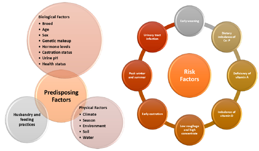
Some predisposing and risk factors associated with obstructive urolithiasis in small ruminants.
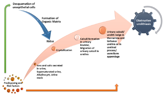
Etiopathogenesis of obstructive urolithiasis in small ruminants.
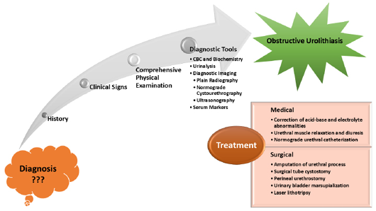
Diagnosis and treatment of obstructive urolithiasis in small ruminants.
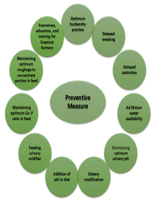
Preventive measures for obstructive urolithiasis in small ruminants.
References
- Abba, Y., Abdullah, F.F.J., Daud, N.H.B.A., Shaari, R. Bin, Tijjani, A., Sadiq, M.A., Mohammed, K., Adamu, L. and Mohd, A.M.L., 2015. Clinical management of dietary induced urolithiasis associated with balanoposthitis in a Boer goat. In: Open Vet. J., 5(1): 30–33. https://doi.org/10.5455/OVJ.2015.v5.i1.p30[CrossRef]
- Ahamed, B., Saha, M., Alam, M. and Alam, M., 2022. Occurrence of urolithiasis and its surgical management in goats. Ann. Bangladesh Agric., 25(2): 1–12. https://doi.org/10.3329/aba.v25i2.62408[CrossRef]
- Alemu, B., Desta, H., Kinati, W., Mulema, A.A., Gizaw, S. and Wieland, B., 2019. Application of mixed methods to identify small ruminant disease priorities in Ethiopia. Front. Vet. Sci., 6: 417. https://doi.org/10.3389/fvets.2019.00417[CrossRef]
- Alexandratos, N., 2009. World food and agriculture to 2030/50: Highlights and views from mid-2009. https://api.semanticscholar.org/CorpusID:127551962
- AlLugami, A., von Pückler, K., Wehrend, A. and Sickinger, M., 2017. Sonography of the distal urethra in lambs. Acta Vet. Scand., 59(1): 16. https://doi.org/10.1186/s13028-017-0283-2[CrossRef]
- Amarpal, K.P., Aithal, H.P., Pawde, A.M., Pratap, K. and Gugjoo, M.B., 2013. A retrospective study on the prevalence of obstructive urolithiasis in domestic animals during a period of 10 years. Adv. Anim. Vet. Sci., 1(3): 88–92.
- Antonelli, A.C., Júnior, R.A.B., Mori, C., Araripe Sucupira, M.C., Marcello, A.C.S. and Ortolani, E., 2012. Effect of different energy sources on predisposition of urolithiasis in male kids. Ciencia Anim. Brasil., 13: 487–493. https://doi.org/10.5216/cab.v13i4.15617[CrossRef]
- Aquilina, G., Bories, G., Chesson, A., Cocconcelli, P. S., De Knecht, J., Dierick, A., Gralak, A., Gropp, J., Halle, I., Hogstrand, C., Leng, L., Puente, S. L., Haldorsen, A.-K. L., Mantovani, A., Martelli, G., Mézes, M., Renshaw, D., Saarela, M., Sejrsen, K., & Westendorf, J. (2012). Scientific Opinion on the safety and efficacy of ammonium chloride for bovines, sheep, dogs and cats. EFSA Journal, 10(6), 2738. https://doi.org/10.2903/J.EFSA.2012.2738[CrossRef]
- Augustinsson, O. and Johansson, K., 1986. Ammonium chloride induced acidosis and aldosterone secretion in the goat. Acta Physiol. Scand., 128(4): 535–540. https://doi.org/10.1111/j.1748-1716.1986.tb08009.x[CrossRef]
- Augustinsson, O., Holst, H., Forsgren, M., Andersson, H. and Andersson, B., 1986. Influence of heat exposure on acid/base and fluid balance in hyperhydrated goats. Acta Physiol. Scand., 126(4): 499–503. https://doi.org/10.1111/j.1748-1716.1986.tb07847.x[CrossRef]
- Bamal, P.S., Kumar, V., John, J.K., Tyagi, S.K., Malik, V. and Shahi, U.P., 2023. High urinary trace element concentrations: A possible indicator of urolithiasis in goats? J. Trace Elem. Miner., 4: 100060. https://doi.org/10.1016/j.jtemin.2023.100060[CrossRef]
- Bashir, M., Athar, H., Ahmad, R., Dar, M. and Fazili, M., 2017. Surgical management of urolithiasis in small ruminants by urethral process amputation.
- Bhattarai, R., 2014. Importance of goat milk. J. Food Sci. Technol. Nepal, pp. 7. https://doi.org/10.3126/jfstn.v7i0.11209[CrossRef]
- Bowen, J.S. and Snyder, J., 2009. Chapter 94, ethical responsibilities of small ruminant veterinarians in selecting and using therapeutics (eds. D.E. Anderson, D.M.B.T.-F.A.P. Fifth E. Rings). pp. 457–460. W.B. Saunders. https://doi.org/10.1016/B978-1-4160-3591-6.10094-6[CrossRef]
- Buarque, V.L.M., Bezerra, H.V.A., Rissi, G.P., Oliveira, R.Í.G. de, Silva, N.M. da, Cônsolo, N.R.B., Ramírez-Zamudio, G.D., Strefezzi, R. de F., Gallo, S.B., Silva, S.L. and Leme, P.R., 2023. The use of additives to prevent urolithiasis in lambs fed diets with a high proportion of concentrate. Vet. Sci., 10(10). https://doi.org/10.3390/vetsci10100617[CrossRef]
- Chigerwe, M., Mavangira, V., Byrne, B.A. and Angelos, J.A., 2017. Antibiotic resistance patterns of bacteria isolated from indwelling Foley catheters following tube cystostomy in goats with obstructive urolithiasis. J. Vet. Diagn. Invest., 29(3): 316–320. https://doi.org/10.1177/1040638717695607[CrossRef]
- Clark, S. and Mora García, M.B., 2017. A 100-Year Review: Advances in goat milk research. J. Dairy Sci., 100(12): 10026–10044. https://doi.org/10.3168/jds.2017-13287[CrossRef]
- Cook, M.J., 2023. Urinary calculi of small ruminants. The veterinary clinics of north America. Food Anim. Pract., 39(2): 355–370. https://doi.org/10.1016/j.cvfa.2023.02.006[CrossRef]
- Darwish, A., 2023. Clinicopathological studies on urine retention in Barki Rams. Adv. Anim. Vet. Sci., 11. https://doi.org/10.17582/journal.aavs/2023/11.10.1673.1680[Full text][CrossRef]
- de Morais, M.V., Filho, A.P.S., de Alencar, S.P., de Mendonça, C.L., de Azevêdo, C.N. and Afonso, J.A.B., 2016. Clinical and epidemiological indicators of urolithiasis in small ruminants met in hospital routine. https://api.semanticscholar.org/CorpusID:79586743
- Dn, M., Sandeep and Mc, A.K. 2023. Tube cystostomy for the treatment of ruptured urinary bladder due to obstructive urolithiasis in a male Jamunapari goat: Case report. Pharma Innov. J., 12(11S): 552–554. https://www.thepharmajournal.com/special-issue?year=2023andvol=12andissue=11SandArticleId=23986
- Eicher, L., Zablotski, Y., Knubben-Schweizer, G. and Voigt, K., 2023. Composition of ovine and caprine uroliths, and short-and long- term treatment success following obstructive urolithiasis a retrospective analysis of clinical records of a Southern German veterinary hospital Open Access CC BY-NC-ND 4.0 2. https://doi.org/10.2376/1439-0299-2022-25[CrossRef]
- Elsheikh, H.A., Taha, A.A.W., Khalafallah, A.I. and Osman, I.A.M., 2002. Disposition kinetics of enrofloxacin (Baytril 5%) in sheep and goats following intravenous and intramuscular injection using a microbiological assay. Res. Vet. Sci., 73(2): 125–129. https://doi.org/10.1016/S0034-5288(02)00020-6[CrossRef]
- Ewoldt, J.M., Jones, M.L. and Miesner, M.D., 2008. Surgery of obstructive urolithiasis in ruminants. Veterinary clinics of North America. Food Anim. Pract., 24(3): 455–465. https://doi.org/10.1016/j.cvfa.2008.06.003[CrossRef]
- Farghali, A.M.H., Khatem, K.A.K. and Shamaa, A.A., 2020. Review on the common surgical affections in sheep and goats. J. Appl. Vet. Sci., 5(3): 40–48. https://doi.org/10.21608/javs.2020.98366[CrossRef]
- Fazili, M.R., Malik, H.U., Bhattacharyya, H.K., Buchoo, B.A., Moulvi, B.A. and Makhdoomi, D.M., 2010. Minimally invasive surgical tube cystotomy for treating obstructive urolithiasis in small ruminants with an intact urinary bladder. Vet. Rec., 166(17): 528–532. https://doi.org/10.1136/vr.b4831[CrossRef]
- Fortier, L.A., Gregg, A.J., Erb, H.N. and Fubini, S.L., 2004. Caprine obstructive urolithiasis: Requirement for 2nd surgical intervention and mortality after percutaneous tube cystostomy, surgical tube cystostomy, or urinary bladder marsupialization. Vet. Surg., 33(6): 661–667. https://doi.org/10.1111/j.1532-950X.2004.04089.x[CrossRef]
- Fritz, S., Chaitow, L. and Hymel, G.M., 2008. Chapter 7 Sanitation (eds. S. Fritz, L. Chaitow and G.M.B.T.-C. M. in the H.S. Hymel). pp. 196–207. Mosby. https://doi.org/10.1016/B978-032303996-3.50014-3[CrossRef]
- Gazi, M. and Makhdoomi, D.M., 2016. Urinalysis a diagnostic factor for urolithiasis and prognostic factor for its recurrence in young ruminants. J. Vet. Sci. Technol., 7. https://doi.org/10.4172/2157-7579.1000336[CrossRef]
- George, J.W., Hird, D.W. and George, L.W., 2007. Serum biochemical abnormalities in goats with uroliths: 107 cases (1992–2003). J. Am. Vet. Med. Assoc., 230(1): 101–106. https://doi.org/10.2460/javma.230.1.101[CrossRef]
- Gofur, M., 2019. Effect of testosterone on penile and urethral development in Black Bengal goat. Bangladesh J. Vet. Med., 17. https://doi.org/10.33109/bjvmjd19rm3[CrossRef]
- Gugjoo, M.B., Zama, M.M.S., Amarpal, A., Mohsina, A., Saxena, A.C. and Sarode, I.P., 2013. Obstructive urolithiasis in buffalo calves and goats: Incidence and management. J. Adv. Vet. Res., 3(3): 109–113.
- Halland, S.K., House, J.K. and George, L.W., 2002. Urethroscopy and laser lithotripsy for the diagnosis and treatment of obstructive urolithiasis in goats and pot-bellied pigs. J. Am. Vet. Med. Assoc., 220(12): 1831–1834. https://doi.org/10.2460/javma.2002.220.1831[CrossRef]
- Hart, S. and Delaney, C.B.T.R.M., 2016. Husbandry of dairy animals goat: Replacement management. Elsevier. https://doi.org/10.1016/B978-0-08-100596-5.00822-2[CrossRef]
- Haven, M.L., Bowman, K.F., Engelbert, T.A. and Blikslager, A.T., 1993. Surgical management of urolithiasis in small ruminants. Cornell Vet., 83(1): 47–55.
- Horst, R.L. and Jorgensen, N.A., 1974. Effect of ammonium chloride on nitrogen and mineral balance in lactating and nonlactating goats. J. Dairy Sci., 57(6): 683–688. https://doi.org/10.3168/jds.S0022-0302(74)84947-7[CrossRef]
- Hussain, T., Javed, I., Muhammad, F. and Rahman, Z., 2014. Disposition kinetics of enrofloxacin following intramuscular administration in goats. Pak. Vet. J., 34: 293–296.
- Jacobs, C.C. and Fecteau, M.E., 2019. Urethrotomy in combination with or after temporary tube cystostomy for treatment of obstructive urolithiasis in male goats. Vet. Surg., 48(3): 315–320. https://doi.org/10.1111/vsu.13170[CrossRef]
- Janke, J.J., Osterstock, J.B., Washburn, K.E., Bissett, W.T., Roussel, A.J. and Hooper, R.N., 2009. Use of Walpole’s solution for treatment of goats with urolithiasis: 25 cases (2001–2006). J. Am. Vet. Med. Assoc., 234(2): 249–252. https://doi.org/10.2460/javma.234.2.249[CrossRef]
- Jones, M.L. and Miesner, M.D., 2009. Chapter 67 Urolithiasis (eds. D.E. Anderson and D.M.B.T.F.A.P. Fifth E. Rings). pp. 322–325). W.B. Saunders. https://doi.org/10.1016/B978-141603591-6.10067-3[CrossRef]
- Jones, M.L., Dominguez, B.J. and Deveau, M.A., 2018. An experimental model for calcium carbonate urolithiasis in goats. J. Vet. Intern. Med., 32(3): 1268–1273. https://doi.org/10.1111/jvim.15061[CrossRef]
- Jones, M.L., Gibbons, P.M., Roussel, A.J. and Dominguez, B.J., 2017. Mineral composition of uroliths obtained from sheep and goats with obstructive urolithiasis. J. Vet. Intern. Med., 31(4): 1202–1208. https://doi.org/10.1111/jvim.14743[CrossRef]
- Kalim, O.R.Z. and Tiwari, S., 2011. Surgical management of obstructive urolithiasis in a male cow calf. Vet. World, 4. https://doi.org/10.5455/vetworld.2011.213-214[CrossRef]
- Kannan, K.V.A. and Lawrence, K.E., 2010. Obstructive urolithiasis in a Saanen goat in New Zealand, resulting in a ruptured bladder. N. Z. Vet. J., 58(5): 269–271. https://doi.org/10.1080/00480169.2010.69302[CrossRef]
- Khan, S., Manjusha, K.M., Kumar, R., Pawde, A.M., Malik, Y., Kinjavdekar, P., Maiti, S. and Amarpal, A., 2021. Prevalence of obstructive urolithiasis in domestic animals: An interplay between seasonal predisposition and dietary imbalance. Iraqi J. Vet. Sci., 35: 227–232. https://doi.org/10.33899/ijvs.2020.126662.1358[CrossRef]
- Khanal, P., Dhakal, R., Khanal, T., Pandey, D., Devkota, N.R. and Nielsen, M.O., 2022. Sustainable livestock production in Nepal: A focus on animal nutrition strategies. Agriculture, 12(5). https://doi.org/10.3390/agriculture12050679[CrossRef]
- Kibria, A.S.M.G., Rahman, M.L., Ahasan, A.S.M.L., Uddin, M. and Quasem, M.A., 2016. Effects of castration on penile and urethral development in Black Bengal Goat. 48: 501–506.
- Kimberling, C.V. and Arnold, K.S., 1983. Diseases of the urinary system of sheep and goats. Veterinary clinics of north America. Large Anim. Pract., 5(3): 637–655. https://doi.org/10.1016/S0196-9846(17)30068-X[CrossRef]
- Kimeli, P., Kipyegon, A.N. and Karanja, D.N., 2014. Fatal obstructive urolithiasis in a 5 months old buck: A case report.
- Kinjavdekar, P., Aithal, H.P., Motiram, A., Krishna, P. and Bashir, M., 2013. Research article a retrospective study on the prevalence of obstructive urolithiasis in domestic animals during a period of 10 Years. 1(3): 88–92.
- Kinjavdekar, P., Aithal, H.P., Pawde, A.M. and Pratap, K., 2005. Management of urolithiasis in goats (capra hircus): A retrospective study of 25 cases. 39(1): 8–13.
- Kinsley, M.A., Semevolos, S., Parker, J.E., Duesterdieck-Zellmer, K. and Huber, M., 2013. Use of plain radiography in the diagnosis, surgical management, and postoperative treatment of obstructive urolithiasis in 25 goats and 2 sheep. Vet. Surg., 42(6): 663–668. https://doi.org/10.1111/j.1532-950X.2013.12021.x[CrossRef]
- Kretsch, C. and Chigerwe, M., 2021. Assessment of preoperative and postoperative l-lactatemia and clinical outcomes in goats undergoing tube cystostomy: A retrospective study of 34 cases (2015-2020). J. Vet. Intern. Med., 35. https://doi.org/10.1111/jvim.16271[CrossRef]
- Kumar, P., Lakshmi, K., Nagaraj, P. and Rani, M., 2024. Occurrence of lower urinary tract affections in goats: A retrospective study. Int. J. Adv. Biochem. Res., 8: 876–879. https://doi.org/10.33545/26174693.2024.v8.i12Sk.3208[CrossRef]
- Kümper, H., 1994. Urolithiasis in male sheep and goats. Clinical picture, therapeutic possibilities and prognostic evaluation. Tierarztliche Praxis, 22(3): 234–241.
- Lai, J., Chang, M.H., Hwang, M.Y. and Wang, J.H., 2016. Urinary retention caused by a botched castration in a goat. Taiwan Vet. J., 43: 1–6. https://doi.org/10.1142/S1682648515720130[CrossRef]
- Liesegang, A., 2008. Influence of anionic salts on bone metabolism in periparturient dairy goats and sheep. J. Dairy Sci., 91(6): 2449–2460. https://doi.org/10.3168/jds.2006-838[CrossRef]
- Lyngset, O., 1968. Studies on reproduction in the goat. II. The genital organs of the pregnant goat. Acta Vet. Scand., 9(3): 242–252. https://doi.org/10.1186/BF03547870[CrossRef]
- Maciel, T., Oliveira, C., Afonso, J., Júnior, R., Fagliari, J., Mathias, L., Oliveira, D., Baraldi-Artoni, S. and Amoroso, L., 2019. Predictive elements of obstructive urolithiasis in sheep. Acta Sci. Vet., 47. https://doi.org/10.22456/1679-9216.97849[CrossRef]
- Maciel, T.A., de Souza Ramos, I.A., da Silva, R.J., Soares, P.C., Carvalho, C.C.D., de Souto Maior Junior, R.J., Amoroso, L., Artoni, S.M.B., Afonso, J.A.B. and Oliveira, D., 2017. Clinical and biochemical profile of obstructive urolithiasis in sheep. Acta Sci. Vet., 15. https://api.semanticscholar.org/CorpusID:91910596
- Mahajan, A., Gupta, A., Bhadwal, M., Bhat, M. and Bhardwaj, H., 2017. Occurrence and management of obstructive urolithiasis in ruminants. J. Anim. Res., 74: 723–731. https://doi.org/10.5958/2277-940X.2017.00111.5[CrossRef]
- Makhdoomi, D.M. and Gazi, M., 2013. Obstructive urolithiasis in ruminants. A review. Vet. World, 6: 233. https://doi.org/10.5455/vetworld.2013.233-238[CrossRef]
- Mavangira, V., Cornish, J.M. and Angelos, J.A., 2010. Effect of ammonium chloride supplementation on urine pH and urinary fractional excretion of electrolytes in goats. J. Am. Vet. Med. Assoc., 237(11): 1299–1304. https://doi.org/10.2460/javma.237.11.1299[CrossRef]
- May, K.A., Moll, H.D., Wallace, L.M., Pleasant, R.S. and Howard, R.D., 1998. Urinary bladder marsupialization for treatment of obstructive urolithiasis in male goats. Vet. Surg., 27(6): 583–588. https://doi.org/10.1111/j.1532-950X.1998.tb00534.x[CrossRef]
- Mejia, S., McOnie, R.C., Nelligan, K.L. and Fubini, S.L., 2022. Small ruminant urinary obstruction: Decision trees for treatment. J. Am. Vet. Med. Assoc., 260(S2): S64–S71. https://doi.org/10.2460/javma.22.02.0071[CrossRef]
- Moald, 2021. Statistical information on nepalese agriculture (2077/78 ). Publicatons of the Nepal in Data Portal, 73: 274. https://nepalindata.com/resource/statistical-information-nepalese-agriculture-207374-201617/
- Muthukumar, S., Vanan, T.T., Sundaram, S.M., Hemalatha, S. and Sughanthi, M., 2020. A case report of cystic calculi in goat kid.
- Nagamine, I. and Sunagawa, K., 2017. Effects of the castration on the growth, meat production and odors in male goats. Anim. Behav. Manage., 53(4): 137–150. https://doi.org/10.20652/abm.53.4_137[CrossRef]
- Navarre, C.B., 2007. Urolithiasis in goats.
- Neal, C.E., Grissett, G.P., Fleming, S., Wills, R.W. and Woolums, A.R., 2024. Effect of D,L methionine and ammonium chloride on urine acidification, urinary fractional excretion of calcium, and blood bicarbonate in clinically healthy goats. Am. J. Vet. Res., 85(11): ajvr.24.04.0097. https://doi.org/10.2460/ajvr.24.04.0097[CrossRef]
- Nuvey, F.S., Mensah, G.I., Zinsstag, J., Hattendorf, J., Fink, G., Bonfoh, B. and Addo, K.K., 2023. Management of diseases in a ruminant livestock production system: A participatory appraisal of the performance of veterinary services delivery, and utilization in Ghana. BMC Vet. Res., 19(1): 237. https://doi.org/10.1186/s12917-023-03793-z[CrossRef]
- Nwaokorie, E.E., Osborne, C.A., Lulich, J.P., Fletcher, T.F., Ulrich, L.K., Koehler, L.A. and Buettner, M.T., 2015. Risk factors for calcium carbonate urolithiasis in goats. J. Am. Vet. Med. Assoc., 247(3): 293–299. https://doi.org/10.2460/javma.247.3.293[CrossRef]
- Oman, R.E., Reppert, E.J., Streeter, R.N. and Jones, M., 2019. Outcome and complications in goats treated by perineal urethrostomy for obstructive urolithiasis: 25 cases (2010-2017). J. Vet. Intern. Med., 33(1): 292–296. https://doi.org/10.1111/jvim.15360[CrossRef]
- Palmer, J.L., Dykes, N.L., Love, K. and Fubini, S.L., 1998. Contrast radiography of the lower urinary tract in the management of obstructive urolithiasis in small ruminants and swine. Vet. Radiol. Ultras. Off. J. Am. Coll. Vet. Radiol. Int. Vet. Radiol. Assoc., 39(3): 175–180. https://doi.org/10.1111/j.1740-8261.1998.tb00335.x[CrossRef]
- Parkinson, T.J. and McGowan, M., 2019. Abnormalities affecting reproductive function of male animals (D.E. Noakes, T.J. Parkinson and G.C.W.B.T.-V.R. and O. (Tenth E. England (eds.); pp. 635–668. W.B. Saunders. https://doi.org/10.1016/B978-0-7020-7233-8.00036-7[CrossRef]
- Parrah, J., Moulvi, B., Gazi, M., Makhdoomi, D.M., Athar, H., Dar, M., Dar, S. and Mir, A.Q., 2013. Importance of urinalysis in veterinary practice. A review. Vet. World, 6: 640–646. https://doi.org/10.14202/vetworld.2013.640-646[CrossRef]
- Patel, J.B., Patel, A.N., Gosai, R.K., Patel, A.M., Sutaria, P.T. and Patel, P.B., 2022. Urethral process excision to relieve urinary obstruction in castrated male goats: A case study. 11(11): 1415–1416.
- Perkins, G.A., Divers, T.J., Smith, M.C. and Callan, R.J., 2017. Chapter 1 examination of the surgical patient (S.L. Fubini and N.G.B.T.F.A.S. (Second E. Ducharme (eds.); pp. 1–22. W.B. Saunders. https://doi.org/10.1016/B978-0-323-31665-1.00001-0[CrossRef]
- Pokharel, S. and Sharma, S., 2019. Case report on use of tube cystotomy for management of obstructive urolithiasis in buffalo calf.
- Pongphitcha, P., Chuchoed, K., Thetsana, T., Dachphun, N., Sukhong, P. and Ratanapob, N., 2024. Factors associated with success rate of oral force-feeding ammonium chloride administration to acidify urine in goats. Open Vet. J., 14: 2310. https://doi.org/10.5455/OVJ.2024.v14.i9.19[CrossRef]
- Prakash, A., Nair, S., Ramankutty, S., Rameshbabu, S., Jishi, D.D.J. and Venugopal, S., 2024. Minimal invasive percutaneous tube cystostomy using three-way foley catheter for management of obstructive urolithiasis in goats. J. Vet. Anim. Sci., 55: 47–52. https://doi.org/10.51966/jvas.2024.55.1.47-52[CrossRef]
- Rafee, M.A. and Baghel, M., 2016. Obstructive urolithiasis in kids: A study on pattern of occurrence, aetiology, age, clinical symptoms and conditions of bladder and urethra. Veterinaria, 4(1): 1–3.
- Raina, R., Prawez, S., Dimitrova, D.J., Pankaj, N.K. and Verma, P.K., 2008. Disposition kinetics and urinary excretion of ciprofloxacin in goats following single intravenous administration. J. Vet. Sci., 9(3): 241–245. https://doi.org/10.4142/jvs.2008.9.3.241[CrossRef]
- Rakestraw, P.C., Fubini, S.L., Gilbert, R.O. and Ward, J.O., 1995. Tube cystostomy for treatment of obstructive urolithiasis in small ruminants. Vet. Surg., 24(6): 498–505. https://doi.org/10.1111/j.1532-950X.1995.tb01361.x[CrossRef]
- Ramankutty, S., Sudheesh, S., Nair, S., Athira, V.S.A., Narayanan, M. and Devanand, C., 2018. Tube cystostomy for management of struvite urolithiasis in male goats: A review of six cases. pp. 895–899.
- Rankins, D.L. and Pugh, D.G., 2012. Chapter 2 Feeding and nutrition (D.G. Pugh and A.N.B.T.S. and G.M. (Second E. Baird (eds.). pp. 18–49. W.B. Saunders. https://doi.org/10.1016/B978-1-4377-2353-3.10002-2[CrossRef]
- Rao, G.S., Ramesh, S., Ahmad, A.H., Tripathi, H.C., Sharma, L.D. and Malik, J.K., 2001. Pharmacokinetics of enrofloxacin and its metabolite ciprofloxacin after intramuscular administration of enrofloxacin in goats. Vet. Res. Commun., 25(3): 197–204. https://doi.org/10.1023/A:1006481625615[CrossRef]
- Raza, A., Muhammad, G., Manzoor, D., Ahmad, M. and Ijaz, D., 2012. Clinical efficacy of allopurinol and ketoprofen in the treatment of urate associated urolithiasis in bucks case report. Int. J. Mol. Vet. Res., 22: 22–24. https://doi.org/10.5376/ijmvr.2012.02.0006[CrossRef]
- Riedi, A., Nathues, C., Knubben-Schweizer, G., Nuss, K. and Meylan, M., 2018. Variables of initial examination and clinical management associated with survival in small ruminants with obstructive urolithiasis. J. Vet. Intern. Med., 32. https://doi.org/10.1111/jvim.15336[CrossRef]
- Riedi, A.K., Knubben-Schweizer, G. and Meylan, M., 2018. Clinical findings and diagnostic procedures in 270 small ruminants with obstructive urolithiasis. J. Vet. Intern. Med., 32(3): 1274–1282. https://doi.org/10.1111/jvim.15128[CrossRef]
- Riet-Correa, F., Simões, S. and Vasconcelos, J., 2008. Urolithiasis in sheep and goats. Pesquisa Vet. Brasil., 28: 319–322. https://doi.org/10.1590/S0100-736X2008000600010[CrossRef]
- Salisi, M. and Saad, M., 2011. Urolithiasis in boer bucks. Pertanika J. Trop. Agric. Sci., 34: 363–366.
- Samal, L., Pattanaik, A., Mishra, C., Maharana, B. and Baithalu, L., 2011. Nutritional stretegies to prevent urolithiasis in animals. Vet. World, 4. https://doi.org/10.5455/vetworld.2011.142-144[CrossRef]
- Saritha, G., 2024. Chapter 29 Common clinical conditions of the urinary system in goats (T.B.T.T. in C.D. Rana production and management of goats (ed.). pp. 387–399. Academic Press. https://doi.org/10.1016/B978-0-443-23697-6.00017-2[CrossRef]
- Sarker, D., Akter, M.A., Rahman, M.S., Yesmin, N. and Alam, M.M., 2020. Clinicopathological consequences of urinary retention due to urolithiasis in indigenous goats. PSM Vet. Res., 5(2): 28–37.
- Schregel, J., Kleinschmidt, S., von Altrock, A., Hoeltig, D., Ganter, M. and Wagener, M.G., 2022. Case report: Urolithiasis, nephrolithiasis and a urinary bladder malformation in a seven-month-old alpaca cria. Front. Vet. Sci., 9. https://doi.org/10.3389/fvets.2022.1038642[CrossRef]
- Scully, C.M., 2021. Management of urologic conditions in small ruminants. Veterinary clinics of north America. Food Anim. Pract., 37(1): 93–104. https://doi.org/10.1016/j.cvfa.2020.10.003[CrossRef]
- Sen, A.R., Akter, N. and Chanda, T., 2018. Prevalence of urolithiasis in goat at Upazilla Veterinary Hospital, Feni, Bangladesh. https://doi.org/10.21276/sjavs.2018.5.6.4[CrossRef]
- Sharma, A., Sarasambi, M.K., Yadav, A., Jaishawal, V., Singh, A. and Jaiswal, S., 2023. Treatment of obstructive urolithiasis by tube cystotomy in goats a report of 4 cases Fig 1: Foleys catheter placed through a stab incision into the bladder Fig 2: Catheter left open for 3-4. 10(11): 470–471.
- Sheikh, F.D., Wani, S.A., Shah, O.S., Khan, H.M. and Beigh, A.B., 2018. Studies on prevalence and epidemiology of obstructive urolithiasis in pashmina goats of Ladakh region in Jammu and Kashmir. J. Pharmacogn. Phytochem., 7(5): 3326–3328.
- Shrestha, S., Thakur, B., Regmi, B. and Shah, M., 2019. Management of obstructive urolithiasis in a male buffalo calf using tube cystostomy technique. Int. J. Appl. Sci. Biotechnol., 7: 120–123. https://doi.org/10.3126/ijasbt.v7i1.22781[CrossRef]
- Sickinger, M. and Windhorst, A., 2022. A systematic review on urolithiasis in small ruminants according to nutrition-dependent prevalence and outcome after surgery. Vet. World, 15(3): 809–817. https://doi.org/10.14202/vetworld.2022.809-817[CrossRef]
- Simões, J. and Gutiérrez, C., 2017. Nutritional and metabolic disorders in dairy goats BT- sustainable goat production in adverse environments: Volume I: Welfare, health and breeding (J. Simões and C. Gutiérrez (eds.); pp. 177–194). Springer International Publishing. https://doi.org/10.1007/978-3-319-71855-2_11[CrossRef]
- Singh, A.K., Kumar, G.A., Sangeeta, D.K. and Singh, H., 2014. Incidence and management of obstructive urolithiasis in buffalo calves and goats. Adv. Anim. Vet. Sci., 2(9): 503–507. https://doi.org/10.14737/journal.aavs/2014/2.9.503.507[Full text][CrossRef]
- Singh, T., Amarpal, K.P., Kinjavdekar, P., Aithal, H.P., Pawde, A.M., Pratap, K. and Mukherjee, R., 2008. Obstructive urolithiasis in domestic animals: A study on pattern of occurrence and etiology. Indian J. Anim. Sci., 78(6): 599.
- Sprake, P., Roussel, A., Stewart, R. and Bissett, W., 2012. The effect of ammonium chloride treatment as a long term preventative approach for urolithiasis in goats and a comparison of continuous and pulse dosing regimes. J. Vet. Intern. Med., 26.
- Stone, W.C., Bjorling, D.E., Trostle, S.S., Hanson, P.D. and Markel, M.D., 1997. Prepubic urethrostomy for relief of urethral obstruction in a sheep and a goat. J. Am. Vet. Med. Assoc., 210(7): 939–941. https://doi.org/10.2460/javma.1997.210.07.939[CrossRef]
- Sutradhar, B., Dey, T., Yadav, S. and Bostami, M., 2018. Surgical management of obstructive urolithiasis in small ruminants by tube cystostomy in Chittagong, Bangladesh. J. Agric. Sci. Technol., A, 8. https://doi.org/10.17265/2161-6256/2018.02.005[CrossRef]
- Talpade, J., Shrman, K., Sharma, R., Sahni, Y.P., Singh, R.P. and Soman, S., 2018. Pharmacokinetic study of single dose intramuscular administration of enrofloxacin in barbari goats. Pharma Innov. J., 7: 609–611. https://api.semanticscholar.org/CorpusID:228744983
- Tamilmahan, P., Mohsina, A., Karthik, K., Gopi, M., Gugjoo, M.B., Rashmi and Zama, M.M.S., 2014. Tube cystostomy for management of obstructive urolithiasis in ruminants. Vet. World, 7(4): 234–239. https://doi.org/10.14202/vetworld.2014.234-239[CrossRef]
- Temple, D. and Manteca, X., 2020. Animal welfare in extensive production systems is still an area of concern. Front. Sustain. Food Syst., 4. https://doi.org/10.3389/fsufs.2020.545902[CrossRef]
- Tharwat, M., 2021. Clinical, ultrasonographic, and postmortem findings in sheep and goats with urinary tract disorders. Vet. World, 14(7): 1879–1887. https://doi.org/10.14202/vetworld.2021.1879-1887[CrossRef]
- Tharwat, M., Hegazy, Y. and Alkheraif, A.A., 2024. Discolored urine in sheep and goats: Clinical, etiological, hematobiochemical, sonographic and postmortem findings. Open Vet. J., 14(4): 1059–1071. https://doi.org/10.5455/OVJ.2024.v14.i4.13[CrossRef]
- Todhunter, P., Baird, A.N. and Wolfe, D.F., 1996. Erection failure as a sequela to obstructive urolithiasis in a male goat. J. Am. Vet. Med. Assoc., 209(3): 650–652. https://doi.org/10.2460/javma.1996.209.03.650[CrossRef]
- Underwood, W.J., Blauwiekel, R., Delano, M.L., Gillesby, R., Mischler, S.A. and Schoell, A., 2015. Biology and diseases of ruminants (sheep, goats, and cattle). Lab. Anim. Med., pp. 623–694. https://doi.org/10.1016/B978-0-12-409527-4.00015-8[CrossRef]
- United Nation, 2017. World population prospects 2017. Data booklet (ST/ESA/SER.A/401). United Nations, Department of Economics and Social Affairs, Population Division, 1–24. https://www.un.org/development/desa/pd/sites/www.un.org.
- Vagg, M.J. and Payne, J.M., 1970. The effect of ammonium chloride induced acidosis on calcium metabolism in ruminants. Br. Vet. J., 126(10): 531–537. https://doi.org/10.1016/S0007-1935(17)48139-5[CrossRef]
- Van Metre, D.C. and Fubini, S.L., 2006. Ovine and caprine urolithiasis: Another piece of the puzzle. Vet. Surg., 35(5): 413–416. https://doi.org/10.1111/j.1532-950X.2006.00168.x[CrossRef]
- van Metre, D.C., 2015. Urolithiasis in small ruminants: Surgical and dietary management. https://api.semanticscholar.org/CorpusID:19569950
- Venkatesh, S.M., Manjunatha, D.R., Nagaraju, N., Balappanavar, B.R., Rajashailesh, N.M., Shankaregowda, A.J., Ramesh, D., Ramya, N.M., Kanteshkumar, M.J. and Dongre, N.A., 2024. Minimally invasive percutaneous tube cystostomy using pigtail catheter for the surgical management of urolithiaisis in bucks. Indian J. Vet. Sci. Biotechnol., 20(4).
- Videla, R. and van Amstel, S., 2016. Urolithiasis. Vet. Clin. North Am. Food Anim. Pract., 32(3): 687–700. https://doi.org/10.1016/j.cvfa.2016.05.010[CrossRef]
- Yohannes, G. and Tesfay, S., 2024. Review on surgical managements of urolithiasis in ruminants. Mathews J. Surg., 7. https://doi.org/10.30654/MJS.10029[CrossRef]
Cited by 1 article
In journals published by ResearchersLinks.
Services
Article usage statistics
Loading…