
Human immunodeficiency virus (HIV) becomes one of the most severe endemic viruses that advance around the world in a deadly manner. It is a serious issue in Pakistan as well and so far 130,000 HIV cases in Pakistan have been identified by the National AIDS Control Programme. Nuclear transcription factor forkhead box P3 (FOXP3) gene is involved in tolerance mechanism so failure of tolerance can lead to mutations in this gene. The main objective of this project was to analyze the possible mutation especially in FOXP3 gene exon 1 that may clarify the reason of reduction of T regulatory cells (Tregs) due to HIV/AIDS. A total of 25 HIV patients were chosen from the Institute of Public Health on the basis of confirm HIV infection and 25 healthy controls as well. First genomic DNA was extracted from the peripheral blood and then amplified by using specific designed primers. Gradient PCR was performed and the product length was 197 bps which was further analyzed on 1% agarose gel. Sequencing was done through genetic analyzer (3500 ABI). No mutation was observed in FOXP3 gene exon 1of Pakistani HIV patients.
Short Communication · Volume 51, Issue 5, 6 August 2019 · Open Access
Mutational analysis of Forkhead box P3 gene in Pakistani Human Immunodeficient Virus Patients
·
Affiliations & Notes ⌄
- 1Department of Microbiology and Molecular Genetics, Quaid-e-Azam Campus, Punjab University, Lahore , * Corresponding author: nageen1704@hotmail.com
Article Info ⌄
- Journal:
- Pakistan Journal of Zoology
- Published:
- 6 August 2019

From the article
- Most of the HIV patients (n=25) lie within the age group of 21-40 years (Fig. 1).
- DNA bands were extracted and Exon 1 of FOXP3 gene was amplified at 57.8°C annealing temperature and the product size of 197bp was achieved (Fig. 3).
- Random purified HIV sequencing PCR products were analyzed, and it was observed that no mutation occurred in exon 1of FOXP3 gene of HIV patients.
Selected from the article's own text.
Abstract
Materials and methods
The total sample size was 50 and the focal point of this study was different cities of the Punjab province of Pakistan. A total of 25 HIV patients were chosen from the Institute of Public Health on the basis of confirm HIV infection and 25 controls were selected. Completely covered Eppendorf’s were further carefully stored at 4°C in an isolated rack of refrigerator before DNA isolation.
Both controls and HIV positive Genomic DNA were extracted from peripheral blood using (Favorgen kit, Catalogue No. FABGK 001, 50 Preps). DNA bands extraction were allowed to check thorough agarose gel electrophoresis. Amplification of exon 1 of FOXP3 gene was performed by using specific designed primers (Table I). The PCR reaction was executed in total volume 20ul using 250 ng of genomic DNA, 2X PCR mixtures and 1ul of each of primer mixture. Gradient PCR was performed and the product length was 197 bps which was analyzed on 1% agarose gel. Thermo Scientific GeneRuler 100bp plus DNA ladder Catalog (# SM0323) was used to identify the results.
Ethanol purification of sequencing PCR products (BigDye Terminator V3.1 Sequencing standard Kit) was done and analyzed through genetic analyzer (3500 ABI).
Results and discussion
Most of the HIV patients (n=25) lie within the age group of 21-40 years (Fig. 1). CD4 and CD8 counts/ul were very lowered as compared to reference range measured through Flow cytometry in Figure 2. DNA bands were extracted and Exon 1 of FOXP3 gene was amplified at 57.8°C annealing temperature and the product size of 197bp was achieved (Fig. 3). Random purified HIV sequencing PCR products were analyzed, and it was observed that no mutation occurred in exon 1of FOXP3 gene of HIV patients. Sequencing results are represented in Figure 4 and 5.
Table I.- Primers designed for exon 1 of FOXP3 gene.
| Oligo name | Sequence (5’-3’) | Length (bp) | MW | TM (°C) | EM | nmols | µg | Area units |
| Forward Primer | CGCACACACTCATCGAAAAA | 20 | 6048 | 53.4 | 230.8 | 42.8 | 258.9 | 9.88 |
| Reverse Primer | CATCTGGTAGGGGAGAGCAG | 20 | 6247 | 59.5 | 228.8 | 45.5 | 284.2 | 10.41 |
FOXP3 is a central controller gene responsible for the production and activity of Tregs cells are defected in IPEX syndrome indicated that mutation has occurred in exon 1 of FOXP3 gene. Deletion mutation was observed that included 1388bp consisted of 5 half exon-1 and segment of 1st intron. This caused the deficiency of Tregs due to low production of degraded mRNA transcribed from FOXP3 gene in IPEX patients. Defective Tregs are present in HIV patients and can be cleared according to one scientific research, when Tregs from HIV patients did not suppress the function of polyclonal autologous CD8+T cells specifying lack of suppressive activity of Treg/HIV+ as compared to HIV negative controls So this clarifies that genetic change in FOXP3 gene cause defective production of Regulatory T cells. This point emphasizes to find out whether the mutations exist or not in FOXP3 gene as the number and function of Tregs are abnormal in HIV patients.
In Pakistan, HIV is mostly prevalent because of the usage of poor management of blood transfusion systems. No health care working institution ponders over this bad and old management of transfusion systems and using constantly resulting most resistant HIV infection advancement. A survey conducted in Faisalabad, Pakistan proved the HIV infection was more in males as compared to females. In this study, the frequency of HIV infection was found to be highest in males (100%). CD4 T cells depletion is an important indicator of HIV infection. It was also confirmed that CD4:CD8 and CD4 counts are inversely related with proviral DNA and both lower values against reference values were confirmation of HIV infection. This study has proved below an average CD4:CD8 ratio (0.3428) in HIV patients . In current scenario, exon 1 having product size of 197bp of FOXP3 gene was amplified by using extracted genomic DNA of both 25 HIV patients and 25 HIV negative controls. PCR bands of HIV samples were sequenced through DNA sequencing Genetic Analyzer. In the present study, no mutation was observed in FOXP3 gene exon 1 of HIV patients by using BLAST and Chromas Pro 2.4.1 software.
Acknowledgements
I would like to acknowledge the Department of Microbiology and Molecular Genetics, University of the Punjab for providing us the working facilities in collaboration with Punjab AIDS control program at the Punjab Institute of Public health, Lahore-Pakistan. I would like to acknowledge HIV patients without whom the study was not possible.
Conflicts of interest
Authors declare no conflict of interest.
Figures
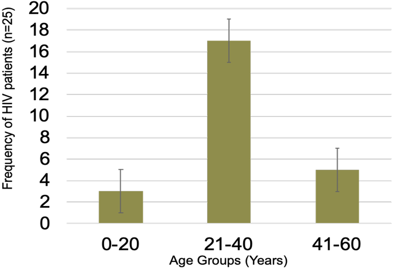
Frequency distribution of HIV patients (n=25) with reference to age.
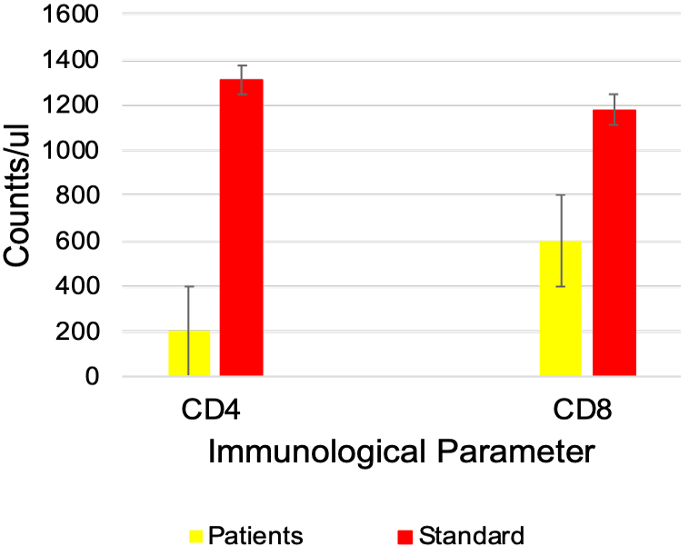
Comparison of patients CD4 and CD8 counts/ul with standards.
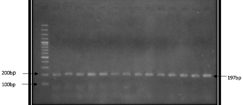
Representation of PCR bands (197bp each) of exon 1 of FOXP 3 gene of both HIV and Control samples. HIV PCR bands from 2nd well to 7th well and control PCR bands from 8th.
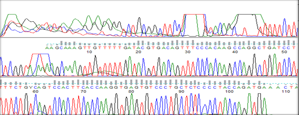
Representation of sequencing of HIV sample 1 from Genetic Analyzer (3500 ABI) Software
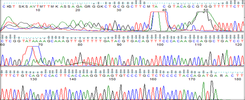
Representation of sequencing of HIV sample 2 from Genetic Analyzer (3500 ABI) Software
References
- Ahmed, K.R., Anwar, M., Waheed, U., Asad, M.J., Abbasi, S. and Abbas, Z.H., 2016. J. Blood Transf., 45: 16-18.
- Apoil, P.A., Puissant, B., Roubinet, F., Abbal, M., Massip, P. and Blancher, A., 2005. FOXP3 mRNA levels are decreased in peripheral blood CD4+ lymphocytes from HIV-positive patients. J. AIDS., 39: 381-385. https://doi.org/10.1097/01.qai.0000169662.30783.2d[CrossRef]
- Becerra, J.C., Bildstein, L.S. and Gach, J.S., 2016. Microb. Cell, 3: 451. https://doi.org/10.15698/mic2016.09.529[CrossRef]
- Chevalier, M.F. and Weiss, L., 2013. Blood, 121:29-37. https://doi.org/10.1182/blood-2012-07-409755[CrossRef]
- Hu, W.S. and Hughes, S.H., 2006. J. Med., 2:a006882.
- Maartens, G., Celum, C. and Lewin, S.R., 2014. The Lancet, 384: 258-271. https://doi.org/10.1016/S0140-6736(14)60164-1[CrossRef]
- Janahi, E.M., Mustafa, S., Alsari, S., Al-Mannai, M. and Farhat, G.N., 2016. J. Infect. Devel. Count., 10:1003-1011. https://doi.org/10.1089/apc.2016.0100[CrossRef]
- Nikolova, M., Wiedemann, A., Lacabaratz, C., 2017. Med. Sci., 33: 723–726. https://doi.org/10.1051/medsci/20173308012[CrossRef]
- Oda, J.M., Hirata, B.K., Guembarovski, R.L. and Watanabe, M.A., 2013. J. Genet., 92:163-171. https://doi.org/10.1007/s12041-013-0213-7[CrossRef]
- Oswald-Richter, K., Grill, S.M., Shariat, N., Leelawong, M., Sundrud, M.S., Haas, D.W. and Unutmaz, D., 2004. PLoS Biol., 2: e198. https://doi.org/10.1371/journal.pbio.0020198[CrossRef]
- Selliah, N., Zhang, M., White, S., Zoltick, P., Sawaya, B.E., Finkel, T.H. and Cron, R.Q., 2008. Virology, 381:161-167. https://doi.org/10.1016/j.virol.2008.08.033[CrossRef]
- Suchard, M.S., Mayne, E., Green, V.A., Shalekoff, S., Donninger, S.L., Stevens, W.S., Gray, C.M. and Tiemessen, C.T., 2010. PLoS one, 5: e11762. https://doi.org/10.1371/journal.pone.0011762[CrossRef]
- Tsunemi, S., Iwasaki, T., Imado, T., Higasa, S., Kakishita. E., Shirasaka, T. and Sano, H., 2005. AIDS, 19:879-886. https://doi.org/10.1097/01.aids.0000171401.23243.56[CrossRef]
- Woodham, A.W., Skeate, J.G., Sanna, A.M., Taylor, J.R., Da Silva, D.M., Cannon, P.M. and Kast, W.M., 2016. AIDS Patient Care STDs, 30:291-306.
Services
Article usage statistics
Loading…