By their relation to the organ, lymphatic vessels are divided into intra- and extraorganic, and by diameter–into small, medium and large, with certain peculiarities of the wall structure (Figure 1).
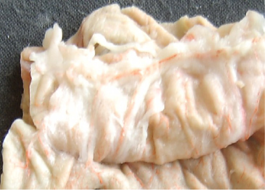
Figure 1: Fragment of the injected part of the abomasum. Vessels of the organ wall are visible.
The intraorganic lymphatic bed of the gastrointestinal tract of sheep is represented by the capillaries that are located in all layers of the organ, postcapillaries, and vessels of the first, second and third-orders. Small lymphatic capillaries transform into postcapillaries, in which the valve apparatus is already present, and the wall consists of endothelial cells. Anatomically postcapillaries are located in the submucous coat, but in the studies, we discovered these capillaries between the layers of the muscle membrane.
The fine-meshed lymphatic network of capillaries of the mucous membrane develops into a large-meshed network of the submucous coat through numerous capillaries perforating the muscular plate of the mucous membrane vertically or at an angle. The peculiarity of a lymphatic vessel wall structure is the presence of smooth muscle cells (myocytes).
Capillaries have an irregular diameter in various parts of the stomach submucous coat. The largest diameter of capillaries is in the region of the lesser curvature of the stomach in the pyloric area. The smallest diameter of lymphatic capillaries is on the side walls of the stomach body, and the smallest diameter was observed in the area of the bottom. Anastomosing with each other, lymphatic capillaries form polygonal, rhomboid, triangular, elongated, or sometimes more rounded loops.
In the muscular layer of the multichamber stomach, including abomasum, we identified three layers of the lymphatic capillaries’ network located in three layers. It means that in the circular muscle layer, lymphatic capillaries are located in various planes (Figure 2).
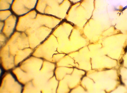
Figure 2: The clarified total preparation. The lymphatic network of the mucosa.
The loops of this network have an elongated shape, and are placed parallel to the muscle bundles of this layer, and transversely to the long axis of the organ. The densest network of lymphatic capillaries was found in the distal half part of the abomasum body, and in the area of the pylorus. The rarest network of lymphatic capillaries in the circular muscle layer was found at the bottom of the abomasum, where this layer was much less developed.
Lymphatic capillaries of the longitudinal muscle layer are located in the connective tissues’ interlayers between bundles of the muscle fibers. Capillaries of this layer are interconnected to form rectangular networks, polygonal and irregular networks are found more rarely. In the abomasum, the densest and the bulkiest network of lymphatic capillaries is found in the pyloric section and in the area of the lesser curvature of the stomach. With age, the size of loops of lymphatic capillaries, and the width of the capillaries increase. Lymphatic capillaries of the longitudinal muscle layer form postcapillaries, which flow into the lymphatic bed of the serous membrane.
Lymphatic capillaries in the intermuscular layer interconnect to form networks of various forms. Lymphatic capillaries of the serous membrane are located in the connective tissues, adhering to the longitudinal muscle layer; by interconnecting, they form in the serous membrane a network of capillaries, the loops of which have an oval or elongated polygonal shape. Longitudinal axes of these loops on the side walls of the abomasum are elongated along its axis.
During the research, we found that the width of the lymphatic capillaries increased in the ontogenesis. The density of the capillary network, the size, and the shape of the capillary cells depend on the type of the tissue and the anatomical part of the studied organ of the abomasum. As a result of the study, it has been found that the mucous and the submucous membranes of the pyloric part and the lesser curvature of the abomasum of sheep have much more tightly spaced lymphatic capillaries than other parts of this organ.
Lymphatic postcapillaries are formed from merging several lymphatic capillaries. Walls of lymphatic postcapillaries, same as in lymphatic capillaries, consist of endothelial cells, however, unlike capillaries, they have valves that determine directed lymph flow. When a lymphatic postcapillary turns into a lymphatic vessel, the wall becomes more complicated: connective tissues and myocytes appear in it.
Lymphatic postcapillaries are contained in the submucous membrane between layers of the muscle coat and the serous coat of abomasum.
In the submucous coat of the abomasum, lymphatic postcapillaries are located between lymphatic capillaries. The number of valves varies from 2 to 5. Lymphatic vessels are formed by merging 2 to 5 lymphatic postcapillaries.
Lymphatic postcapillaries of the muscle coat of abomasum are in the connective tissues between its layers. Here the number of valves reaches 3 to 7, or even 8. Lymph outflow from these plexuses is directed toward the serous membrane to a greater extent, and insignificantly – to the side of the submucous coat (Figure 3).
In the serous membrane, the number of valves in the lymphatic postcapillaries ranges from 5 to 15. Plexuses of lymphatic postcapillaries are located closer to the longitudinal muscle layer; 3–5 postcapillaries merge to form a first order lymphatic vessel of the serous membrane.
Lymphatic postcapillaries in the abomasum differ from lymphatic capillaries by the presence of valves, which are in fact folds of the endothelium. By merging, lymphatic postcapillaries form lymphangions.
A lymphangion is a section of a lymphatic vessel between two valves. Alternating of narrowing and expanding sections gives the lymphatic vessel a peculiar beaded appearance and distinguishes it from blood vessels. In a lymphangion, the valve and the wall are distinguished. Three parts have been identified in the wall of lymphangion: the muscular cuff, the wall of the valve sinus, and the area of valve attachment. The structure of the walls of intraorganic lymphangions in the abomasum of ruminants becomes more complex toward extraorganic vessels. The valves of lymphangion are its key elements that determine the direction of lymphatic drainage from the organ to the thoracic duct and prevent lymph backflow.
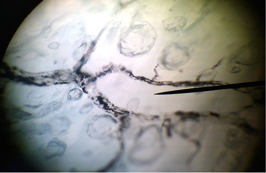
Figure 3: The clarified total preparation. Lymphocapillary net of the longitudinal muscular layer of the abomasum.
The number of lymphangions in a lymphatic vessel varies widely and depends on the type of the lymphatic vessel, its length and the age of the animal.
The presence of smooth muscle cells called myocytes in the wall is a characteristic feature of the lymphatic vessels. The lymphatic vessels in the abomasum of sheep are located in the submucous coat and the serous membrane. On the contours of these vessels, nodes are clearly visible, which correspond to the valves’ locations. The submucous coat contains only lymphatic vessels of the first order, which have been formed by merging of 2–5 capillaries. Anastomosing between themselves, lymphatic vessels form submucous plexuses of lymphatic vessels that are located in deeper layers of the submucous coat than the network of lymphatic capillaries. During the research, we have found that the width of first-order lymphatic vessels in various parts of the submucous coat of abomasum varies.
The largest loops formed by the first order lymphatic vessels were found in the arch of the abomasum, and the smallest ones–in the folds of the submucous coat, in the pyloric part, and on the side walls of abomasum body adjacent to the lesser curvature of the organ. The shape of the loops in the plexuses of first-order lymphatic vessels is most often oval and polygonal. Passing the muscle coat at an angle, first order lymph vessels accept its lymphatic channel and flow into lymphatic vessels of the serous membrane close to the smaller and large curvature of the abomasum.
Lymphatic vessels in the serous membrane were divided into three orders. Vessels of the first order, merging in the quantities from 2 to 5, form second-order lymphatic vessels. In turn, 2–3 second-order lymph vessels form third-order vessels, which reach the small and big curvatures. Next, third-order lymphatic vessels turn to extraorganic vessels (the vessels of the fourth order) that reach the regional lymph nodes.
As a result of studying intraorganic lymphatic vessels of the gastrointestinal tract, including abomasum of small ruminants, it should be noted that abomasum wall, which is thick, rich in glands has an intricate lymphatic system. Formation of intraorganic lymphatic vessels starts in the submucous coat and ends in the serous membrane. We have found that the width of intraorganic lymphatic vessels increases in the proximal direction, and is directly proportional to the age of the animal.
At the same time, some small lymph vessels usually located near the capillaries in networks do not always have clear signs of lymph vessels. Depending on the localization (above or below superficial fascia), lymphatic vessels are divided into superficial and deep ones.
Lymphatic vessels located in the areas with well-developed fatty tissue have a greater number of valves, compared to the vessels in other areas. The valves are intended for ensuring the centripetal direction of lymph flow through a lymphatic vessel and preventing backflow.
Extraorganic afferent lymphatic vessels of the abomasum are formed near the edges of the organ from intraorganic third-order lymphatic vessels in the serous network. Afferent extraorganic lymphatic vessels formed near the lesser curvature follow between the layers of the lesser omentum to the regional lymphatic nodes of the lesser curvature (cardiac part, midpart, and pyloric part), as well as the lymph nodes in the omasum and the forestomach. Lymphatic vessels formed near the greater curvature, passing between the layers of the greater omentum, proceed to the lymphatic nodes in the fundal part of the abomasum. Afferent lymphatic vessels follow the course of blood vessels or cross them.
In the cardiac part, 1–2 afferent lymphatic vessels in this part of abomasum are directed to the lymphatic nodes in the forestomach.
From the midpart of the right wall in the region of curvature, 7 to 15 afferent lymphatic vessels flow into the right midpart group of lymphatic vessels.
From the pyloric part of the right wall in the area of lesser curvature, 1–6 afferent lymphatic vessels originate, which are interrupted in the right pyloric group of lymphatic nodes.
From the cardial part of the lesser curvature area of the abomasum, 2–3 afferent lymphatic vessels originate, which join the left-side cardiac group of lymph nodes.
During the research, it has been found that from the median part of the abomasum wall, 1–2 afferent lymphatic vessels in 15% of cases fall into the median right-sided lymphatic nodes.
From the pyloric part of the lesser curvature area, 1–2 afferent vessels originate, which spread to the lymphatic nodes in the duodenum at the beginning of the right gastro-omental, and then the gastro-duodenal arteries.
From the cardiac part of the left wall in the area of large curvature, 1–3 afferent lymphatic vessel originate, which enter the lymphatic nodes in abomasum – forestomach – omasum.
Afferent lymphatic vessels (1–4) in the fundal part of the left wall of abomasum are accompanied by short branches of the left gastro-omental artery and vein, merging together to flow into the fundal lymphatic node.
From the cardial part of the greater curvature area of the abomasum, 1–4 afferent lymphatic vessels originate, which join the left-side cardiac group of lymph nodes. In addition, in 65% of cases, 1–3 lymphatic vessels from this part extend to the cranial nodes in the omasum.
From the middle of the left wall in the area of lesser curvature, 4 to 8 afferent lymphatic vessels originate, which flow into the left median group of lymphatic nodes.
During the research, it has been found that from this part of the abomasum wall, 1–2 afferent lymphatic vessels in 15% of cases fall into the median right-sided lymphatic nodes. From the caudal segment of this part, in 7% of cases, 1–3 afferent vessels extend to the lymph node above the pyloric stomach region.
From the pyloric part of the left wall in the area of lesser curvature, 1–2 afferent lymphatic vessels originate, which extend to the left-hand pyloric group of lymphatic nodes. It has been found that in 15% of cases, 1–2 afferent lymphatic vessels extend to the lymphatic nodes in the duodenum along the flow first in the right gastro-omental, and then the gastro-duodenal arteries. Along with that, in 13% of cases, 1–3 afferent lymphatic vessels depart from this segment and flow into the lymphatic node above the pyloric stomach region.
From the pyloric part of the right wall in the area of the greater curvature of the abomasum, perpendicular to the edge of the organ, 1–2 afferent lymphatic vessels depart, which are interrupted in the lymphatic node below the pyloric stomach region. During the research, we have found that in 45% of cases, 1–2 afferent lymphatic vessels extend to the lymphatic nodes in the duodenum along the flow of the right gastro-omental artery and veins.
Outgoing extraorganic efferent lymphatic vessels are different from afferent lymphatic vessels. The number of efferent lymphatic vessels outgoing from a lymphatic node is several times less than the number of afferent ones.
Efferent lymphatic vessels from the lymphatic nodes of the lesser curvature extend in various directions parallel to the lesser curvature between layers of the omentum accompanying first the right and then the left gastric artery.

