Melamine is a widely used chemical in the industry. There is a paucity of data pertaining to acute and chronic exposure of melamine in humans and animals (Skinner et al., 2010). Melamine (MA) exposure causes reproductive toxicity in males; however, the underlying mechanism is yet unclear (Chang et al., 2018). On the other hand, Moringa oleifera has actively been used in the treatment of certain diseases such as hepatic steatosis, insulin resistance, and cardiovascular disease (Almatrafi et al., 2017). Moringa oleifera encompasses certain plant pigments that possess a powerful antioxidant ability (Maida et al., 2005). It has anti-hypertensive, antioxidant, anti-inflammatory, anti-diabetic, cholesterol-lowering (Sharma et al., 2012) and hepatoprotective activities (Huang et al., 2012). Therefore, our objective was to assess the potential ameliorative effect of Moringa oleifera leaves extract (M/MOLE) on melamine-induced testicular toxicity in rats.
Evaluation of several semen parameters, including sperm count, motility, and abnormality index, is essential for proper assessment of male fertility (Komiya et al., 2013). These parameters reflect any physical or chemical alterations that may affect testicular tissue (Atli et al., 2016). Our data showed that sperm motility and count were significantly decreased in the melamine group than the control. Melamine showed reproductive toxicity against male mice since it damaged testicular tissue structure, induced abnormal formation, and maturation of sperms and lowered semen quality and quantity leading to the enhanced apoptotic activity of spermatogenic cells (Huang et al., 2018). Our data indicated that the decrease in sperm motility and count returned to the normal level in M/MOLE group. These results are consistent with those reported previously (Sadek, 2014), where the author reported significant improvement in testicular toxicity effects upon concurrent administration of chromium and Moringa oleifera extract. The results indicated significantly increased sperm abnormalities in the melamine treated group than the control. However, M/MOLE group significantly improved these abnormalities compared to the melamine group. These results may be attributed to the antioxidant’s rich characteristics of moringa leaves, which might influence the epididymal antioxidant system and improve the spermatogenic process. Following proper treatment with moringa or its isolated phytochemicals, several studies have shown improved levels of several detoxication biomarkers and antioxidant enzymes previously (Ashok and Pari, 2003; Faizi et al., 1994). Another study reported an improved morphology of sperms upon concurrent administration of chromium and Moringa oleifera extract than chromium induced-testicular toxicity group (Sadek, 2014).
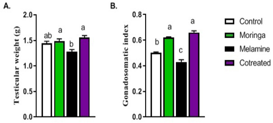
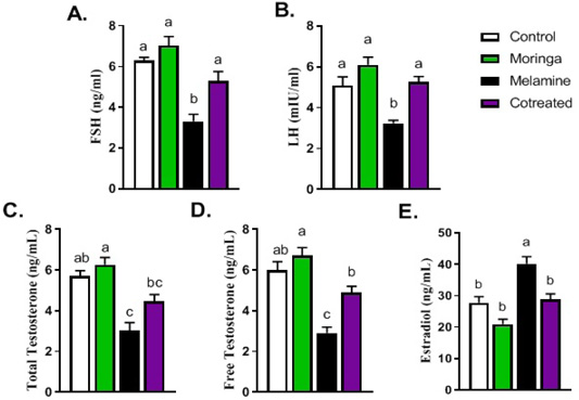
Testicular weight and gonadal-somatic index were significantly decreased in the melamine group than the control. This decrease was significantly increased in M/MOLE group compared to melamine. Testosterone synthesized and produced by testicular interstitial cells is controlled via a feedback mechanism under the hypothalamic-pituitary-gonadal axis control (Chang et al., 2014). Regarding hormonal measurements in our experiment, FSH, LH, free testosterone, and total testosterone were significantly decreased in the melamine group than the control. However, it was found otherwise in M/MOLE group compared to melamine treated group. These results are in agreement with those reported previously (El-Sheikh et al., 2016), who revealed an increase in the level of serum testosterone, LH and FSH in male Wister rats after administration of moringa. Treatment with moringa significantly increased the levels of three sex hormones to the normal values compared to diabetic group (Ebong, 2014). An increased testosterone level in M/MOLE group may be attributed to increasing serum LH levels. LH binds to its receptor in the testicular interstitial cells to improve testosterone synthesis and subsequent elevation in the serum (Huang et al., 2018). On the other hand, upon administration of hexane fraction from the leaves of M. oleifera, (Cajuday and Pocsidio, 2010a) reported an enhancement in testis, epididymis, and seminal vesicle with no alteration in serum FSH and LH levels. Our results indicated a significant increase in estradiol levels in the melamine treated group than the control. However, M/MOLE group revealed otherwise where it was returned to a normal physiological level. These observations are consistent with those reported previously (Zeng et al., 2019), where no change in serum estradiol level was observed in female mice fed with Moringa oleifera leaves.
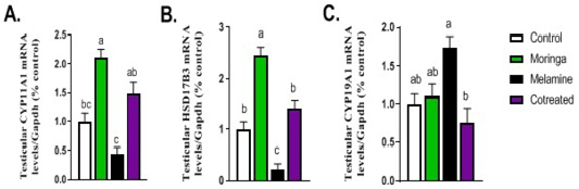
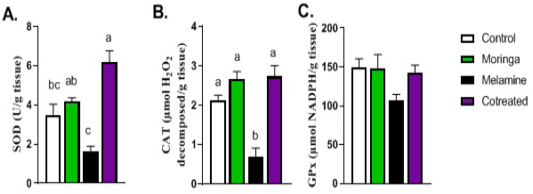
To our knowledge, we are the first who reported the effect of M/MOLE on the expression of mRNAs (CYP11A1, HSD17B3, and CYP19A1). The expression of steroidogenic hormones, including CYP11A1 and HSD17B3 genes, showed a significant decrease in the expression of testicular mRNA in the melamine group than the control. These decreased levels were increased significantly in the M/MOLE group compared to the melamine treated group. This result was further confirmed by an increase in expression of both genes in testicular tissue of male rats that were administered MOLE alone. These results indicate that MOLE may increase the synthesis of steroidogenic hormones in the testis of male rats; however, further studies are needed to ascertain our understanding of the underlying mechanism. The expression of the aromatase gene (CYP19A1) showed a significant increase in the melamine group than the control. This increased expression was decreased significantly in the M/MOLE group compared to the melamine treated group. This indicates the potential role of Moringa oleifera leaves in maintaining the normal level of aromatization of testosterone into estrogen in testis. Regarding antioxidant status in the testis, the levels of SOD and catalase antioxidant enzymes showed a significant decrease in the melamine group than the control. (Huang et al., 2018) reported the reproductive toxicity of melamine in male mice with a significant reduction in sperm quality and antioxidant capacity. Such a decreased antioxidants level increased in M/MOLE group compared to the melamine treated group. Contrary to this, (Zeng et al., 2019) reported that no change in serum superoxide dismutase (SOD) level in mice fed with Moringa oleifera leaves. Low concentrations of cytoplasmic scavenging enzymes in the sperm have been reported where, following moringa administration, elevated levels of the antioxidant enzymatic system can improve the reproductive process (D’Cruz S and Mathur, 2005). No significant alterations in the glutathione peroxidase level were detected between the different experimental groups. These observations are consistent with those reported previously where no change in serum glutathione peroxidase was observed in mice fed with Moringa oleifera leaves (Zeng et al., 2019).
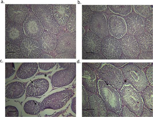
Histopathological examination showed abnormal architecture, structural changes in the seminiferous tubules, and a focal separation of the basement membrane from the overlying disorganized and vacuolized germinal epithelium in the melamine group. Melamine has been reported to impact mouse testicles and induce pathological damage (Crissman et al., 2004). Another study revealed melamine induced reproductive toxicity, abnormal testicular tissue structure, abnormal formation and maturation of sperms, and a lowered semen quality and quantity (Huang et al., 2018). On the other hand, testes of the M/MOLE treated group showed moderate inter-tubular edema and mild disorganized and vacuolized germinal epithelium.