
Ehrlichia canis is an important zoonotic tick-borne pathogen transmitted by the tick Rhipicephalus sanguineus (Latrielle) that causes canine monocytic ehrlichiosis (CME) in dogs. We have investigated the prevalence of R. sanguineus and E. canis infection in pet dogs in Wenzhou, China. Serum samples were obtained from animal hospitals were examined for antibodies by commercial rapid in-clinic ELISA kits. Ticks were collected from dogs and molecular detection methods were used to identify the tick species and CME by amplification of the cytochrome oxidase subunit I and disulfide oxidoreductase gene, respectively. The results indicated that 1.29% of the serum samples were positive for E. canis, and 5.50% of dogs were infested with ticks. The Wenzhou samples of R. sanguineus exhibited a high homology (99.7%–99.8%) and these parasites showed a 99.1%–100% homology to previously reported isolates. The E. canis derived from R. sanguineus in the current study showed a similarity of 98.7%–99.7% to previously published isolates. Our results indicate that precaution should be employed for the potential threat posed by these ticks, especially to pet owners.
Research Article · Volume 53, Issue 6, 12 September 2021 · Open Access
Molecular Identification and Prevalence of Ehrlichia canis and Rhipicephalus sanguineus (Acari: Ixodidae) Infecting Pet Dogs in Wenzhou, China
· · · · · · · ·
Affiliations & Notes ⌄
- 1College of Animal Science, Wenzhou Vocational College of Science and Technology, Wenzhou 325006, People’s Republic of China 2Wuhan Business University, Wuhan 430056, People’s Republic of China 3Jiangxi Animal Disease Prevention and Control Center, Nanchang 330096, People’s Republic of China 4Qilu Animal Health Products Co., LTD., Jinan 250100, People’s Republic of China 5Agriculturral Service Center of Street Office of Datang affiliated to The People’s Government of Zhuji Municipality, Zhuji 311
Article Info ⌄
- Journal:
- Pakistan Journal of Zoology
- Published:
- 12 September 2021

From the article
- The prevalence was 2.80% and 1.41% in ≤ 1 and 1–3-year-old animals, respectively, with no clear difference among the dogs of the different ages (p = 0.539, i.e. > 0.05) (Table I).
- Therefore, it is necessary to monitor tick infections in pet dogs from the Wenzhou region with an average temperature of 17.3–19.4°C and rainfall of 1113–2494 mm. In the current research, it was seen that the prevalence of tick infection in dogs was 5.50%.
- To confirm E. canis infection, we amplified the dsb genes of E. canis from the pooled tick samples; we obtained one E. canis sequence, which had a high similarity of 98.7%–99.7% to previously reported isolates, as revealed by the analysis of pair-wise distance of the COI gene from R. sanguineus (Fig. 2).
- The current study found that R. sanguineus infects pet dogs in Wenzhou, which has a subtropical climate, contributing to the spread of ticks.
Selected from the article's own text.
Abstract
INTRODUCTION
The Ixodidae ticks are obligate hematophagous ectoparasites, which are distributed globally . These ticks are considered the most important vectors of animal or wild lives lives’ pathogens, and the second most important vector (next to mosquitoes) of human pathogens for the transmission of various kinds of tick-borne diseases (TBDs) caused by these pathogens (bacteria, viruses and parasites) ; ; . Emerging and re-emerging TBDs such as spotted fever, Lyme disease, babesiosis, forest encephalitis, Crimean-Congo hemorrhagic fever, anaplasmosis, and severe fever with thrombocytopenia syndrome are ubiquitous and easily disseminated ; . Also pathogenic and non-pathogenic tick-borne bacteria including Babesia spp., Anaplasma spp., Theileria spp. among others have been observed in animals .
Ehrlichia canis can cause an important severe zoonotic TBD named canine monocytic ehrlichiosis (CME) in dogs ; . This gram-negative intracellular bacterium is mainly transmitted by a common tick species named Rhipicephalus sanguineus . The symptoms of CME in dogs are high fever, depression, lethargy, myalgia, anorexia, lymphadenomegaly, anemia, splenomegaly, and thrombocytopenia ; . Human E. canis infection was first isolated from an asymptomatic human in Venezuela in 1996 and detected as clinical ehrlichiosis in Venezuela in 2006 ; . Since then, E. canis has been identified as a causative agent of human ehrlichiosis in Brazil, USA and Costa Rica ; ; .
Previously, 36 tick species from more than 119 tick species were documented and demonstrated to transmit one or more diseases in China ; ; . The prevalence of E. canis infection in dogs was found to be 2.04%–40% in some regions in China . However, until now, there is very little information about the tick species and E. canis infection in pet dogs in Wenzhou (northern latitude: 27°3´-28°36´; east longitude: 119°37´-121°18´), which is situated to the southeast of the Zhejiang province in China. The present study was undertaken to determine the prevalence and molecular identification of Ehrlichia canis and the vector hipicephalus sanguineus in pet dogs in Wenzhou, China.
MATERIALS AND METHODS
Ethics statement
Samples were obtained after due permission from the relevant institutions. All procedures were performed under the instructions and approval of the Laboratory Animals Research Centre of Zhejiang province in China and the Ethics Committee of the Wenzhou Vocational College of Science and Technology.
Serum samples and ticks collection
A total of 309 blood samples were collected from pet dogs (male 173, female 136) from the animal hospitals in Wenzhou, China during 2017–2018. The gender and age information were recorded in detail. Each blood sample was centrifuged at 1000×g for 15 min, and serum was separated and stored at -70°C for further analysis. Ticks were obtained from pet dogs without obvious symptoms visiting the University Veterinary Hospital, and ticks were washed by double distilled water. Then these ticks were identified morphologically by using a light microscope . Ticks from the same dogs were pooled together. All the ticks were stored at -70°C until further testing.
Determination of antibodies against E. canis
Serums from pet dogs were examined for antibodies against E. canis by commercial rapid in-clinic ELISA (enzyme-linked immunosorbent assay) kits (SNAP® 4Dx®, IDEXX Laboratories, Westbrook, Maine, USA), according to the manufacturer’s instructions. Each serum sample (150 µL) was mixed with 200 µL of the testing reagent and loaded into the device detection well.
DNA isolation, amplification and sequencing
The pooled adult ticks were washed thrice using distilled water and then cut into pieces using sterilized scissors, as reported previously . The genomic DNA (gDNA) of ticks was prepared by using a DNA extraction reagent kit (TIANamp Genomic DNA Kit, Tiangen Biotech CO., LTD, Wuhan, China) according to the manufacturer’s instructions. The eluted DNA was kept at -20°C prior to PCR analysis.
The cytochrome oxidase subunit I of the mitochondrial gene of ticks was amplified by utilizing the forward primer pairs LCO1490, 5′-GGTCAACAAATCATAAAGATATTG G-3′ and reverse primer HCO2198, 5′-TAAACTTCAGGGTGACCAAAAAAT CA-3′ . The disulfide oxidoreductase gene of E. canis was amplified by using the forward primer ECAN5, 5′-gatgatgtctgaagatatgaaacaaat-3′ and reverse primer HE3, 5′-ctgctcgtctattttacttcttaaagt-3′) . The PCR mixture contained 10 µl of autoclaved and distilled water, 8.5 µl of PCR buffer (10×), 2.5 µl of dNTPs (2.5 mM), 1 µl of DNA, 1 µl of Taq, 1 µl of each forward and reverse primer (working concentration: 10 µmol/L), to attain a total reaction volume of 25 µl. Each of the 32 PCR cycles consisted of 94°C for 45 s, 48°C for 30 s, and 72°C for 1 min after an initial hot start at 94°C for 3 min, and ended with 72°C for 5 min. The PCR products were analyzed using electrophoresis on a 1.0% agarose gel, followed by staining with ethidium bromide. The PCR electrophoresis products were purified using a Hi-TIANgel Midi Purification Kit (Tiangen Biotech CO., LTD, Beijing, China) according to the manufacturer’s instructions; they were then sent for commercial sequencing by a company (Tsingke, Wuhan, China).
Sequence alignment and phylogenetic analysis
Multiple alignments were performed for the COI and dsb sequences relative to reference sequences available at NCBI (http://blast.ncbi.nlm.nih.gov/Blast.cgi) by DNAMAN (5.2.9 Demo version). The phylogenetic analysis was conducted by the NJ (neighbor-joining) algorithm to determine the ticks and rickettsial species, respectively, using the MEGA (version 6.0) software, and the distances were computed using the Tajima-Nei means. The stability of branches was assessed after bootstrapping with 1000 replicates.
Statistical analysis
Statistical analysis was performed by employing the chi-square test with the IBM SPSS (Statistical Analysis System, Version 20.0.) software. The differences were considered significant for P < 0.05.
RESULTS
In the current study, 1.29% (4/309) serum samples were found to be positive for E. canis (Table I). The seroprevalence was 1.73% and 0.74% in male and female pet dogs, respectively, with no obvious difference in the two genders (p = 0.441, i.e. > 0.05) (Fig. 1). The prevalence was 2.80% and 1.41% in ≤ 1 and 1–3-year-old animals, respectively, with no clear difference among the dogs of the different ages (p = 0.539, i.e. > 0.05) (Table I).
Table I. Prevalence of R. sanguineus and E. canis infection in pet dogs in Wenzhou, China.
| Variable | Category | Seroprevalence (%)/(No. positive/total samples) | |
| R. sanguineus | E. canis | ||
| Gender | Male | 5.20% (9/173) | 1.73% (3/173) |
| Female | 5.88% (8/136) | 0.74%a (1/136) | |
| Age (years) | ≤ 1 | 9.35%d (10/107) | 2.80%b (3/107) |
| > 1 and ≤ 3 | 0 (0/71) | 1.41% (1/71) | |
| > 3 and ≤ 5 | 0 (0/29) | 0 (0/29) | |
| < 5 | 6.86%e (7/102) | 0c | |
| Total | 5.50% (17/309) | 1.29% (4/309) | |
aDifference between the prevalence of R. sanguineus and E. canis infection in female dogs was found to be statistically significant (p= 0.018, i.e. < 0.05, χ2= 5.631); bA significant difference between the prevalence of R. sanguineus and E. canis infection in dogs with an age ≤ 1 year was observed (p= 0.045, i.e. < 0.05, χ2= 4.013); cA significant difference between the prevalence of R. sanguineus and E. canis infection in dogs with an age > 5 years was observed (p= 0.007, i.e. < 0.01, χ2= 7.249); dA noteworthy difference was observed between the prevalence of R. sanguineus infection in dogs with an age ≤ 1 year and those with an age > 1 year and ≤ 3 years (p= 0.008, i.e. < 0.01, χ2= 7.030); eA conspicuous difference was observed between the prevalence of R. sanguineus infection in dogs with an age > 5 years and those with an age > 1 year and ≤ 3 years (p= 0.024, i.e. < 0.05, χ2= 5.078).
A total of 17 out of 309 (5.5%) dogs were found to be infected by ticks (Table I). The prevalence was 5.20% and 5.88% in male and female dogs, respectively. The prevalence was 9.35% and 6.86% in dogs ≤ 1 and ≥ 5 years of age, respectively. The prevalence of tick infection was significantly higher in dogs with an age ≤ 1 year (p < 0.01) and < 5 years (p < 0.05) than in dogs with an age > 1 year and ≤ 3 years, respectively (Table I). There were noteworthy differences between the prevalence of R. sanguineus and E. canis infection in female dogs (p < 0.05). Difference between the prevalence of R. sanguineus and E. canis infection in dogs with an age ≤ 1 year (p < 0.05) and > 5 years (p < 0.01) was found to be significant (Table I).
COI genes of 17 pooled tick samples and dsb genes from only one sample were amplified. For testing dogs whose tick pool samples were not analyzed for dsb genes, we utilized the commercial Canine Distemper Virus/Canine parvovirus Antigen Test Kits (Korea); it was found that these dogs were infected with either CDV or CPV. The COI gene and dsb gene sequences were submitted to the GenBank of NCBI with accession numbers of MG969504–MG969508.
The sequence alignment phylogenetic analysis showed that the tick infecting pet dogs in the Wenzhou region was R. sanguineus (Fig. 1). The Wenzhou isolates had a high homology, i.e. a 99.7%–99.8% homology to one other, while the homology of the present isolates to previously reported isolates was 99.1%–100% (Fig. 1). The rickettsia derived from R. sanguineus in the current study were identified as E. canis via phylogenetic analysis (Fig. 2). The Wenzhou E. canis isolate presented a high similarity of 98.7%-99.7% to the previously published isolates (Fig. 2).
The available reference sequences of COI are shown in Figure 1. The reference sequences of dsb are shown in Figure 2.
DISCUSSION
The importance of the potential distribution of the ticks and their species lays a foundation for possible threat prediction and implementation of suitable epidemiologic strategies using DNAMAN (5.2.9 Demo version) . The diversity of ticks is mainly affected by biotic (vegetation, host) and abiotic (climate, temperature) factors . Therefore, it is necessary to monitor tick infections in pet dogs from the Wenzhou region with an average temperature of 17.3–19.4°C and rainfall of 1113–2494 mm. In the current research, it was seen that the prevalence of tick infection in dogs was 5.50%. The molecular characterization of parasites is crucial for understanding the epidemiology of their infections and their control . Previously, genomic regions such as COI, COII, NADH dehydrogenase subunit 1, and the internal transcribed spacers of nuclear ribosomes have been used for phylogenetic studies of different parasitic species , 2017). In the present study, R. sanguineus, with a high homology to previously reported isolates with regards to the COI gene was detected (Fig. 1). This is consistent with results from a previous study, where R. sanguineus was reported in an imported dog .
E. canis causes CME, which severely affects domestic dogs . In the present study, the serum prevalence of E. canis infection in dogs was 1.29%, which is in line with its prevalence in dogs (1.3%) in south-eastern China . However, the current prevalence of infection was notably lower than the prevalence of E. canis infection in dogs from Italy (7.6%), Thailand (9.88%), and Costa Rica (38.2%) ; ; . This difference may be because of the difference in climate, temperature, and host abundance . To confirm E. canis infection, we amplified the dsb genes of E. canis from the pooled tick samples; we obtained one E. canis sequence, which had a high similarity of 98.7%–99.7% to previously reported isolates, as revealed by the analysis of pair-wise distance of the COI gene from R. sanguineus (Fig. 2).
A previous study indicated that dogs in subtropical climates may be at higher risk for infection by R. sanguineus . The current study found that R. sanguineus infects pet dogs in Wenzhou, which has a subtropical climate, contributing to the spread of ticks. From the present results, we can demonstrate that these ticks may pose a threat to public health, as they carry zoonotic TBD pathogens such as E. canis. Therefore, efficient precautions should be taken against the potential threat posed by the ticks, especially to pet owners.
ACKNOWLEDGMENTS
This study was supported by Wenzhou Association for Science and Technology of “industry-university-research” Integration innovation projects in 2008, Wenzhou Zhejiang Province, China (No.201806), and “Supervisor + Project + Team” of Wenzhou Vocational College of Science and Technology in 2020, Zhejiang, China, and Wenzhou Engineering Research Center of Pet (Grant No:WP04).
Statement of conflict of interest
The authors have declared no competing interest.
Figures
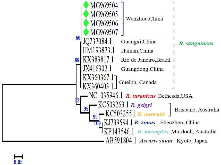
Phylogenetic tree for R. sanguineus based on partial COI gene sequences. Phylogenetic tree based on partial COI gene sequences of R. sanguineus was constructed using the neighbor-joining method with Kimura two-parameter analysis and bootstrap analysis of 1000 replicates. The numbers on the branches indicate the percentage of replicates that reproduced the topology for each clad. The green rhombi indicate the sequences acquired from the current study (Ascaris suum was used as an outgroup).
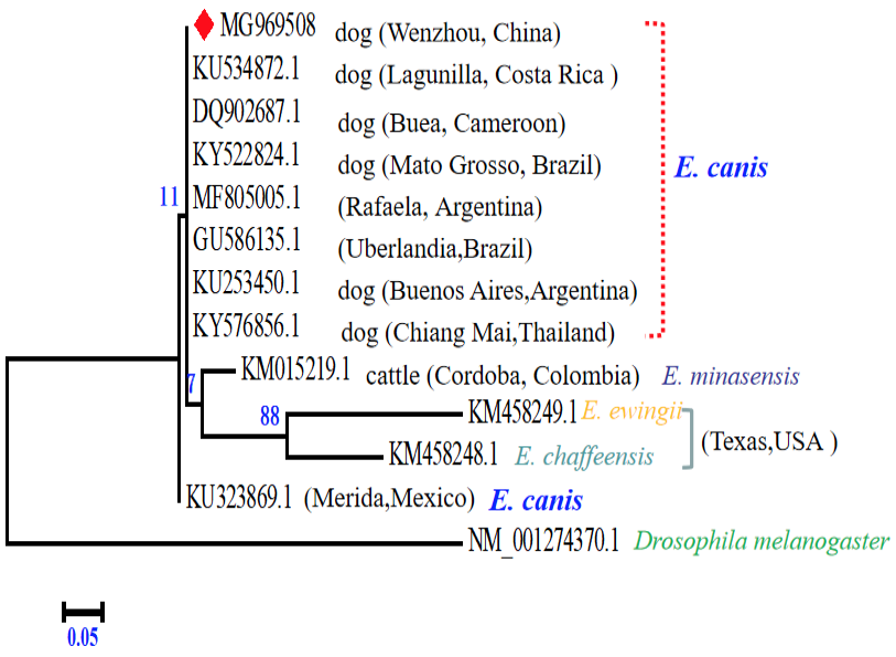
Phylogenetic tree for E. canis based on partial dsb gene sequences. Phylogenetic tree based on partial dsb gene sequences of E. canis was constructed by employing the neighbor-joining method with Kimura two-parameter analysis and bootstrap analysis of 1000 replicates. The numbers on the branches indicate the percentage of replicates that reproduced the topology for each clads. The red rhombi indicate the sequences acquired from the current study (Drosophila melanogaster was used as an outgroup).
References
- Aktas, M. and Ozubek. 2017. Molecular survey of haemoplasmas in shelter dogs and associations with Rhipicephalus sanguineus sensu lato. Med. Vet. Ent., 31: 457-461. https://doi.org/10.1111/mve.12244[CrossRef]
- Benedicto, B., Ceylan, O., Moumouni, P.F.A. Lee, S.H., Tumwebaze, M.A., Li, J., Galon, E.M., Liu, M., Li, Y., Ji, S., Ringo, A., Rizk, M., Sevinc, F. and Xuan, X., 2020. Molecular detection and assessment of risk factors for tick-borne diseases in sheep and goats from Turkey. Acta Parasit., 65: 723-732. https://doi.org/10.2478/s11686-020-00207-0[CrossRef]
- Bouza-Mora, L., Dolza, G., Solórzano-Morales, A., Romero-Zuňiga, J.J., Salazar-Sánchez, L., Labruna, M.B. and Aguiard, D.M. 2017. Novel genotype of Ehrlichia canis detected in samples of human blood bank donors in Costa Rica. Ticks Tick-borne Dis, 8: 36-40. https://doi.org/10.1016/j.ttbdis.2016.09.012[CrossRef]
- Campbell-Lendrum, D., Manga, L., Bagayoko, M. and Sommerfeld, J., 2015. Climate change and vector-borne diseases: what are the implications for public health research and policy? Philos. Trans. R. Soc. Lond. B Biol. Sci., 370: 20130552. https://doi.org/10.1098/rstb.2013.0552[CrossRef]
- Campos-Calderón. L., Ábrego-Sánchez, L., Solórzano-Morales, A., Alberti, A., Tore, G., Zobb, R., Jiménez-Rocha, A.E. and Dolz, G., 2016. Molecular detection and identification of Rickettsiales pathogens in dog ticks from Costa Rica. Ticks Tick-borne Dis, 7: 1198-1202. https://doi.org/10.1016/j.ttbdis.2016.07.015[CrossRef]
- Diniz, P.P., Schwartz, D.S., de Morais, H.S. and Breitschwerdt, E.B. 2007. Surveillance for zoonotic vector-borne infections using sick dogs from southeastern Brazil. Vector Borne Zoon. Dis., 7: 689-697. https://doi.org/10.1089/vbz.2007.0129[CrossRef]
- Escribano, D., Cihan, H., Martínez-Subiela, S., Levent, P., Kocaturk, M., Aytug, N., Cerón, J.J., Tvarijonaviciute, A. and Yilmaz, Z., 2017. Changes in serum proteins in dogs with Ehrlichia canis infection. Microb. Pathog., 113: 34-39. https://doi.org/10.1016/j.micpath.2017.10.024[CrossRef]
- Kaewmongkol, G., Lukkana, N., Yangtara, S., Kaewmongkol, S., Thengchaisri, N., Sirinarumitr, T., Jittapalapong, S. and Fenwick, S.G., 2017. Association of Ehrlichia canis, Hemotropic Mycoplasma spp. and Anaplasma platys and severe anemia in dogs in Thailand. Vet. Microbiol., 201: 195-200. https://doi.org/10.1016/j.vetmic.2017.01.022[CrossRef]
- Licari, E., Takács, N., Solymosi, N. and Farkas R., 2017. First detection of tick-borne pathogens of dogs from Malta. Ticks Tick-borne Dis., 8: 396-399. https://doi.org/10.1016/j.ttbdis.2017.01.002[CrossRef]
- Li, K., Lan, Y.F., Luo, H.Q., Zhang, H., Liu, D.Y., Zhang, L.H., Gui, R., Wang, L., Shahzad, M., Sizhu, S.Z. Li, J.K. and Chamba, Y.M., 2016. Prevalence, associated risk factors, and phylogenetic analysis of Toxocara vitulorum infection in yaks on the Qinghai Tibetan Plateau, China. Korean J. Parasitol., 54: 645-652. https://doi.org/10.3347/kjp.2016.54.5.645[CrossRef]
- Li, K., Luo, H.Q., Zhang, H., Lan, Y.F., Han, Z.Q., Shahzad, M., Wang, X.Q., Qiu, G., Huang, S.C., Jiang, W.T. and Li, J.K., 2016. First report of Metastrongylus pudendotectus by the genetic characterization of mitochondria genome of cox1 in pigs from Tibet, China. Vet. Parasitol., 223: 91-95. https://doi.org/10.1016/j.vetpar.2016.04.036[CrossRef]
- Li, K., Lan, Y.F., Luo, H.Q., Shahzad, M., Zhang, H., Wang, L., Zhang, L.H., Liu, D.Y., Liu, X.Y., Hao, Y.N., Sizhu, S.L. and Li, J.K., 2017. Prevalence of three Oesophagostomum spp. from Tibetan Pigs analyzed by genetic markers of nad1, cox3 and ITS1. Acta Parasitol., 62: 90-96. https://doi.org/10.1515/ap-2017-0010[CrossRef]
- Lu, M., Tian, J.H., Yu, B., Guo, W.P., Holmes, E.C. and Zhang, Y.Z., 2017. Extensive diversity of rickettsiales bacteria in ticks from Wuhan, China. Ticks Tick-borne Dis., 8: 574-580. https://doi.org/10.1016/j.ttbdis.2017.03.006[CrossRef]
- Malheirosa, J., Costa, M.M., do Amaral, R.B., de Sousa, K.C.M., André, M.R., Machado, R.Z. and Vieira, M.I.B., 2016. Identification of vector-borne pathogens in dogs and cats from Southern Brazil. Ticks Tick-borne Dis., 7: 893-900. https://doi.org/10.1016/j.ttbdis.2016.04.007[CrossRef]
- Mircean, V., Dumitrache, M.O., Gyorke, A., Pantchev, N., Jodies, R., Mihalca, A.D. and Cozma V., 2012. Seroprevalence and geographic distribution of Dirofilaria immitis and tick-borne infections (Anaplasma phagocytophilum, Borrelia burgdorfei sensu lato and Ehrlichia canis) in dogs from Romania. Vector Borne Zoon. Dis., 7: 595-604. https://doi.org/10.1089/vbz.2011.0915[CrossRef]
- Montenegro, V.M., Bonilla, M.C., Kaminsky, D., Romero-Zúňiga, J.J., Siebert, S. and Krämer, F., 2017. Serological detection of antibodies to Anaplasma spp., Borrelia burgdorferi sensu lato and Ehrlichia canis and of Dirofilaria immitis antigen in dogs from Costa Rica. Vet. Parasitol., 236: 97-107. https://doi.org/10.1016/j.vetpar.2017.02.009[CrossRef]
- Nicholson, W.L., Allen, K.E., Mc Quiston, J.H., Breitschwerdt, E.B. and Little, S.E., 2010. The increasing recognition of rickettsial pathogens in dogs and people. Trends Parasitol., 26: 205-212. https://doi.org/10.1016/j.pt.2010.01.007[CrossRef]
- Papa, A., Tsioka, K., Kontana, A., Papadopoulos, C. and Giadinis, N., 2017. Bacterial pathogens and endosymbionts in ticks. Ticks Tick-borne Dis., 8: 31-35. https://doi.org/10.1016/j.ttbdis.2016.09.011[CrossRef]
- Pére, M., Bador, M., Zhang, C., Xiong, Q. and Rikihisa, Y., 2006. Human infection with Ehrlichia canis accompanied by clinical signs in Venezuela. Annls. N. Y. Acad. Sci., 1078: 110-117. https://doi.org/10.1196/annals.1374.016[CrossRef]
- Perez, M., Rikihisa, Y. and Wen, B., 1996. Ehrlichia canis-like agent isolated from a manin Venezuela: Antigenic and genetic characterization. J. clin. Microbiol., 34: 2133-2139. https://doi.org/10.1128/JCM.34.9.2133-2139.1996[CrossRef]
- Piantedosi, D., Neola, B., D’Alessio, N., Prisco, F.D., Santoro, M., Pacifico, L., Sgroi, G., Auletta, L., Buch, J., Chandrashekar, R., Breitschwerdt, E.B. and Veneziano, V., 2017. Seroprevalence and risk factors associated with Ehrlichia canis, Anaplasma spp., Borrelia burgdorferi sensu lato, and D. immitis in hunting dogs from southern Italy. Parasitol. Res, 116: 2651-2660. https://doi.org/10.1007/s00436-017-5574-z[CrossRef]
- Wright, I., Wildgoose, W.H., Wall, R. and McGarry, J. 2018. Rhipicephalus sanguineus in an imported dog. Vet. Rec., 182: 25-26. https://doi.org/10.1136/vr.k3[CrossRef]
- Yu, Z., Wang, H., Wang, T., Sun, W., Yang, X. and Liu, J., 2015. Tick-borne pathogens and the vector potential of ticks in China. Parasit. Vector, 14: 24. https://doi.org/10.1186/s13071-014-0628-x[CrossRef]
- Zhao, Li., Lv, J.Z., Li, F., Li, K.R., He, B., Zhang, L.Y., Han, X.Q., Wang, H.Y., Johnson, N., Lin, X.M., Wu, S.Q. and Liu, Y.H., 2020. Identification and molecular analysis of ixodid ticks (Acari: Ixodidae) infesting domestic animals and tick-borne pathogens at the Tarim Basin of Southern Xinjiang, China. Korean J. Parasitol., 58: 37-46. https://doi.org/10.3347/kjp.2020.58.1.37[CrossRef]
- Zhang, R.L., Zhao, A.H., Wang, X.J. and Zhang, Z., 2017. Diversity of tick species on domestic animals in Shandong Province, China, using DNA barcoding. Exp. appl. Acarol., 73: 79-89. https://doi.org/10.1007/s10493-017-0161-7[CrossRef]
Services
Article usage statistics
Loading…