
Avian Hepatitis E Virus (aHEV) is a single-stranded positive-sense RNA virus causing hepatitis-splenomegaly syndrome (HSS) in chickens. To the best of our knowledge the circulation of aHEV in chicken has not been studied in Pakistan so far. Therefore, in the present study, we aimed to isolate and identify aHEV from layer chickens of Pakistan. The bile fluids, liver and spleen tissues were collected from overnight dead layer chickens (n = 8) from Pattoki region of Punjab province, Pakistan, during July to August 2016. The RT-PCR showed the amplifications of selected regions of helicase (186 bps, 2769-2954 nt) and capsid (280 bps, 5461-5741 nt) domain in the bile fluids of two birds. The histological data demonstrated pathological changes in liver and spleen tissues of layer chickens positive for viral RNA. The extensive phylogenetic analysis on the basis of partial helicase domain (ORF1) and capsid protein (ORF2) revealed clustering of Pakistani aHEV (Pak aHEV) strains with members of Orthohepevirus B species. However, Pak aHEV strains were highly divergent from other known members within Orthohepevirus B suggesting as novel aHEV strains circulating in the population of layer chickens in the country. Detection of HEV in layer chickens may pose public health risk in context of zoonosis and food borne transmission if aHEV emerges as zoonotic HEV.
Research Article · Volume 51, Issue 6, 14 September 2019 · Open Access
Molecular Detection of Hepatitis E Virus in Layer Chickens: A Possible Public Health Risk in Pakistan
· · · · ·
Affiliations & Notes ⌄
- 1Department of Biochemistry and Biotechnology, University of Gujrat, Gujrat (50700), Pakistan 2School of Biological Sciences, University of the Punjab, Lahore (54000), Pakistan 3Department of Zoology, University of Gujrat, Gujrat (50700), Pakistan 4Hazara University, Mansehra, Khyber Pakhtunkhwa, Pakistan , * Corresponding author: umer.rashid@uog.edu.pk
Article Info ⌄
- Journal:
- Pakistan Journal of Zoology
- Published:
- 14 September 2019

From the article
- Molecular identification through phylogenetic analysis demonstrated Pakistani strains as member of Orthohepevirus B species but distantly divergent as novel aHEV strains as compared to other aHEV genotypes.
- Moreover, Pakistani aHEV strains showed diverged clustering as compared to other aHEV genotypes and they may have different cross-species infection behavior.
- The data presented in this study suggested that aHEV strains reported in this study are novel strains.
- Furthermore, comparison with each other showed close sequence homology which further suggests that single aHEV is circulating within the layer chickens’ population in Pattoki region of Punjab Province, Pakistan.
Selected from the article's own text.
Abstract
INTRODUCTION
Hepatitis E virus (HEV) is a positive-sense, single-stranded RNA virus which causes self-limiting infection. However, HEV infection in individuals with weakened immune system is linked with developing chronic hepatitis having higher death rates . The HEV clinical symptoms include nausea, fever and followed by anorexia, vomiting, abdominal pain, jaundice and hepatitis . The HEV mortality rate ranges from 0.5 % - 4.0 % in overall population but it could reach up to 30 % in pregnant women particularly in third trimester . The water-borne transmission of HEV causing repeated outbreaks of HEV genotype (gt) 1 and 2 is considered as emerging public health risk, especially in Asia and Africa . The estimated HEV infections caused by gt-1 and 2 in 2005 were almost 20.1 million, out of which 3.4 million were found symptomatic with 70,000 fatalities while 3000 still births were recorded . The outbreaks burden of HEV is relatively higher in Asian countries including Pakistan, as compare to rest of the world. In Pakistan, so far, four major outbreaks were reported where prevalence rate ranged from 10.4 % to 20 % ; . Some small outbreaks were also reported times to times .
The HEV belongs to Hepeviridae family and members of which, on the basis of host range and phylogeny, are classified into two genotypes; Orthohepevirus and Piscihepevirus . Orthohepevirus reported to contain four species (A-D) while Piscihepevirus consists of single species only (A). The HEV infecting human, deer, wild boar, pig and rabbit were placed in Orthohepevirus A and those isolated from bats were placed in Orthohepevirus D. Orthohepevirus B contains HEV isolated from chicken (layer and broiler breeders) while Orthohepevirus C consists of HEV identified in ferret and rats .
The human HEV genome size is approximately 7.2 kb which encode three open reading frames (ORFs). The ORF1 encodes non-structural polyprotein having different functional domains like, methyl transferase, Y, protease, hypervariable region, X, helicase and RNA polymerase (RNA dependent). Whereas, ORF2 and ORF3 code for capsid protein and a multifunctional protein, respectively. In most of the strains, ORF2 and ORF3 are overlapped with each other but separated from ORF1 through intergenic junction region . On the other hand, the aHEV genome is bit short (6.6 kb) but contains components and protein products same as other HEV strains mentioned above. Almost 600 bp putative short region, as compare to other HEVs, is located between methyl transferase and helicase domain in ORF1 .
Identification of many animals HEV strains suggests cross species infection which poses a big public health challenge in term of zoonosis and food borne transmission. So far, only genotype 3 and 4 are reported to be zoonotic which putatively caused infection in human through food borne transmission ; . The cross-species infection of aHEV is reported within birds but no attempt has been successful in describing infection in non-human primates or other mammals ; . So, the trend of cross-species infection of aHEV within birds may be an indication of zoonosis in future. Moreover, the risk of food borne transmission cannot be underestimated as chicken based food stuff, especially the semi-cooked or under-cooked, may play important role in this regard. To the best of our knowledge no data about the circulation of aHEV is reported from Pakistan till date, so the present study was designed to isolate and characterize aHEV strain circulating in layer chickens from Pakistan.
MATERIALS AND METHODS
Samples collection and processing
The bile fluid, liver and spleen tissue samples were collected from overnight dead layer chickens (n = 8) through necropsy from Pattoki region of Punjab province, Pakistan. The chicken specimens were designated as PT7, PT9, PT10, PT11, PT12, PT13, PT14 and PT16. Similarly, bile samples obtained were designated as PT7B, PT9B, PT10B, PT11B, PT12B, PT13B, PT14B and PT16B. Notably, no outbreak was reported in the area during sampling. The tissues (liver and spleen) were fixed in 10 % formalin for histological studies while bile fluid samples collected in sterilized 1.5 ml tubes were stored at -80°C till future analyses.
Tissue samples processing and histological examination
For histological examination, formalin fixed paraffin embedded block of liver and spleen tissues were made. Briefly, formalin-fixed tissues were cut into cubical shape and dipped into different percentages of ethanol for 16 hours followed by xylene treatment. The tissues were then embedded in paraffin wax at 58-60°C and placed into embedding cassettes. The paraffin embedded tissues were sectioned through microtome and proceeded for hematoxylin and eosin (H&E) staining according to the described procedure .
Viral genome detection
Total RNA was isolated from bile samples by using Trizol reagent (Invitrogen). The RNA was converted into cDNA through Reverse Transcription PCR (RT-PCR) using VILO 2X master mix kit (Invitrogen), according to the protocol described previously . The described method was used for viral genome detection in bile samples through amplification of partial helicase (ORF1) 186 bps and partial capsid protein (ORF2) 280 bps fragments using PerfeCta SYBR Green Fast Mix 2X kit (Quanta Biosciences). The PCR strategy was modified to two steps-PCR and amplification was carried at; initial denaturation 95 °C for 2 min after which 40 cycles of 95 °C for 30 secs, 50 °C for 30 secs, 72 °C for 1 min and final extension 72 °C for 10 min.
Sequencing and phylogenetic analysis
The PCR products were purified and sequenced through Sanger method using dideoxynucleotides (Big dye Invitrogen) while obtained sequences were processed and compiled through DNASTAR (Lasergene). Phylogenetic analysis was performed through Molecular Evolutionary Genetics Analysis version 6 (MEGA6) using Neighbor-Joining method with 1000 bootstrapping replicates . The obtained sequences were submitted in Gen Bank. The accession numbers of sequences used in this study are given in Fig. 3 and 4.
RESULTS
Molecular detection of aHEV in bile samples
The bile samples of total eight specimens were screened for aHEV RNA and two samples (PT12B and PT16B) were found positive for aHEV RNA. The 186 bps (2769-2954 nt, reference AM943647) from helicase domain (ORF1) and 280 bps (5461-5741 nt) from capsid protein (ORF2) were amplified from bile fluid of infected layer chickens (PT12 and PT16) (Fig. 1) while complete ORF3 of these two aHEV isolates was amplified in our earlier study .
Histological analysis of aHEV positive liver and spleen tissues
Histological analyses of infected/viral genome (RNA) positive layer chickens (PT12, PT16) revealed lymphocytic infiltration causing portal phlebitis and periphlebitis in liver tissue (Fig. 2A). Additionally, granulocytes and multifocal necrosis was also observed in infected liver tissue (Fig. 2B). Similarly, spleen tissues demonstrated infiltration of monocytes, granulocytes, basophils and other large cells (Fig. 2C and 2D).
Identification and phylogenetic analysis
The obtained PCR products (ORF1=186 bps and ORF2=280 bps) were sequenced and sequences were submitted to Gen Bank database with following accession numbers; helicase domain (ORF1) MH243320, MH243321 and capsid protein (ORF2) MG692744, MH094852. These sequences were further subjected to the phylogenetic analysis. Our results revealed that on the basis of partial nucleotides sequence of both helicase and capsid proteins, Pak aHEV strains made monophyletic clade with Orthohepevirus B species of Orthohepevirus genus upon comparison with Orthohepevirus A species (HEV members from mammals including human), Orthohepevirus B species (HEV strains from chickens), Orthohepevirus C species (HEV only from Rats), Orthohepevirus D species (HEV specifically from Bats) and Piscihepevirus (HEV from cutthroat trout) (Fig. 3 and 4). On other hand, the results of this study manifested that Pak aHEV strains showed distant clustering within Orthohepevirus B species as compared to other member strains belonging to gt1, gt2, gt3 and gt4 which is evident from low bootstrap values on the branch point within monophyletic clade of Orthohepevirus B species showing that Pak aHEV strains may represent a novel genotype. Interestingly, phylogenetic analysis of Pak HEV strains with each other showed 100% bootsrap value for helicase domain and 99% for capsid protein gene showing that same HEV is circulating in layer chickens’ population in the area.
DISCUSSION
The infectivity of HEV in human and other animal species poses a concern of cross species infection which further extends to zoonosis. There are many factors associated to virus itself, potential host and environment which may promote its transmission and infection (Sooranarain and Meng, 2019). Human HEV gt1 and 2, infecting only human, are endemic to developing countries and their transmission is associated with sanitary contaminated drinking water through oral-fecal route . On the other hand, gt3 and 4 are zoonotic and have been reported from industrialized and developing countries as well and commonly follow foodborne transmission route The main foodborne transmission of gt3 and 4 is associated with consumption of contaminated animal meats like pig and deer ; . Pig has been reported as a reservoir of HEV multi-genotypes including gt3, 4, 5, and 6 while gt7, gt8 primarily infect camel and these genotypes possibly cause infection in human too ; . Recently cow has been identified as another potential reservoir for HEV (gt4) in China . Similarly, rabbit HEV is also known to cause infection in human ; . In this scenario the handler and people in close contact with these animals are at high risk of HEV infection which is evident by high rate of anti-HEV seropositivity among these peoples from different countries . In this context the characterization and monitoring of HEV animal strains is needed to understand HEV epidemiology.
Little is known about the pathogenesis and replication of HEV due to the lack of a cell culture system and a practical animal model for the propagation of this virus. In the present study, we selected layer’s flock with a history of signs and symptoms associated with aHEV that mainly include reduced egg production and HSS ; . We cannot say with surety that selected birds died because of aHEV infection. The reason of death in selected birds could be the HEV or any other infection sharing the signs of hepatitis, peritonitis and airsacculitis, for instance, Fasciola hepatica or Escherichia coli. Presence of virus in few (2 out of 8) birds itself is an indication that HEV was not the sole reason behind death of birds. In fact, HEV infection is an essential but not only the one factor for the development of HS syndrome in poultry .
Molecular identification through phylogenetic analysis demonstrated Pakistani strains as member of Orthohepevirus B species but distantly divergent as novel aHEV strains as compared to other aHEV genotypes. The molecular identification of aHEV in layer chickens in this research work seemed to be sporadic infection as no outbreak was reported in the area. So far, global distribution of aHEV is reported from United States of America (USA), Australia, China, Hungary, South Korea and Taiwan and this data may be the first report from Pakistan. The association of aHEV infection with histopathalogical lesions in liver and spleen tissue causing hepatitis-splenomegaly syndrome (HSS) in layer chickens and broiler breeders has been reported in different parts of the world ; which are evident in the present study and same have been reported in many animal species as well .
Phylogenetic analysis has revealed that avian HEV is genetically related to, but distinct from, other known HEV strains . It has similar genomic organization and share 60% nucleotide sequence with human HEV viruses . Likewise, the target organ (liver) and receptors (Heparan Sulfate) used by aHEV and human HEV are same . Our data suggests that Pakistani aHEV described in this study is a novel strain because of its clustering in a separate clade upon comparison with previously known genotypes. Further studies will be required in order to describe pathogenic and zoonotic potential of this newly described strain.
Zoonosis of HEV is a big public health risk not only in developing countries but also in developed world. Foodborne transmission of zoonotic HEV gt3 and gt4 in developed countries is well established. In this regard under-cooked or un-cooked pork liver sausages, wild boar and shellfish have been recognized as potential risk factor . Poultry industry is well established all over the world and full fills a large portion of human food needs. The poor hygiene of poultry farms and substandard poultry wastes disposal play very important role in aHEV transmission among chicken flocks . However, aHEV has not been yet reported as zoonotic HEV but its cross-species infection within birds and sharing of conserved antigenic epitopes in capsid protein gene with swine HEV may be an indication of zoonosis in future as virus evolve ; . Having this assumption, the foodborne transmission of aHEV through under-cooked or un-cooked chicken meat and other derived products may emerge as a potential public health risk in future. Moreover, Pakistani aHEV strains showed diverged clustering as compared to other aHEV genotypes and they may have different cross-species infection behavior. So, further investigations are needed on zoonosis and cross-species infection in this regard. Keeping in view the above scenario, detection of aHEV in layer chickens in Pakistan may pose a potential public health challenge.
CONCLUSION
The data presented in this study suggested that aHEV strains reported in this study are novel strains. Furthermore, comparison with each other showed close sequence homology which further suggests that single aHEV is circulating within the layer chickens’ population in Pattoki region of Punjab Province, Pakistan.
ACKNOWLEDGEMENT
This research work was partially funded by Higher Education Commission (HEC) Pakistan under International Research Support Initiative Program (IRSIP). The positive aHEV control samples were kindly provided by X. J. Meng, Center for Molecular Medicine and Infectious Diseases, Virginia Tech VA, USA.
Statement of conflict of interest
All authors declared that no conflict of interest.
Figures
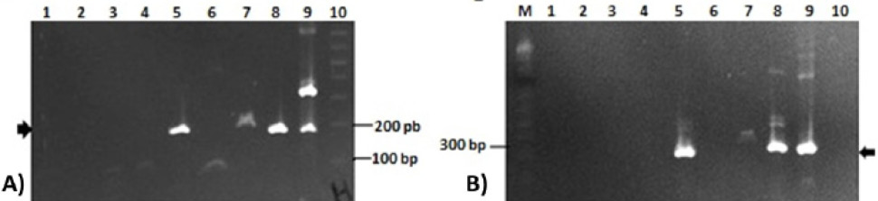
2% agarose gel showing amplifications of ORF1 and ORF 2 of aHEV genome in bile samples. (A) Helicase (ORF1) amplification (186 bp, arrow); Lane 1: PT7B; lane: PT9B; lane PT10B; lane 4: PT11B; lane 5: PT12B; lane 6: PT13B; lane 7: PT14B; lane 8: PT16B; lane 9: positive control; lane 10: 1 kb plus DNA marker; (B) ORF2 amplification (280 bp, arrow); Lane 1: PT7B; lane 2: PT9B; lane PT10B; lane 4: PT11B; lane 5: PT12B; lane 6: PT13B; lane 7: PT14B; lane 8: PT16B; lane 9: positive control; lane 10: negative control; M: 1 kb plus DNA marker.
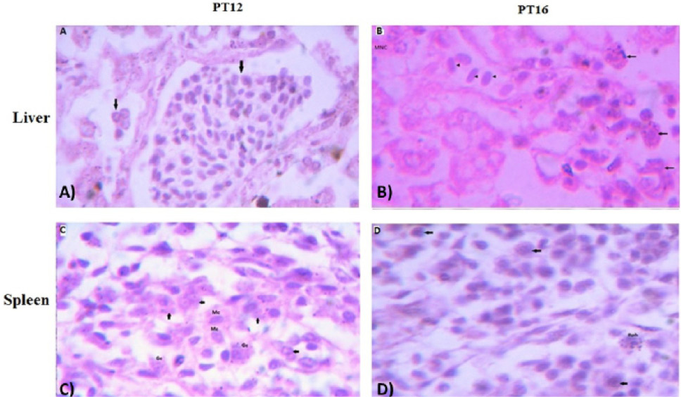
The histological examination of liver (upper panel) and spleen (lower panel) tissues of aHEV positive layer chickens PT12 and PT16; (A) PT12 liver histology showing lymphocytic infiltration in liver tissues causing portal phlebitis and periphlebitis (arrows) ; (B) PT16 liver histology with lymphocytic infiltration (arrow heads) ; granulocytes (arrows) and multifocal necrosis (MNC); (C) PT12 spleen histology showing infiltration of monocytes (Mc); granulocytes (arrows); (D) PT16 spleen histology showing infiltration of basophils (Bph) and other large cells (arrows). The images were observed under 100X magnifications.
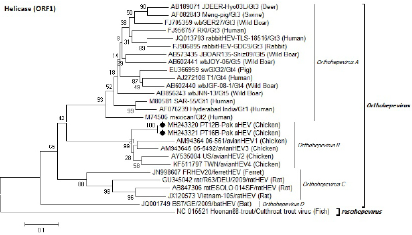
Identification of Pak aHEV strains (♦) on the basis of partial helicase domain (ORF1) nucleotides sequence. Neighbor-Joining method with 1000 bootstrapping replicates was used. GenBank accession number isolate/genotype (host) is shown for each member.
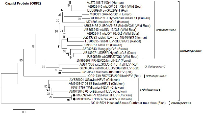
Identification of Pak aHEV strains (♦) on the basis of partial capsid protein (ORF2) nucleotides sequence. Neighbor-Joining method with 1000 bootstrapping replicates was used. GenBank accession number isolate/genotype (host) is shown for each member.
References
- Abravanel, F., Lhomme, S., El Costa, H., Schvartz, B., Peron, J.M., Kamar, N. and Izopet, J., 2017. Rabbit hepatitis E virus infections in humans, France. Emerg. Infect. Dis., 23: 1191-1193. https://doi.org/10.3201/eid2307.170318[CrossRef]
- Billam, P., Huang, F.F., Sun, Z.F., Pierson, F.W., Duncan, R.B., Elvinger, F., Guenette, D.K., Toth, T.E. and Meng, X.J., 2005. Systematic pathogenesis and replication of avian hepatitis E virus in specific-pathogen-free adult chickens. J. Virol., 79: 3429-37. https://doi.org/10.1128/JVI.79.6.3429-3437.2005[CrossRef]
- Capai, L., Charrel, R. and Falchi, A., 2018. Hepatitis E in high-income countries: What do we know? And what are the knowledge gaps? Viruses, 10: 285. https://doi.org/10.3390/v10060285[CrossRef]
- Cardiff, R.D., Miller, C.H. and Munn, R.J., 2014. Manual hematoxylin and eosin staining of mouse tissue sections. Cold Spring Harbor Protocol. https://doi.org/10.1101/pdb.prot073411[CrossRef]
- Clemente-Casares, P., Ramos-Romero, C., Ramirez-Gonzalez, E. and Mas, A., 2016. Hepatitis E virus in industrialized countries: the silent threat. Biomed. Res. Int., 9838041. https://doi.org/10.1155/2016/9838041[CrossRef]
- Crespo, R., Opriessnig, T., Uzal, F. and Gerber, P.F., 2015. Avian hepatitis E virus infection in organic layers. Avian Dis., 59: 388–393. https://doi.org/10.1637/11070-032215-Reg.1[CrossRef]
- Doceul, V., Bagdassarian, E., Demange, A. and Pavio, N., 2016. Zoonotic hepatitis E virus: Classification, animal reservoirs and transmission routes. Viruses, 8: 270. https://doi.org/10.3390/v8100270[CrossRef]
- Hakim, M.S., Wang, W., Bramer, W.M., Geng, J., Huang, F., de Man, R.A., Peppelenbosch, M.P. and Pan, Q., 2017. The global burden of hepatitis E outbreaks: a systematic review. Liver Inter., 37: 19–31. https://doi.org/10.1111/liv.13237[CrossRef]
- Haqshenas, G., Huang, F.F., Fenaux, M., Guenette, D.K., Pierson, F.W., Larsen, C.T., Shivaprasad, H.L., Toth, T.E. and Meng, X.J., 2002. The putative capsid protein of the newly identified avian hepatitis E virus shares antigenic epitopes with that of swine and human hepatitis E viruses and chicken big liver and spleen disease virus. J. Gen. Virol., 83: 2201–2209. https://doi.org/10.1099/0022-1317-83-9-2201[CrossRef]
- Haqshenas, G., Shivaprasad, H.L., Woolcock, P.R., Read, D.H. and Meng, X.J., 2001. Genetic identification and characterization of a novel virus related to human hepatitis E virus from chickens with hepatitis–splenomegaly syndrome in the United States. J. Gen. Virol., 82: 2449–62. https://doi.org/10.1099/0022-1317-82-10-2449[CrossRef]
- Huang, F., Li, Y., Yu, W., Jing, S., Wang, J., Long, F., He, Z., Yang, C., Bi, Y., Cao, W., Liu, C., Hua, X. and Pan, Q., 2016. Excretion of infectious hepatitis E virus into milk in cows imposes high risks of zoonosis. Hepatology, 64: 350–359. https://doi.org/10.1002/hep.28668[CrossRef]
- Huang, F.F., Sun, Z.F., Emerson, S.U., Purcell, R.H., Shivaprasad, H.L., Pierson, F.W., Toth, T.E. and Meng, X.J., 2004. Determination and analysis of the complete genomic sequence of avian hepatitis E virus (avian HEV) and attempts to infect rhesus monkeys with avian HEV. J. Gen. Virol., 85: 1609-1618. https://doi.org/10.1099/vir.0.79841-0[CrossRef]
- Iqbal, M., Ahmed, A., Qamar, A., Dixon, K., Duncan, J.F., Islam, N.U, Rauf, A., Bryan, J.P., Malik, I.A. and Legter, L.J., 1989. An outbreak of enterically transmitted non-A, non-B hepatitis in Pakistan. Am. J. trop. Med. Hyg., 40: 438–443. https://doi.org/10.4269/ajtmh.1989.40.438[CrossRef]
- Iqbal, T., Idrees, M., Ali, L., Hussain, A., Ali, M., Butt, S., Yousaf, M.Z. and Sabar, M.F., 2011. Isolation and characterization of two new hepatitis E virus genotype 1 strains from two mini-outbreaks in Lahore, Pakistan. Virol. J., 8: 94. https://doi.org/10.1186/1743-422X-8-94[CrossRef]
- Iqbal, T., Rashid, U. and Idrees, M., 2018. Structure prediction of ORF3 encoded protein of a novel Pakistani avian hepatitis E virus strain. Biologia (Pakistan), 64: 211-220.
- Kaiser, M., Delaune, D., Chazouilleres, O., Blumel, J., Roque-Afonso, A.M. and Baylis, S.A., 2018. A World Health Organization human hepatitis E virus reference strain related to similar strains isolated from rabbits. Genome Announc., 6: e00292-18. https://doi.org/10.1128/genomeA.00292-18[CrossRef]
- Kalia, M., Chandra, V., Rahman, S.A., Sehgal, D. and Jameel, S., 2009. Heparan sulfate proteoglycans are required for cellular binding of the hepatitis E virus ORF2 capsid protein and for viral infection. J. Virol., 12714–12724. https://doi.org/10.1128/JVI.00717-09[CrossRef]
- Khuroo, M.S., 2011. Discovery of hepatitis E: the epidemic non-A, non- B hepatitis 30 years down the memory lane. Virus Res., 161: 3-14. https://doi.org/10.1016/j.virusres.2011.02.007[CrossRef]
- Kmush, B.L., Nelson, K.E. and Labrique, A.B., 2015. Risk factors for hepatitis E virus infection and disease. Expert. Rev. Anti. Infect. Ther., 13: 41-53. https://doi.org/10.1586/14787210.2015.981158[CrossRef]
- Kwon, H.M., Sung, H.W. and Meng, X.J., 2012. Serological prevalence, genetic identification and characterization of the first strains of avian hepatitis E virus from chickens in Korea. Virus Genes, 45: 237–245. https://doi.org/10.1007/s11262-012-0761-6[CrossRef]
- Liu1, B., Sun, Y., Chen, Y., Du, T., Nan, Y., Wang, X., Li, H., Huang, B., Zhang, G., Zhou, E.M. and Zhao, Q., 2017. Effect of housing arrangement on fecal-oral transmission of avian hepatitis E virus in chicken flocks. BMC Vet. Res., 13: 282. https://doi.org/10.1186/s12917-017-1203-4[CrossRef]
- Mansuy, J.M., Abravanel, F., Miedouge, M., Mengelle, C., Merviel, C., Dubois, M., Kamar, N., Rostaing, L., Alric, L., Moreau, J., Peron, J.M. and Izopet, J., 2009. Acute hepatitis E in south-west France over a 5-year period. J. clin. Virol., 44: 74–77. https://doi.org/10.1016/j.jcv.2008.09.010[CrossRef]
- Meng, X.J., 2013. Zoonotic and foodborne transmission of hepatitis E virus. Semin. Liver. Dis., 33: 41–49. https://doi.org/10.1055/s-0033-1338113[CrossRef]
- Meng, X.J., 2016. Expanding host range and cross-species infection of hepatitis E virus. PLoS Pathog., 12: e1005695. https://doi.org/10.1371/journal.ppat.1005695[CrossRef]
- Meng, X.J., Dea, S., Engle, R.E., Friendship, R., Lyoo, Y.S., Sirinarumitr, T., Urairong, K., Wang, D., Wong, D., Yoo, D., Zhang, Y., Purcell, R.H. and Emerson, S.U., 1999. Prevalence of antibodies to the hepatitis E virus in pigs from countries where hepatitis E is common or is rare in the human population. J. med. Virol., 59: 297-302. https://doi.org/10.1002/(SICI)1096-9071(199911)59:3<297::AID-JMV6>3.0.CO;2-3[CrossRef]
- Moon, H.W., Lee, B.W., Sung, H.W., Yoon, B.I. and Kwon, H.M., 2016. Identification and characterization of avian hepatitis E virus genotype 2 from chickens with hepatitis-splenomegaly syndrome in Korea. Virus Genes, 52:738–742. https://doi.org/10.1007/s11262-016-1351-9[CrossRef]
- Navaneethan, U., Al Mohajer, M. and Shata, M.T., 2008. Hepatitis E and pregnancy: Understanding the pathogenesis. Liver Int., 28: 1190–1199. https://doi.org/10.1111/j.1478-3231.2008.01840.x[CrossRef]
- Park, W.J., Park, B.J., Ahn, H.S., Lee, J.B., Park S.Y., Song, C.S., Sang-Won Lee, S.W., Yoo, H.S. and Choi, I.S., 2016. Hepatitis E virus as an emerging zoonotic pathogen. J. Vet. Sci., 17: 1-11. https://doi.org/10.4142/jvs.2016.17.1.1[CrossRef]
- Purdy, M.A., Harrison, T.J., Jameel, S., Meng, X.J. and Okamoto, H., 2017. ICTV virus taxonomy profile: Hepeviridae. J. Gen. Virol., 98: 2645–2646. https://doi.org/10.1099/jgv.0.000940[CrossRef]
- Rab, M.A., Bile, M.K., Mubarik, M.M., Asghar, H., Sami, Z., Siddiqi, S., Dil, A.S., Barzgar, M.A., Chaudhry, M.A. and Burney, M.I., 1997. Water-borne hepatitis E virus epidemic in Islamabad, Pakistan: a common source outbreak traced to the malfunction of a modern water treatment plant. Am. J. trop. Med. Hyg., 57: 151–157. https://doi.org/10.4269/ajtmh.1997.57.151[CrossRef]
- Rein, D.B., Stevens, G.A., Theaker, J., Wittenborn, J.S. and Wiersma, S.T., 2012. The global burden of hepatitis E virus genotypes 1 and 2 in 2005. Hepatology, 55: 988–997. https://doi.org/10.1002/hep.25505[CrossRef]
- Reuter, G., Boros, A., Mátics, R., Kapusinszky, B., Delwart, E. and Pankovics, P., 2016. A novel avian-like hepatitis E virus in wild aquatic bird, little egret (Egretta garzetta), in Hungary. Infect. Genet. Evol., 46: 74–77. https://doi.org/10.1016/j.meegid.2016.10.026[CrossRef]
- Sooryanarain, H. and Meng, X.J., 2019. Hepatitis E virus: reasons for emergence in humans. Curr. Opin. Virol., 34: 10–17. https://doi.org/10.1016/j.coviro.2018.11.006[CrossRef]
- Sun, Z.F., Larsen, C.T., Huang, F.F., Billam, P., Pierson, F.W., Toth, T.E. and Meng, X.J., 2004. Generation and infectivity titration of an infectious stock of avian hepatitis E virus (HEV) in chickens and cross-species infection of turkeys with avian HEV. J. clin. Microbiol., 42: 2658–2662. https://doi.org/10.1128/JCM.42.6.2658-2662.2004[CrossRef]
- Tamura, K., Nei, M. and Kumar, S., 2004. Prospects for inferring very large phylogenies by using the neighbor-joining method. Proc. natl. Acad. Sci., (USA), 101: 11030-11035. https://doi.org/10.1073/pnas.0404206101[CrossRef]
- Tamura, K., Stecher, G., Peterson, D., Filipski, A. and Kumar, S., 2013. MEGA6: Molecular evolutionary genetics analysis version 6.0. Mol. Biol. Evol., 30: 2725-2729. https://doi.org/10.1093/molbev/mst197[CrossRef]
- Zhao, Q., Liu, B., Sun, Y., Du, T., Chen, Y., Wang, X., Li, H., Nan, Y., Zhang, G. and Zhou, E.M., 2017. Decreased egg production in laying hens associated with infection with genotype 3 avian hepatitis E virus strain from China. Vet. Microbiol., 203: 174-180. https://doi.org/10.1016/j.vetmic.2017.03.005[CrossRef]
Services
Article usage statistics
Loading…