
Mineral supplementation to livestock enhances their health, immunity, and behavior. We aimed to investigate the influence of mineral supplementation on diseased buffalo calves on health, blood parameters, skin, and behavioral repertoire, and to record the association influencing animals before, during and after minerals supplementation. Twenty buffalo calves suffering from skin lesions were enrolled in this study and the diet was supplemented with mineral mixture for 60 days. Skin conditions and behavioral modifications were monitored alongside blood and plasma samples were harvested at days 0, 30, and 60 of the study for hematological and biochemical analysis. Results revealed an overall improvement in animal health, skin conditions, and liver and kidney functions. Significant (P < 0.05) increases in plasma proteins, zinc, and copper were recorded, while malondialdehyde, TNF-α, and MMP-9 significantly (P < 0.05) decreased. Furthermore, buffalo calves showed significant ( P < 0.05) increases in both ingestive and locomotor activities, while grooming and lateral recumbency significantly (P < 0.05) declined. Mineral supplementation had a favorable effect on animal skin, health conditions, and antioxidant activity, as well as it has a minimizing influences on TNF-alpha and MMP-9.
Research Article · Volume 11, Issue 8, 10 July 2023 · Open Access
Mineral Supplementation in Diseased Buffalo Calves and Impact on Health, Behavior, and Clinical Blood Profiles
· ·
Affiliations & Notes ⌄
- 1Department of Animal Medicine, Faculty of Veterinary Medicine, Suez Canal University, Ismailia, 41522, Egypt
- 2Department of Clinical Pathology, Faculty of Veterinary Medicine, Suez Canal University, Ismailia, 41522, Egypt
- 3Department of Animal, Poultry and Fish Behavior and Management, Faculty of Veterinary Medicine, Suez Canal University, Ismailia, 41522, Egypt. *Correspondence | Ibrahim M Hegab, Department of Animal, Poultry and Fish Behavior and Management, Faculty of Veterinary Medicine, Suez Canal University, Ismailia, 41522, Egypt
- 4Email: ibrahim_hegab@vet.suez.edu.eg
Article Info ⌄
- Journal:
- Advances in Animal and Veterinary Sciences
- Published:
- 10 July 2023

From the article
- GOT and GPT activities and creatinine levels were significantly reduced after supplementation with mineral mixture.
- Buffalo calves showed increased locomotor activities and reduced resting behaviors post-supplementation.
- Mineral supplementation of diseased buffalo calves, improved animal health, skin, and behavior.
- Mineral supplementation also modulated creatinine levels, liver enzyme activities, lipid peroxidation, TNF-α, and MMP-9 levels, thus proving to possess antioxidant and anti-inflammatory impacts.
Selected from the article's own text.
Abstract
INTRODUCTION
Improving animal health and production requires careful attention to nutritional status and nutrition composition, which is not only concerned with protein, carbohydrates, and major elements but it is dependent on trace minerals. Trace minerals are essential for growth and development, immunity, and reproduction; they act as a co-factor in the physiological functions of cells, cell metabolism, and the oxidation process .
Zinc is a trace element that must be given periodically to maintain adequate immuno-physiological functions . It regulates more than 300 enzymes and involved in DNA, RNA, and protein synthesis ; which makes it essential for the physiological cellular process. Copper has numerous antioxidant activities; therefore, it plays a crucial role in cell metabolism and development. It is a component of numerous metalloproteins and metalloenzymes such as collagen formation, cytochrome C oxidase (Cox), superoxide dismutase (SOD), and ceruloplasmin (Cp) production. Furthermore, copper plays a role in the regulation of processes such as cellular respiration and carbohydrates and lipids metabolism . Iron is an important component of hemoglobin and myoglobin that are responsible for oxygen transport to cells and muscles and selenium which acts as an antioxidant through involvement in the biosynthesis of glutathione peroxidase to protect cells from lipid peroxidation . Although trace minerals are required in trace amounts in diet according to the nutrition research council NRC any deficiency can negatively influence animal health, growth, and reproduction. Zinc deficiency is reported to affect animal performance and fertility as Engle et al. (1997) found that protein accretion was decreased when Zn supplementation was removed from basal forage for 21 days. Copper deficiency is responsible for unthiftness, depigmented rough hair, and peat scours in calves ,while in lambs copper deficiency can produce a demyelinating syndrome known as swayback or enzootic ataxia . However, skin diseases in livestock animals are attributed to parasitological or fungal agents, but also it is important to refer to the dermatological lesions which may result from nutritional deficiency of some trace elements such as Zn, Cu or vitamin A deficiency.
Therefore, adding mineral mixture to diet has become a basic routine work in preparing animal feed formula not only to improve their health and production but also to avoid several diseases that arise from their deficiencies. The mineral mixture has proven to give a favorable effect on mitigating oxidative stressors and enhancing the anti-oxidative status. We aimed in this study to evaluate the effect of mineral mixture on animals that are suffering from skin lesions and their impact on health conditions, behavioral activities, and blood clinicopathological profiles.
MATERIALS AND METHODS
Animals And Diet
The current study was conducted at the educational farm of the Faculty of Veterinary Medicine, Suez Canal University, Ismailia, Egypt. The experimental animals were housed in free stall barns- open door system-, The study was applied from May to August, so the average environmental temperature was about 34°C, relative humidity about 68%, animals were fed on separate feeding forage and commercially concentrated pellets. Forage (green barseem (Trifolium alexandrinum), sorghum, and wheat straw) were provided ad libitum, while concentrates (corn, soybean meal, rice bran, and corn gluten feed) were offered twice daily at 8:00 and 16:00. Water was available ad libitum. The chemical composition of feedstuff is shown in Table (1). All experimental procedures were approved by the Animal Ethics Committee of the Faculty of Veterinary Medicine, Suez Canal University with approval No. (2023011).
Table 1: Diet biochemical composition.
| Chemical composition of diet | g/kg |
| Dry matter | 920 |
| Organic matter | 865.7 |
| Ash | 63.3 |
| Crude protein | 164 |
| NDF | 102.8 |
NDF= neutral detergent fibers
Study Design And Treatment
Twenty buffalo calves at 3-6 months with an average body weight of 120 kg were enrolled in the current study. Mineral mixture (Star Pharma Co. -5409- Egypt) consisted of 12500 mg Mn, 12500 mg Zn, 12500 mg Iron, 3750 mg Cu, 62.5 mg Se, 125 mg Co, and CaCo3 added until 1 kg. The mineral mixture was thoroughly mixed with animals’ concentrates daily at a rate of 8 g/kg D.M diet. The study was designed to last for 60 days. All animals were exposed to full clinical examination with the dermatological examination, as well as skin scrapes were collected from animals ( twenty samples) . Animals were monitored at D0 (day zero, pre-mineral supplementation), D30 (30 days post-mineral supplementation), and D60 (60 days post-mineral supplementation). Blood samples were harvested at the D0, D30, and D60 from the jugular vein using vacutainer tubes with EDTA (Hebei Xinle Sci& Tech Co, LTD) . The blood samples were divided into two parts one for complete blood count (CBC) and the other part was centrifuged at 1000×g for 15 min at 4°C for plasma separation and kept at -20°C for further analysis.
Hematological Analysis
Automatic cell counter (Methyc 18vet, Orphee, France) was used to determine the red blood cell count (RBCs), hemoglobin concentration (Hb), hematocrit value (Hct), mean corpuscular volume (MCV), mean corpuscular hemoglobin (MCH), mean corpuscular hemoglobin concentration (MCHC), total Leucocyte count (TLC), neutrophils, lymphocytes, monocytes, eosinophils, and platelet counts in a total of 28 samples (10 Samples at D0, 9 at D30 and 9 at D60).
Biochemical Analysis
Total proteins, albumin, creatinine, glutamic-oxaloacetic transaminase (GOT), and glutamic pyruvic transaminase (GPT) were assayed using commercial kits (Biodignostics CO, Giza, Egypt. Zinc levels in plasma were assessed by Spectrum assay kits (The Egyptian Company for biotechnology S.A.E., Cairo, Egypt). Copper level in plasma was assessed by assay kits (SPINREACT CO, Santa Coloma, Spain). Plasma malondialdehyde (MDA) and glutathione- S- transferase (GST) activities were measured by assay kits (Biodignostics CO, Giza, Egypt). Biochemical analyses were performed using Photometer 5010 V5+, Germany. Plasma TNF-α concentration was measured by a bovine TNF-α ELISA kit (Cusabio, USA). Bovine matrix metalloproteinase MMP-9 was measured with an ELISA kit (Wuhan Fine Biotech. Co, Wuhan, China). The optical density of the sample was determined at 450 nm using the ELX800® microplate reader (BioTek Instruments, USA).
Measurement Of Behavioral Activities
We assessed some behavioral activities such as ingestive (feeding and drinking), standing idle, lateral recumbency, grooming, and locomotion behaviors. Behavioral observations were conducted for 60 days; at D0, D30, and D60. Recording sessions were distributed across days starting from 6 am to 5 pm. All testing sessions were video recorded with a digital camera (Sony hxr-mc 2500, Sony, Japan). Scan sampling technique was used at 5 min intervals during the analysis of the recordings and the number of animals that were displayed each behavioral element was recorded. Later, these numbers were expressed as a percentage of individuals in each behavioral category. Each testing session was analyzed using the Behavioral Observation Research Interactive Software (BORIS, v. 2.95, University of Torino, Torino, Italy .
Statistical Analysis
The SPSS 22.0 software (Armonk, NY: IBM Corp. SPSS, Inc., Chicago, IL, USA) was used for all the analysis. One-way Analaysis of Variance (ANOVA) was used to investigate the effect of mineral supplementation on the hematological, biochemical, and behavioral modifications with a single independent variable investigated [The time of supplementation: Before (D0), Mid (D30) and at the end (D60) of supplementation]. The level of significance (α) at which the null hypothesis was rejected was 0.05, and the results were expressed as mean ± standard error. Dimension reduction by Principal Component Analysis (PCA) was applied to the plasma biochemical variables at different time points (D0, D30, and D60) including Zinc (Zn), Protein, Albumin, Creatinine, GOT, GPT, MDA, GST, TNF-alpha, MMP9, and Copper (Cu). Principal component analysis (PCA) was done using the prcomp command in R. PCA biplots were generated using the gg biplot package in R. The PCA output typically consists of five to ten uncorrelated principal components, each explaining a proportion of the total dataset variance. The first principal component typically explains most of the variance, but successive principal components may also detect relevant variance. In the current work, principal components were described in terms of a) proportion of explained variance and b) Eigenvalue (i.e. the principal component standard deviation squared). Only principal components with an Eigenvalue above 1.0 were considered significant. In terms of cumulative variance, the extracted components accounted for at least 70% of the variance of the dataset.
RESULTS
Clinical Findings
At D0, clinical examination revealed that buffalo calves were alert and show a normal act of urination, normal posture, gait, weak appetite, and easy act of defecation. Only two claves showed stunted growth, dullness weakness, and prefered to sit than be active. Skin examination revealed alopecia on the dorsum and lateral abdomen, dull, lusterless hair coat, limited areas of erosions, skin ulceration, thick dry skin surface, hyperkeratosis, loss of pigmentation on different areas of skin, and rusty red with discoloration of hair (Fig.1A). Two weeks post-mineral supplementation, skin pigmentation started to return to normal, and hair grows in areas of alopecia for 2-4 cm long. Skin became elastic and keratinization was regressed in most of the animals except for four calves who showed keratinization in limited areas on the dorsum. At D30, all calves’ skin pigmentation returned to the normal color, hair coat growed in the affected areas and no keratinization was observed (Fig.1B). At D60, the skin was completely covered by glossy shiny hair and animals were mentally alert and with a good appetite (Fig.1C).
Hematological Profile
There were non-significant variations in the erythrocytic counts, hemoglobin concentrations, hematocrit values, total and differential leukocytic counts (Table 2) among all groups throughout the experimental period with a significant (P ≤ 0.05) increment in MCV and a significant (P ≤ 0.05) decrement in MCHC at D30. Platelets were significantly (P ≤ 0.05) decreased at both D30 and D60 when compared with D0.
Table 2: Hemogram of buffalo calves at D0, D30, and D60 post-supplementation with the mineral mixture.
| Hematological parameters | D0 | D30 | D60 | F | P |
| Hb (g/dl) | 11.94±0.47 | 10.91±0.41 | 11.06±0.21 | 2.15 | 0.145 |
| RBCs (×106/µl) | 7.56±0.46 | 7.03±0.39 | 7.22±0.17 | 0.57 | 0.577 |
| Hct (%) | 35.36±1.39 | 35.46±1.19 | 34.37±0.64 | 0.29 | 0.753 |
| MCV (fl) | 47.12±1.19b | 50.78±1.07a | 47.69±1.07ab | 3.13 | 0.068 |
| MCH (pg) | 15.94±0.54 | 15.61±0.26 | 15.33±0.25 | 0.65 | 0.532 |
| MCHC (%) | 33.80±0.61a | 30.76±0.17c | 32.18±0.31b | 13.98 | 0.001 |
| T.L.C. (×103/µl) | 13.13±0.83 | 14.50±0.62 | 14.54±0.86 | 1.07 | 0.363 |
| Neutrophils (×103/µl) | 5.53±0.38 | 6.34±0.37 | 6.07±0.41 | 1.12 | 0.350 |
| Lymphocytes (×103/µl) | 6.04±0.58 | 6.74±0.61 | 6.99±0.62 | 0.67 | 0.524 |
| Monocytes (×103/µl) | 1.53±0.14 | 1.41±0.11 | 1.30±0.06 | 1.15 | 0.340 |
| Eosinophils (×103/µl) | 0.19±0.03 | 0.18±0.02 | 0.17±0.03 | 0.07 | 0.934 |
| Platelets (×103/µl) | 422.43±13.21a | 304.57±10.13b | 384.43±17.27a | 18.86 | 0.001 |
a, b, c different superscripts in the same row indicate significance at P≤0.05
Table 3: Plasma biochemical Profile of buffalo calves at D0, D30, and D60 post-supplementation with the mineral mixture.
| Biochemical parameters | D0 | D30 | D60 | F | P |
| Zn (µg/dl) | 12.43±0.86c | 33.30±1.47a | 23.76±4.27b | 11.8 | 0.001 |
| Cu (µg/dl) | 100.21±2.10c | 107.90±2.42b | 118.96±2.32a | 17.06 | 0.003 |
| Total protein (g/dl) | 6.77±0.15b | 6.95±0.16b | 7.43±0.14a | 5.06 | 0.02 |
| Albumin (g/dl) | 3.77±0.10 | 3.69±0.21 | 3.82±0.08 | 0.21 | 0.82 |
| Creatinine (mg/dl) | 1.49±0.24a | 1.55±0.14a | 0.91±0.07b | 4.23 | 0.03 |
| GOT (unit/ml) | 77.67±7.92a | 59.92±5.29b | 56.37±0.60b | 4.60 | 0.03 |
| GPT (unit/ ml) | 24.21±3.04a | 16.94±1.78b | 18.09±1.13ab | 3.35 | 0.06 |
| MDA (nmol/ml) | 41.82±4.79a | 29.87±2.17b | 24.69±1.86b | 4.77 | 0.006 |
| GST (U/L) | 651.68±40.87 | 757.31±72.70 | 615.13±57.43 | 1.60 | 0.255 |
| TNF-α (ng/ml) | 0.79±0.02a | 0.69±0.01b | 0.63±0.01c | 25.40 | 0.001 |
| MMP-9 (ng/ml) | 1.05±0.03a | 0.95±0.01b | 0.84±0.02c | 20.02 | 0.002 |
a, b, c different superscripts in the same row indicate significance at P≤0.05
Effect Of Mineral Mixture On Blood Biochemistry, Trace Minerals, Antioxidants, Tnf-Αlpha And Mmp-9
A significant (P ≤ 0.05) rise in Zn and Cu plasma levels was recorded at D30 and D60 in correspondence to D0. A significant (P ≤ 0.05) increment in plasma total proteins concentration coupled with a significant (P ≤ 0.05) decrement in plasma creatinine level was recorded at D60. Plasma activities of GPT and GOT, levels of MDA, TNF-α, and MMP-9 showed a significant (P ≤ 0.05) decline at D30 and D60 compared to D0. GST results showed insignificant alteration among all groups, although there is a numerical increase in GST value at D30 (Table 3).
Principal component (PCA) analysis for the plasma concentration of different elements at D0 showed that four principal components (PCs) had eigenvalues >1 and accounted for a considerable proportion (89.9%) of the total variance. In PC1, the concentrations of Cu, TNF-α, MMP9, Albumin, and Zn describe 40.9% of the total variance. The second PC (PC2) is described by protein, GST, GPT, and GOT, with 21.2 % of the total variance. The third PC describes 15.2% of the total variance and is explained by the concentration of creatinine, GOT, GST, Zn, and GPT. Finally, GPT, MDA, and GOT concentrations are described by PC4. Figure 2A shows the scores and loadings plots for the combination between PC1 (40.9%) vs PC2 (21.2%) and between PC2 (21.2%) vs PC3 (15.2%) and it showed the positive correlation between TNF-α and MMP-9 and the negative one between them and copper. PCA of the plasma concentrations at D30 showed that three PCs explained 84.1% of the total variance and had
Table 4: Behavioral patterns of buffalo calves at D0, D30, and D60 post-supplementation with the mineral mixture.
| Behavioral parameters | D0 | D30 | D60 | F | P |
| Feeding (%) | 21.3 ± 0.8a | 30.1 ± 1.3b | 33.5 ± 1.4b | 31.69 | <0.001 |
| Drinking (%) | 4.7 ± 0.2a | 5.7 ± 0.4b | 6.4 ± 0.4b | 7.25 | 0.001 |
| Standing idle (%) | 19.5 ± 0.7a | 16.3 ± 0.7b | 17.3 ± 0.8b | 5.13 | 0.007 |
| Lateral recumbency (%) | 34.3 ± 1.6 a | 24.4 ± 1.9b | 18.1 ± 1.9 c | 19.30 | <0.001 |
| Grooming (%) | 6.2 ± 0.3a | 3.2 ± 0.2b | 3.3 ± 0.3b | 28.84 | <0.001 |
| Locomotion (%) | 14.0 ± 0.9a | 20.3 ± 0.8b | 21.4 ± 0.9b | 21.64 | <0.001 |
a, b, c different superscripts in the same row indicate significance at P≤0.05
eigenvalues >1. PC1 described 42.1% of the total variance was mostly influenced by TNF-α, MDA, Albumin, GST, and protein, while PC2 MMP9, Cu, and AST concentrations explained 21.9% of the total variance. Creatinine, Zn, and protein concentrations explained 20.1% of the total variance. Fig. 2B showed the scores and loadings plots for the combination between PC1 (42.1%) vs PC2 (21.9%) and between PC2 (21.9%) vs PC3 (20.1%). Even though copper and MMP-9 were the second principal affecting elements, they showed strong contributions during this stage and yet they still negatively correlated (Fig. 2B). PCA of the serum concentrations at D60 showed that three PCs explained 81.4% of the total variance and had eigenvalues >1. PC1 described 42.7% of the total variance and explained by MMP9, GOT, and TNF-alpha concentration components affecting and also copper which was on the opposite side as shown in Figure 2C. PC2 is explained by protein, Zn, Albumin, GST, and creatinine concentrations and constituted 21.9% of the total variance. Finally, GPT, MDA, and creatinine concentrations explained 16.2% of the total variance. Figure 2C showed the scores and loadings plots for the combination between PC1 (42.7%) vs PC2 (21.9%) and PC2 (21.9%) vs PC3 (16.2%).
Behavioral Profile
An overall significant (P < 0.05) increase in ingestive behaviors as shown in Table 4 was observed as the percentages of time spent feeding and drinking significantly increased 30 and 60 days post-treatment. On the other hand, the resting activities revealed that the animal stand idle and lateral recumbency were significantly (P < 0.05) lower pre-treatment and increased significantly (P < 0.05) at 30 and 60 days. Animals spent less time in grooming activities at 30 and 60 days post-treatment. The locomotor activities significantly (P < 0.05) increased at 30 and 60 days post-treatment (Table 4).
DISCUSSION
Clinical examinations of calves revealed skin lesions such as alopecia, hyperkeratinization, and loss of pigmentation concerning copper deficiency and severe zinc deficiency ; Hosned Lová et al., 2007). Post-supplementation, skin conditions significantly improved as lesions healed, keratinization regressed then completely disappeared, and skin elasticity restored normal conditions besides the hair growth on the affected areas. Copper and Zn supplementations at different concentrations increased the collagen fibrogenesis rate in rat’s tail on mica . Zinc supplementation improved protein synthesis and enhanced wound healing as it is involved in the structure of more than 300 enzymes including protein synthesis enzymes, thus its deficiency slows down the process ; . The biochemical results in the current study revealed that plasma total proteins were significantly increased at D60.
Zinc is crucial in the management of iron metabolism, control of erythropoiesis, and immunity regulation against some infectious diseases . The current work showed insignificant changes in the hemogram between groups except for a significant increment in MCV and a significant decrement in MCHC at D30. Yanagisawa et al. (2009) reported that rats administered a high Zn diet showed reticulocytosis and extra-medullary erythropoiesis in the spleen which could be responsible for the maintenance of RBC levels. Mattioli et al. (2018) found no change in the assessed hematological parameters in pre-weaning calves supplemented with Cu and Zn. Moreover, Cope et al. (2009) denoted that erythrocytes and leukocytes were unaffected by the dietary zinc levels. Chide et al. (2015) noted the negative correlation between platelet count and copper serum level and attributed this to copper-induced oxidant cellular damage that selectively destroys platelets.
Mineral supplementation to calves’ diet improved liver and kidney functions. GOT and GPT activities and creatinine levels were significantly reduced after supplementation with mineral mixture. Liver transaminases represent markers for hepatic damage, even if the animals did not exhibit icterus or hepatitis. Mineral supplementation protected the hepatocytes’ integrity and prevented any further damage or release of GOT and GPT enzymes. These results were speculated by Humer et al. (2019); Ruttkay- Nedecky et al. (2013). Mineral supplementation could reduce the reactive liberated oxygen from the metabolism and suppress cellular apoptosis by scavenging the free radicals which are responsible for lipid peroxidation .
Reactive oxygen species (ROS) can be produced by the single-electron and oxygen transfer processes. Iron and ROS are important initiators and mediators of cell death and in the pathogenesis of diseases The production of ROS during cellular activity may exceed and can initiate peroxidation to the cell membrane causing cellular damage. Malondialdehyde (MDA) arises from the peroxidation of the polyunsaturated fatty acids in the cell, it is considered as a marker of oxidative stresses and an indicator of antioxidant status. In the current study, there was a significant reduction in MDA level by the D30 and continued to D60, this comes in agreement Manimaran et al. (2022) and with Shen et al. (2020) who recorded similar findings in goats. Shanigaram et al. (2015) noted the antioxidant actions of zinc in buffalo calves by diminishing MDA levels with raised glutathione peroxidase (GPx) and glutathione (GSH) activities. At D30 post-treatment, numerical increase in protein glutathione transferase (GST) value.. GST plays key roles in prostaglandin synthesis, intracellular transport of hydrophobic substances, and oxidative stress responses . On the other hand, dietary copper or zinc deficiency contributes to the development of oxidative stress and raised levels of lipid peroxidation due to decreased activity of the antioxidant cuproenzymes such as ceruloplasmin and Cu, Zn-superoxide dismutase and GST , .
Trace minerals have anti-inflammatory properties, thus protecting against TNF via the enhancement of heat shock protein 70 (HSP70) gene expression especially in the small intestine and liver of mice . TNF levels were elevated in Zn-deficient rats . Our results similar to Pandey et al. (2022) who explained that tumor necrosis factor-α (TNF-α) plasma concentrations were decreased by the dietary supplementation of nano-Cu and nano-Zn to dairy calves. Prasad et al. (2007) also found that the zinc-supplemented animals had significantly greater plasma zinc levels and lower tumor necrosis factor and oxidative stress indicators when compared to the placebo group. Han et al. (2001) stated that TNF-α enhanced pro-MMP-9 activity in cultured human skin resulting in aberrant healing or tumor cell invasion.
Matrix metalloproteinases (MMPs) are proteolytic metalloenzymes that are zinc-dependent. Several diseases are linked to MMP-9 dysregulation and overexpression. Controlling and inhibiting MMP-9 is an important therapeutic strategy for treating many diseases . Matrix metalloproteinase MMP-2 and MMP-9 levels were significantly higher in the lungs of cu-deficient rats . Zn deficiency contributed to an increase in MMP activity, while, increasing Zn levels contributed to a decrease in MMP activity and their destructive effects .
Principal component analysis results revealed a positive correlation between MMP-9 and TNF-alpha and attributed this correlation to the ability of TNF- α to selectively up-regulate the macrophage expression of MMP-9 . Also, the results recorded the negative correlation between copper and matrix metalloproteinase-9 at all-time points as the copper deficiency induced skin lesion and MMP-9 and TNF-α become overexpressed when tissue injury. MMP-9 and TNF-α were downregulated post-mineral supplementation and copper and zinc were progressively increased in plasma, which is attributed to the inhibitory effect of copper on gene expression of TNF-α and MMP-9 .
Animal behavior is strongly linked to macroclimatic conditions such as housing, management, and feeding practices and it’s an important indicator of animal welfare status . In the current study, we observed a wide array of behavioral changes including increased appetite, increased locomotor activities, and decreased resting and grooming behaviors post- mineral supplementation. On the other hand, animals suffering from trace mineral deficiencies (at D0) showed abnormal behaviors including lethargy and illthriftiness, becoming uninterested in feeding, and developing locomotor disturbances . This decreased feeding time before mineral supplementation could be explained via several mechanisms. Cobalt deficiency may alter propionate metabolism contributing to higher blood propionate and inversely reduced feed intake . Similarly, Abou-Zeina et al. (2008) reported that trace element deficiency may reduce or even stop feeding in lambs. Zinc supplementation increased animal’s appetite as zinc-deficient animals may experience an impaired taste . Buffalo calves showed increased locomotor activities and reduced resting behaviors post-supplementation. Buffalo calves suffered from zinc deficiency suffered showed clear social isolation and less exploring than apparently healthy ones . Iron-deficient animals were hypoactive with impaired skeletal motor activity which may be due to altered dopaminergic function . We observed a decrease in grooming activities post-supplementation. Contrary to Ebrahim et al. (2016) who observed that mineral deficiency led to a decrease in the body care behavior in sheep. Buffalo calves in the current study showed patchy alopecia characterized by rough skin surface and crusts contributing to animal itch more to relieve the desire to scratch. The grooming duration was lower post-supplementations confirming that before mineral supplementation the grooming activities functioned to relieve the itchy sensations of the skin lesions rather than a body care activity.
CONCLUSION
Mineral supplementation of diseased buffalo calves, improved animal health, skin, and behavior. Mineral supplementation also modulated creatinine levels, liver enzyme activities, lipid peroxidation, TNF-α, and MMP-9 levels, thus proving to possess antioxidant and anti-inflammatory impacts.
ACKNOWLEDGEMENTS
We are grateful to Prof. Dr. Dalia Mansour for supporting and providing us with all facilities to collect data.
CONFLICT OF INTERESTS
The authors declare no conflict of interest.
Novelty Statement
To the best of our knowledge, this is the first report to conduct a study mineral supplementation using buffalo calves (Bubalus bubalis) to determine its effect on health, behavioural, biochemical and hematological profiles.
AUTHOR CONTRIBUTION
W. I. performed the animal examination, collection of samples, and analysis of the studied samples. H.A. performed hematological and plasma biochemical analysis and interpretation of data. I.H. performed the collection, analysis, and interpretation of the behavioral data. All authors contributed equally to the statistical analysis and writing the manuscript. All authors read and approved the final version of the manuscript and the revision. All authors agree with the content of the manuscript.
Figures

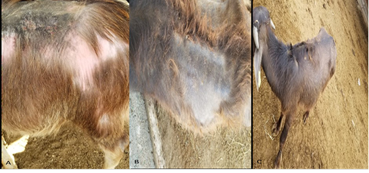
Skin lesion in buffalo calves under study. (A) Alopecia, loss of pigmentation, and excessive keratinization on the dorsum of animals at D0. (B) Skin is pigmented and hair started to grow on animals at D30. (C) Skin is completely cured and all alopecic areas are covered with hair at D60.
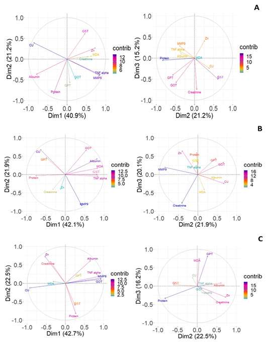
Principal component (PC) biplot for PC1 (Dim1), PC2 (Dim2), and PC3 (Dim3) of plasma biochemical parameters in buffalo calves. (A) At D0. (B) At 30 days post-supplementation. (C) At 60 days post-supplementation.
References
- Abou-Zeina HAA, Zaghawa A, Nasr S, Keshta H, (2008). Effects of dietary cobalt deficiency on performance, blood and rumen metabolites and liver pathology in sheep. Glob. Vet., 2 (4): 182-191.
- Albena A, Elena B, Anton K, Marián K, Daniel K, (2007). Copper decreases gene expression of TNF α, IL10, and of matrix metalloproteinases MMP-2 and MMP-9 in isolated perfused rat livers. Biologia. 62 (3): 365-369. https://doi.org/10.2478/s11756-007-0061-0[CrossRef]
- Anil SV, Venkata Seshaiah Ch, Ashalatha P, Sudhakar K, (2020). Effect of Dietary Nano Zinc Oxide Supplementation on Haematological Parameters, Serum Biochemical Parameters and Hepato-Renal Bio-Markers in Crossbred Calves. Int. J. Curr. Microbiol. Appl. Sci., 9(4): 2034-2044. https://doi.org/10.20546/ijcmas.2020.904.243[CrossRef]
- Chide O, Aneke J, Manafa P, Samuel N, Nancy I, Onah C, (2015). Serum copper level significantly influences platelet count, lymphocyte count and mean cell hemoglobin in sickle cell anemia. Zahedan J. Res. Med. Sci., 18 (12): e4117. https://doi.org/10.17795/zjrms-4117[CrossRef]
- Cope CM, Mackenzie AM, Wilde D, Sinclair LA, (2009). Effects of level and form of dietary zinc on dairy cow performance and health. J. Dairy Sci., 92 (5): 2128-2135. https://doi.org/10.3168/jds.2008-1232[CrossRef]
- Dixon S J, Stockwell B R, (2014). The role of iron and reactive oxygen species in cell death. Nat. Chem. Biol. 10(1): 9-17. https://doi.org/10.1038/nchembio.1416[CrossRef]
- Ebrahim Z, Ahmed A, Lebda M, (2016). Behavioral and Biochemical Alterations in Sheep with Trace Elements Deficiency: A Trial for Treatment. Am. J. Life Sci. Res., 4(2):93-103. https://doi.org/10.20286/ajlsr-040292[CrossRef]
- Engle TE, Nockels C F, Kimberling C V, Weaber D L, Johnson A B, (1997). Zinc repletion with organic or inorganic forms of zinc and protein turnover in marginally zinc-deficient calves. J. Anim. Sci., 75 (11): 3074-3081. https://doi.org/10.2527/1997.75113074x[CrossRef]
- Friard O, Gamba M, (2016). BORIS: A free, versatile open-source event-logging software for video/audio coding and live observations. Methods Ecol. Evol., 7: 1325-1330 https://doi.org/10.1111/2041-210X.12584[CrossRef]
- Gooneratne S R, Buckley W T, Christensen D A, (1989). Review of copper deficiency and metabolism in ruminants. Can. J. Anim. Sci., 69 (4), 819-845. https://doi.org/10.4141/cjas89-096[CrossRef]
- Han YP, Tuan TL, Wu H, Hughes M, Garner WL, (2001). TNF-alpha stimulates activation of pro-MMP2 in human skin through NF-(kappa) B mediated induction of MT1-MMP. J. Cell Sci., 114(1):131-139. https://doi.org/10.1242/jcs.114.1.131[CrossRef]
- Hegab I M, Mohamaden W I. (2023). Behavioral Changes Related to Zinc Deficiency in Buffalo Calves (Bubalus bubalis). Int. J. Vet.Sci. In press.
- Holanda A O, Oliveira A R, Cruz K J, Severo J S, Morais J B, Silva B B, Marreiro D D, (2017). Zinc and metalloproteinases 2 and 9: What is their relation with breast cancer? Rev. Assoc. Med. Bras., 63(1): 78-84. https://doi.org/10.1590/1806-9282.63.01.78[CrossRef]
- Hosnedlová B T, Rávníček J, Šoch M, (2007). Current view of the significance of zinc for ruminants: A review. Agricultura Tropica et Subtropica, 40: 57-64.
- Humer E, Kröger I, Neubauer V, Reisinger N, Zebeli Q, (2019). Supplementation of a clay mineral-based product modulates plasma metabolomic profile and liver enzymes in cattle fed grain-rich diets. Anim., 13 (6): 1214–1223. https://doi.org/10.1017/S1751731118002665[CrossRef]
- Kudo H, Doi Y, Nishino T, Nara S, Hamasaki K, Fujimoto S. (2000) Dietary zinc deficiency decreases glutathione S-transferase expression in the rat olfactory epithelium. J Nutr. ; 130(1) :38-44. https://doi.org/10.1093/jn/130.1.38[CrossRef]
- Lee V E, Arnott G, Turner SP, (2022). Social behavior in farm animals: Applying fundamental theory to improve animal welfare. Front.Vet. Sci., 12: 932217. https://doi.org/10.3389/fvets.2022.932217[CrossRef]
- Lentsch AB, Kato A, Saari JT, Schuschke DA, (2001). Augmented metalloproteinase activity and acute lung injury in copper-deficient rats. Am. J. Physiol. Lung cellular and molecular physiology, 281: L387-393. https://doi.org/10.1152/ajplung.2001.281.2.L387[CrossRef]
- Lozoff B, (2011). Early iron deficiency has brain and behavior effects consistent with dopaminergic dysfunction. J. Nutr., 141(4):740S-746S. https://doi.org/10.3945/jn.110.131169[CrossRef]
- Macpherson A, (1982). Recent Research on the Vitamin Requirements of Ruminants. In: PH-LRC AG, editor. Roche Vitamin Symposium: Basel, Switzerland.
- Manimaran S, Kekan P, Daware SB, Wankar A, Munde VK, Khose K, Bhagade PM, (2022). Effect of Copper and Zinc Supplementation on Antioxidants and Biochemical Status of Osmanabadi Goats. Indian J. Anim, Res., DOI: 10.18805/IJAR.B-4926 https://doi.org/10.18805/IJAR.B-4926[CrossRef]
- Martin P, Bateson P, (2007). Measuring Behaviour: An Introductory Guide Cambridge University Press, 3rd Edition https://doi.org/10.1017/CBO9780511810893.[CrossRef]
- Mattioli G A, Rosa D E, Turic E, Relling A E, Galarza E, Fazzio L E, (2018). Effects of Copper and Zinc Supplementation on Weight Gain and Hematological Parameters in Pre-weaning Calves. Biol. Trace Elem. Res., 185: 327-331. https://doi.org/10.1007/s12011-017-1239-0[CrossRef]
- Mondal S, dhikari N, Banerjee S, Amin SA, Jha T, (2020). Matrix metalloproteinase-9 (MMP-9) and its inhibitors in cancer: A minireview. Eur. J. Med. Chem., 194: 112260. https://doi.org/10.1016/j.ejmech.2020.112260[CrossRef]
- Nosrati R, Kheirouri S, Ghodsi R, Ojaghi H, (2019). The effects of zinc treatment on matrix metalloproteinases: A systematic review. J. Trace Elem. Med. Biol., 56: 107-115. https://doi.org/10.1016/j.jtemb.2019.08.001[CrossRef]
- Pandey P, Kumar M, Kumar V, Kushwaha R, Vaswani S, Kumar A, Singh Y, Shukla PK, (2022). The Dietary Supplementation of Copper and Zinc Nanoparticles Improves Health Condition of Young Dairy Calves by Reducing the Incidence of Diarrhoea and Boosting Immune Function and Antioxidant Activity. Biol. Trace Elem. Res., PMID: 36370333. https://doi.org/10.21203/rs.3.rs-2098195/v1[CrossRef]
- Pathak A, Mahmood A, Pathak R, Dhawan D, (2004). Role of zinc on lipid peroxidation and antioxidative enzymes in intestines of ethanol-fed rats. Biol. Trace Elem. Res., 100 (3): 247-257. https://doi.org/10.1385/BTER:100:3:247[CrossRef]
- Prasad A S, Beck F W, Bao B, Fitzgerald J T, Snell D C, Steinberg J D, Cardozo L J, (2007). Zinc supplementation decreases incidence of infections in the elderly: effect of zinc on generation of cytokines and oxidative stress. Am. J. Clin. Nutr., 85: 837-844. https://doi.org/10.1093/ajcn/85.3.837[CrossRef]
- Prohaska J R, (1991). Changes in Cu, Zn-superoxide dismutase, cytochrome c oxidase, glutathione peroxidase and glutathione transferase activities in copper-deficient mice and rats. J. Nutr., 121(3):355-363. https://doi.org/10.1093/jn/121.3.355[CrossRef]
- Ranson H, Hemingway J, (2005). 5.11-Glutathione Transferases, in: Gilbert, L.I. (Ed.), Comprehensive Molecular Insect Science. Elsevier, Amsterdam, pp. 383-402. https://doi.org/10.1016/B0-44-451924-6/00074-0[CrossRef]
- Reeves M A, Hoffmann P R (2009). The human selenoproteome: Recent insights into functions and regulation. Cell. Mol. life Sci., 66 (15): 2457–2478. https://doi.org/10.1007/s00018-009-0032-4[CrossRef]
- Ruttkay-Nedecky B, Nejdl L, Gumulec J, Zitka O, Masarik M, Eckschlager T, Stiborova M, Adam V, Kizek R., (2013). The role of metallothionein in oxidative stress. Int. J. Mol. Sci., 14: 6044-6066. https://doi.org/10.3390/ijms14036044[CrossRef]
- Schubert C, Guttek K, Reinhold A, Grüngreiff K, Reinhold D, (2015). The influence of the trace element zinc on the immune system. Laboratoriums Medizin, 39. https://doi.org/10.1515/labmed-2015-0060[CrossRef]
- Shanigaram P, Nagalakshmi D, Srinivasa Rao D, Kumar K, Swain P, (2015). Effect of Zinc Supplementation on Antioxidant Status and Immune Response in Buffalo Calves. Anim. Nutr. Feed Technol., 15 (2):179. https://doi.org/10.5958/0974-181X.2015.00020.7[CrossRef]
- Shen X, Song C, Wu T, (2020). Effects of Nano-copper on Antioxidant Function in Copper-Deprived Guizhou Black Goats. Biol. Trace Elem. Res, 199(6): 2201 - 2207. https://doi.org/10.1007/s12011-020-02342-1[CrossRef]
- Spears J W, Kegley E B, (2002). Effect of zinc source (zinc oxide vs zinc proteinate) and level on performance, carcass characteristics, and immune response of growing and finishing steers. J. Anim. Sci., 80: 2747-2752. https://doi.org/10.2527/2002.80102747x[CrossRef]
- Suzuki H, Asakawa A, Jiang BL, Tsai M, Amitani H, Ohinata K, Komai M, Inui A, (2011). Zinc as an appetite stimulator - the possible role of zinc in the progression of diseases such as cachexia and sarcopenia. Recent Pat. food, Nutr. Agric., 3 (3): 226-231. https://doi.org/10.2174/2212798411103030226[CrossRef]
- Suzuki T, Katsumata S, Matsuzaki H, Suzuki K, (2016). Dietary zinc deficiency induces oxidative stress and promotes tumor necrosis factor-α- and interleukin-1β-induced RANKL expression in rat bone. J. Clin. Biochem. Nutr., 58: 122-129 https://doi.org/10.3164/jcbn.15-87.[CrossRef]
- Van Molle W, Van Roy M, Van Bogaert T, Dejager L, Van Lint P, Vanlaere I, Sekikawa K, Kollias G, Libert C, (2007). Protection of Zinc against Tumor Necrosis Factor–Induced Lethal Inflammation Depends on Heat Shock Protein 70 and Allows Safe Antitumor Therapy. Cancer Res., 67:7301-7307. https://doi.org/10.1158/0008-5472.CAN-06-4010[CrossRef]
- Watson J, Angelos J A, Clothier K A, Estell K E, Madigan J, Smith B P, Spier S J, Sponseller B A, Norris J W, Tabin F, (2020). Diseases of the Hematopoietic and hemolymphatic systems In book: Large Animal Internal Medicine. Elsevier, (6th Edition), 1151-1196.e11. https://doi.org/10.1016/B978-0-323-55445-9.00037-9[CrossRef]
- Wysocka D, Sobiech P, Snarska A, (2018). Copper – An essential micronutrient for calves and adult cattle. J. Elementol., 24 (1): 101-110. https://doi.org/10.5601/jelem.2018.23.2.1645[CrossRef]
- Yanagisawa H, Miyakoshi Y, Kobayashi K, Sakae K, Kawasaki I, Suzuki Y, Tamura J (2009). Long-term intake of a high zinc diet causes iron deficiency anemia accompanied by reticulocytosis and extra-medullary erythropoiesis. Toxicol. Lett., 191: 15-19. https://doi.org/10.1016/j.toxlet.2009.07.024[CrossRef]
- Yue C, Ding C, Su J, Cheng B, (2022). Effect of copper and zinc ions on type I collagen self-assembly. Int. J. Polym. Anal. Charact., 27(6): 394-408. https://doi.org/10.1080/1023666X.2022.2093569[CrossRef]
- Zhang Y, McCluskey K, Fujii K, Wahl L M, (1998). Differential regulation of monocyte matrix metalloproteinase and TIMP-1 production by TNF-alpha, granulocyte-macrophage CSF, and IL-1 beta through prostaglandin-dependent and -independent mechanisms. J. Immunol., 161 (6):3071–3076 https://doi.org/10.4049/jimmunol.161.6.3071.[CrossRef]
Services
Article usage statistics
Loading…