Research Article · Volume 12, Issue 2, 27 January 2024 · Open Access
Male Reproductive System Cytotoxicity and Immunotoxicity Evaluation Following Folcord Exposure
· ·
Affiliations & Notes ⌄
1Department of Pathology and Poultry Diseases, University of Baghdad, College of Veterinary Medicine, Iraq
2Department of Microbiology, University of Baghdad, Collage of Veterinary Medicine, Iraq. *Correspondence | Salema Lafta Hassan, Department of Pathology and Poultry Diseases, University of Baghdad, College of Veterinary Medicine, Iraq
In our work sought to assess the immunotoxicity of Folcord as well as the histological status of male mice testes. Eighty adult Swiss Albino mice were separated into four groups at the age of two months. The first group (G1) was given Folcord (500 mg/kg) orally daily for four weeks, whereas the second group (G2) was given Folcord as well as I/P inoculation with 0.1 ml oil adjuvant vaccine (OAV) of Pasteurella multocida twice a week for two weeks. The third group was vaccinated by I/P with 0.1 ml oil adjuvant vaccine (OAV) of Pasteurella multocida in two doses separated by two weeks. The fourth group (G4) was assigned the role of negative control at this time, the 28th day after immunization for work, serum samples were taken for passive hemagglutination test (PHA); Immunoglobulin G(IgG) detection using a chemical immunosorbent assay test. the outcomes of PHA test revealed that Folcord inhibited humoral immune response, with a considerable rise in antibody titer in the third and second groups, but decreased serum antibody levels in the first group. Furthermore, the (IgG) titer in serum is measured using the Enzyme linked immunosorbent assay (ELISA) method. G1 revealed a significant decrease (P≤0.05) in the first group when compared to G2, G3, and G4. Histopathological examination of the testes revealed testicular degeneration, together with the absence of spermatids, no epididymal sperm in the epididymal tubules. In conclusion, Folcord has a severe toxic effect on mice, as evidenced by histological and immunological changes in the male reproductive mice, according to the results of the current study.
Many insect and mammalian pollinators are at danger of exposure due to the use of synthetic insecticides. In general, pollinators are exposed to insecticides through a variety of pathways, such as drinking polluted water, feeding on contaminated plant tissues, and inhaling volatile insecticides . Folcord’s reproductive toxicity has been evaluated in our lab . When immunogenic proteins can activate T cells along with co-receptors CD4 and CD8, which both contribute to immune system activation, they are most effective as vaccines. While CD8+ T cells focus on intracellular pathogens, CD4+ T cells support B cells’ ability to make antibodies and engage in phagocytosis to get rid of bacteria that have been ingested . It is known that the lymphocyte’s toll-like receptor (TLR) regulates the production of antibodies, the presentation of antigens, and cell activation and proliferation. To move tagged virions to the cytosolic proteasome, IgG binds with TRIM21, the human IgG receptor with the highest affinity, where it participates in intracellular antibody-mediated proteolysis and antibody-dependent cell-mediated cytotoxicity (ADCC) . Immunoglobulin IgG . Characterizing the humoral immune response of mice recipients of the Pasteurella multocida oil adjuvant vaccine (OAV) was the study’s main goal. Folcord exposure has been studied to see how it affects histological structures Moreover, a humoral immune response is indicated by an increase in antibody titer immunoglobulin (IgG). Folcord is comparable to toxicities and neurobehavioral consequences observed in chicks and other animals when exposed to other toxicants with distinct mechanisms of action, according to Khaerea et al. (2020). The current study therefore concentrated on the, histological and immunological effects seen in the male reproductive mice treated with. Folcord.
MATERIALS AND METHODS
Chemicals
Folcord (C22H19Br2NO3) from Syngenta Company (Switzerland) is present in the form of solution dissolved in distal water.
Oil adjuvant vaccine (OAV) of Pasteurella multocida
This Ag was employed in the vaccination of animals. prepared in accordance with .
Experimental animals
Eighty adult Swiss Albino mice (7-8) weeks age and the weight between (25-30 gm) were obtained from animal house of the College Vet. Med. University of Baghdad for adaptation. Animals were housed in plastic cages in an air-conditioned room with temperature maintained at 25±2˚C, the plastic cages contained hard-wood chip as bedding and the bedding was changed continuously to ensure a clean environment. mice were given food pellets and water ad libitum.
Immunological tests
Passive hemagglutination test (PHA) test was performed on the 28th day after inoculation . The serum Immunoglobulin G (IgG) content in mouse sera was determined using a commercially by assay Enzyme linked immunosorbent assay (ELISA) Kit.
Experimental design
A total number of 80 mice 7-8 weeks, were arbitrarily separated straight as an arrow four primary style and similarly thought-about in that fashion: first group (G1) received Folcord (500 mg kg) according to (The Agrochemicals Handbook, 1983) orally daily for four weeks, whereas the second group (G2) received Folcord as well as I/P inoculation with 0.1 ml oil adjuvant vaccine (OAV) of Pasteurella multocida twice a week for two weeks. The third group was vaccinated by I/P with 0.1 ml oil adjuvant vaccine (OAV) of Pasteurella multocida in two doses separated by two weeks. The 4th group was classified as the unfavorable control group. Day 28 following immunization, serum samples were taken for passive hemagglutination test (PHA); Immunoglobulin G (IgG) detection using an enzyme-linked immunosorbent assay test.
Histopathology
All animals were slaughtered at the end of the experiments (4 weeks), and samples of testes and epididymis were collected. We fixed the tissues in 10% formaldehyde solution and processed them normally using a histokinete after that. Paraffin blocks with implanted tissue slices were sectioned by Hematoxylin and eosin staining and microtome, then investigated with a light microscope .
Statistical analysis
One-way ANOVA was used, and SPSS determined that the mean difference was significant at the (P≤0.05) level (statistical package for social sciences) (SPSS, 2008).
RESULTS AND DISCUSSION
Hemagglutination test (PHA)
The serum Abs titers in the group three (G3) were (97.32±0.90) significantly (P≤0.05) greater than in the group two (G2) (60.15±0.81), group one (G1) (11±10.12), and control group, according to the findings of the PHA investigation at the 4 weeks mark, as shown in Table 1.
Table 1: shows the antibody titers against oil adjuvant vaccine (OAV) of Pasteurella multocida, vaccinated and control groups of mice at 4 weeks.
Groups
Abs tests at four weeks PHA (mean ±SE)
Group one (G1)
11±10.12 C
Group two(G2)
60.15±0.81 B
Group three (G3)
97.32±0.90 A
Group four (G4)
0
Different superscripted marks in the same column indicate differences that are statistically significant (P≤0.05).
The group three G3 group’s serum Abs titers were significantly (P≤0.05) higher than those of the group two G2 (52.34±1.08), group one G1 (14.30±1.42), and control groups, according to the findings of the Immunoglobulin G (Gg) testing at the 4 weeks, as shown in Table 2.
Table 2: Shows the average serum immunoglobulin G (IgG) levels 30 days after immunization for the various mouse groups immunized at 4 weeks using the enzyme linked immunosorbent assay (ELISA) test.
Groups
IgG (g/L) at 4 weeks (Mean± SE)
Group one (G1)
14.30±0.42 C
Group two (G2)
52.34±3.08 B
Group three (G3)
143.54±2.11 A
Group four (G4)
0
Significant differences (P≤0.05) are shown by different salutation smirch in the identical column.
Clinical signs and symptoms
During the trial period, all of the treated mice displayed depression and decreased appetite. While the control group showed normal consumption, the anorexia and weight loss were caused by pathological lesions that caused indigestion in the stomach and malabsorption in the intestine .
Histopathological alterations
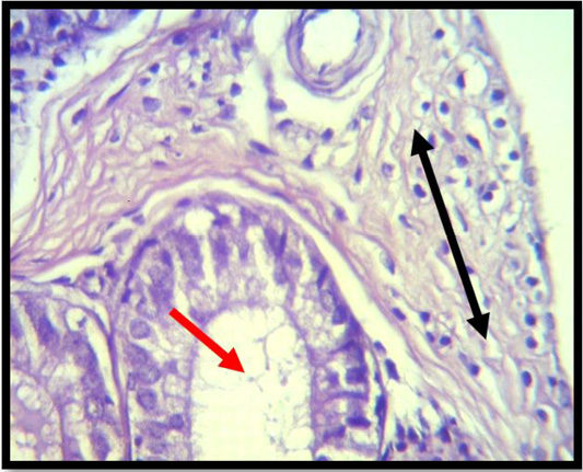
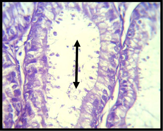
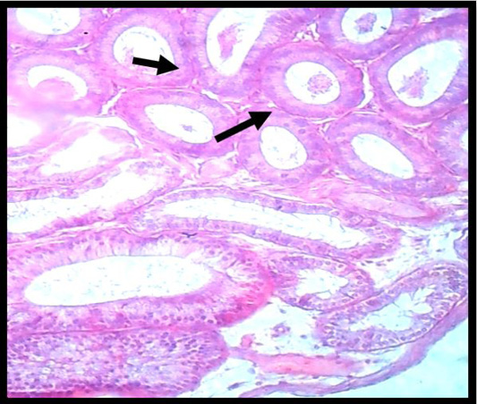
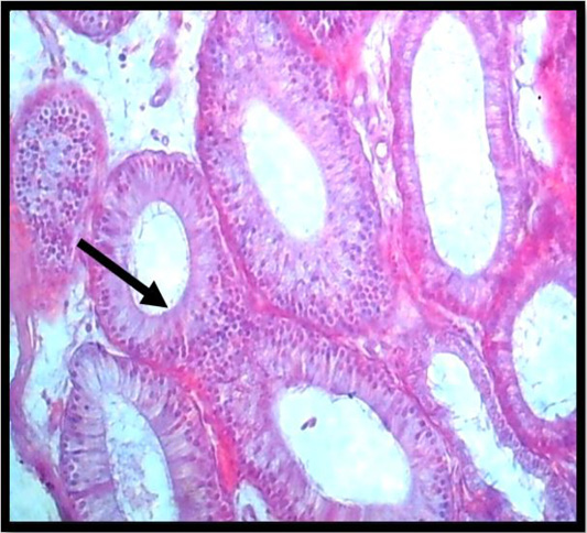
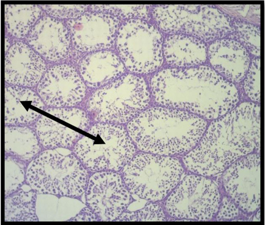
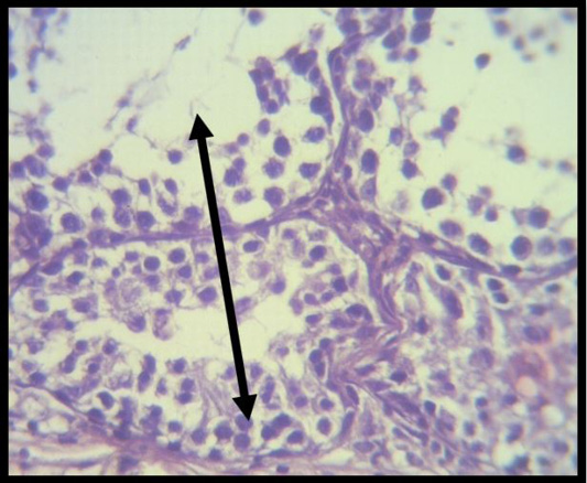
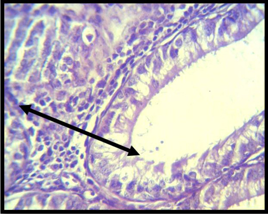
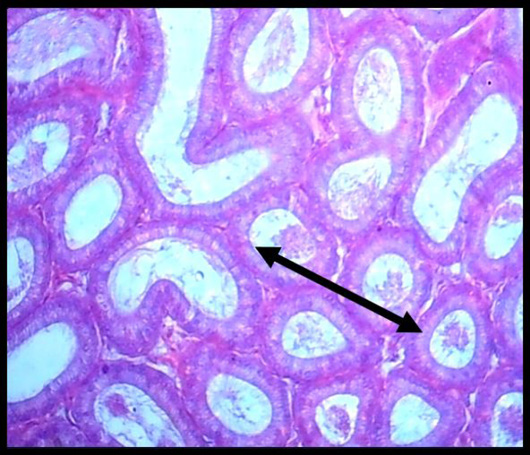
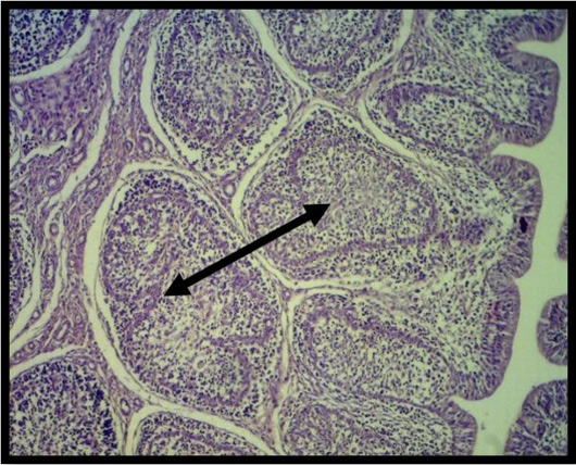
G1Histopathological alterations in the testes of animals treated with Folcord revealed vacuolated cells, poorly differentiated spermatogenic cells and interstitial edema. with inflammatory cell infiltration in Interstial tissue (Figure 1), also marked vacuolations of seminiferous tubular epithelium alongside absence of spermatogenic cells (Figure 2), in other section showed complete absence of epididymal sperm reserve in tubules in the lumen, homogeneous material and cellular detritus were seen (Figure 3), moreover exhibit granuloma establishment (Figure 4).
Group two (G2) Histopathological alterations in rats treated with Folcord and inoculated with oil adjuvant vaccine (OAV) of Pasteurella multocida in two doses separated by two weeks. Showed moderate degeneration and absence of sperm (Figure 5), Sertoli cells are heavily vacuolated, and the spermatogenic cell layer is not continuous as large cells (Figure 6), inflammatory cell aggregation between seminiferous tubules in other areas, which is fairly vacuolated, and a discontinuity in the spermatogenic cell layer (Figure 7), also showed Hyperplasia of clear cells highly vacuolated, homogen (Figure 8),
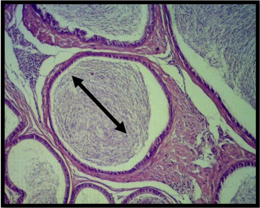
Group three (G3) Histopathological changes of animals immunized with oil adjuvant vaccine (OAV) of Pasteurella multocida two doses, two weeks Throughout the various stages of spermatogenesis, seminiferous tubules develop; Leydig cells can be found in the interstitial region, and the lumen is filled with fully developed spermatozoa (Figure 9), also showed complete epididymal sperm reserve in the tubules (Figure 10). The fourth group of animals’ histopathological abnormalities were designated as the control negative group. There were no noteworthy macroscopic discoveries.
This study found that vaccinated mice administered with Folcord had considerably lower serum antibody titers than immunized animals alone. This finding might imply that Folcord suppressed the humoral immune response. Based on this observation, it was proposed that Folcord may inhibit macrophage and lymphocyte proliferation and attraction onward to the examination location; this concept is consistent with . Phenotyping and cytokine tests are crucial indicators of immunological response. We have discovered that folcord therapy causes a concentration-dependent reduction in the number of T and B cells in the spleen (Kumar et al., 2013).
The toxicants cause a decrease in cytokine production. folcord was also shown to reduce cytokines (IL-2, IL-4, and IFN) and alter immunological processes . The activation of memory cells on the 30th day after initial immunization causes an increase in antibody titer Immunoglobulin G (IgG), and when exposed to antigens, the adaptive immune system can learn similarly to a neurological system. The vaccination results showed that the clostrdium vaccine protein may stimulate a humoral immune response in inoculated mice, resulting in the production of antibodies Immunoglobulin G (IgG). According to the principles of the immunization approach, which, by creating memory responses for specific pathogens using non-virulent or non-toxic antigens, increases immunity and provides protective protection .
When an experimental animal is vaccinated with one of the antigens, an immunological response (antibody) or cell-mediated reaction manifests within a short period of time, increases quickly and exponentially, this is the defining feature of the primer immune response .
Tareq et al. (2012) when reported Anti-Brucella antibodies were tested using an indirect immunofluorescent method, it was shown that animals treated with immunomodulators and those who had received vaccinations had much higher levels of these antibodies than the animals in the negative and positive groups.
Reactive oxygen species-induced oxidative stress may be responsible for the degenerative damages in the examined organs in the Folcord-treated group (ROS). These Radicals are the culprits behind continuous irreversible damage and tissue oxidative stress .
In another study, Jasim et al. (2022) found that reactive oxygen species (ROS), which are thought to be the main cause of oxidative stress and the primary contributing factor to the etiology of infertility, react with macromolecules. They also found that different clinical conditions are the main cause of ROS production.
In addition to , who reported pesticides, Sertoli cell destruction, germ cell modification, cellular deprivation accumulation, as well as the identification of enormous cells within seminiferous tubule lumen revealed that an increase in basal membrane thickness in seminiferous tubules, coupled by defective spermatogenesis, might be related to an increase in reactive oxygen species (ROS) generation, which injury normal reproductive cell.by triggering lipid peroxidation (LPO) and DNA damage.
The symptoms documented were comparable to those reported in prior investigations on testicular toxicity in rats . Histological examination of testicular tissue sections revealed that only the basal germ cells, primary and secondary spermatocytes underwent apoptosis. Indicating the reason for the suppression of spermatogenesis, sertoli cell vacuoles were also observed. . These outcomes correspond to those of Abou-Donia et al. (2003). The current study found that vaccinated and immunized- Folcord mice performed much better than mice treated alone with Folcord. This finding suggests that the immune response may promote enzymatic antioxidants to protect against the harmful effects of Folcord. According to , birds may be able to avoid stress brought on by a temporary rise in reactive species as a result of immune activation. Their findings are comparable to those of our study.
The histopathological lesions in the testes of animals treated with clostrdium vaccination were characterized by mild lesions caused by the clostrdium vaccine’s innate immune response, which offered partially protective immune responses against Folcord ; Ciabattini et al., 2016).
CONCLUSIONS AND RECOMMENDATION
We showed, based on the experimental results of this work, that Folcord has a deleterious toxic effect on mice, which is reflected in immunological and histological alterations in the male reproductive system. Our recommendation in the current study is that the use of Folcard should be restricted to a specific program.
ACKNOWLEDGEMENTS
The authors are thankful to the head College of Veterinary Medicine, University of Baghdad for their assistance during work.
NOVELTY STATEMENT
The novelty of the study is focus on the immunotoxicity of Folcord as well as the histological status of male mice testes.
AUTHOR’S CONTRIBUTION
Salema Lafta Hassan, Taghred Jabbar Humadai, Sabrin Ibraheem Mohsin: Designed and Performed the experiments, analyzed the data, contributed reagents, materials, analysis tools and wrote the paper.
Conflict of interest
The authors have declared no conflict of interest.
Figures
Figure 1:.
A histopathological slice of the first group’s epididymes at 4 weeks demonstrates vacuolated cells, poorly differentiated spermatogenic cells (red arrow), and interstitial edema. Interstial tissue with inflammatory cell infiltration (black arrow) (H & E stain X 40).
Figure 2:.
A histopathological slice of the first group’s testes at 4 weeks reveals severe vacuolations of the seminiferous tubular epaithelium with the lack of spermatogenic cells (black arrow) (H & E stain X 40).
Figure 3:.
A histopathological slice of the first group’s epididymis at 4 weeks demonstrates that there is no epididymal sperm reserve in the tubules. The lumen included homogeneous material and cellular detritus. (black arrow) (H & E stain X 10).
Figure 4:.
Histopathological slice of the first group’s epididymis at 4 weeks demonstrates granuloma development (black arrow) (H & E stain X 40).
Figure 5:.
A histopathological slice of the second group’s testes after 4 weeks indicates considerable deterioration and the absence of testes sperm (black arrow) (H&E stain X 10).
Figure 6:.
Sertoli cells are somewhat vacuolated in the second group’s testes at 4 weeks, and the creation of big cells does not continue with the spermatogenic cell layer (H & E stain X 40).
Figure 7:.
Histopathological slice of the second group’s testes at 4 weeks shows inflammatory cell aggregation in moderately vacuolated interstial tissue, and the layer of spermatogenic cells is discontinuous. (black arrow) (H & E stain X 40).
Figure 8:.
Histopathological slice of the second group’s testes at 4 weeks exhibits hyperplasia of clear cells that are strongly vacuolated, homogeneous material, and cellular debris (black arrow) in the lumen (H & E stain X 40).
Figure 9:.
Histopathological section of testes at 4 weeks of 3rd group shows at various phases of spermatogenesis, seminiferous tubules form, and the Leydig cells and mature spermatozoa fill the lumen. (black arrow) (H&E stainX10).
Figure 10:.
A histopathological slice of the third group’s testes at 4 weeks demonstrates full epididymal sperm reserve in the tubules (black arrow) (H & E stain X 10).
References
Abbas AK, Litchman AH (2005). Cellular and molecular immunology. 5th ed. International Edition. Elsevier Saunders Inc., Philadelphia, PA, Pennsylvania, pp. 41-105.
Abdollahi M, Mostafalou S, Pournourmohammadi S, Shadnia S (2004). Oxidative stress and cholinesterase inhibition in saliva and plasma of rats following subchronic exposure to malathion. Comp. Biochem. Physiol. C Toxicol. Pharmacol., 137: 29–34. https://doi.org/10.1016/j.cca.2003.11.002[CrossRef]
Abdulla JM, Al-Okaily BN (2022). Histomorphometric and histopathological alterations of rat testis following exposure to hydrogen peroxide: Protective role of resveratrol supplement. Iraqi J. Vet. Med., 46(1): 17-23. https://doi.org/10.30539/ijvm.v46i1.1313[CrossRef]
Abou-Donia M, Suliman H, Khan W, Abdel-Rahman A (2003). Testicular germ cells apoptosis in stressed rats following combined exposure to pyridostigmine bromide, N, N-diethyl m-toluamide (DEET), and permethrin. J. Toxicol. Environ. Health, 66(1): 57-73. https://doi.org/10.1080/15287390306463[CrossRef]
Agrochemicals Handbook. 1983. The Royal Society Of Chemistry, The University of Nottingham, England
Alvarez JG, Sharma RK, Ollero M, Saleh RA, Lopez MC (2002). Increased DNA damage in sperm from leukocytospermic semen samples as determined by the sperm chromatin structure assay. Fertil. Steril., 78: 319-329. https://doi.org/10.1016/S0015-0282(02)03201-6[CrossRef]
Casais R, Millan J, Rosell JM, Dalton KP, Prieto JM (2015). Evaluation of an ELISA using recom- binant Ssλ20ΔB3 antigen for the serological diagnosis of Sarcoptes scabiei infestation in domestic and wild rabbits. Vet. Parasitol., 214(3-4): 315-321. https://doi.org/10.1016/j.vetpar.2015.07.011[CrossRef]
Ciabattini A, Pettini E, Fiorino F, Pastore G, Andersen P, Pozzi G and Medaglini, D (2016). Modulation of primary immune response by different vaccine adjuvants. Front. Immunol., 7: 427. https://doi.org/10.3389/fimmu.2016.00427[CrossRef]
El-Gohary M, Awara WM, Nassar S, Hawas S (1999). Deltamethrin-induced testicular apoptosis in rats: The protective effect of nitric oxide synthase inhibitor. Toxicology, 132: 1-8. https://doi.org/10.1016/S0300-483X(98)00114-0[CrossRef]
Herbert WJ (1978). Passive haemagglutination with special reference to the tanned cell technique. Cellular immunology. In: Weir D.M, editor. Handbook of experimental immunology. 3rd ed. Ch. 20. II. Oxford: Blackwell Scientific Publication, pp. 1–20.
Hernandez A, Gomez M, Perez V, Gracialario J and Pla A (2006). Influence of exposure to pesticides on serum components and enzyme activities of cytotoxicity among intensive agriculture farmers. J. Environ. Res., 102: 70-76.
Janeway CA, Jr PT, Walport M, Shlomchik MJ (2001). Ch3 antigen recognition by B-Cell and T-cell Receptors. Immunobiology: The immune system in health and disease (5th ed.). New York: Garland Science, pp. 78-98.
Jasim MA and Baraa NAO (2022). Histomorphometric and histopathological alterations of rat testis following exposure to hydrogen peroxide: protective role of resveratrol supplement. Iraqi J. Vet. Med., 46(1): 17-23.
Khaerea A, Mustafa, Banan Kh, Al-Baggou (2020). Toxicological and neurobehavioral effects of chlorpyrifos and deltamethrin insecticides in mice. Iraqi J. Vet. Sci., 34(1): 189-196. https://doi.org/10.33899/ijvs.2019.125738.1144[CrossRef]
Kumar A, Sasmal D, Sharma N (2013). Deltamethrin induced an apoptogenic signalling pathway in murine thymocytes: Exploring the molecular mechanism. J. Appl. Toxicol., 34: 1303-1310. https://doi.org/10.1002/jat.2948[CrossRef]
Lastuti NDR, Rantam FA, Hastutiek P, Chrismanto D (2017). Toll-like receptor (TLRs) play a role in adaptive immunity in rabbits immunized by Sarcoptes scabiei proteins. VMIC Conf. Proc. KnE Life Sci., 1: 1-9. https://doi.org/10.18502/kls.v3i6.1100[CrossRef]
Luna LG (1968). Manual of histologic staining methods of the armed forces institute of pathology, 3rd edn. McGrawHill, New York, NY, pp. 55-78.
Mallery DL, McEwan WA, Bidgood SR, Towers GJ, Johnson CM, James LC (2010). Antibodies mediate intracellular immunity through tripartite motif-containing 21 (TRIM21). Proc. Natl. Acad. Sci., 107(46): 19985-19990. https://doi.org/10.1073/pnas.1014074107[CrossRef]
Marri V, Richner H (2015). immune response, oxidative stress and dietary antioxidants in great tit nestlings. Comp. Biochem. Physiol., 179: 192-196. https://doi.org/10.1016/j.cbpa.2014.10.013[CrossRef]
Mohr E, Siegrist CA (2016). Vaccination in early life: Standing up to the challenges. Curr. Opin. Immunol., 41: 1–8. https://doi.org/10.1016/j.coi.2016.04.004[CrossRef]
Muhammad M, Hossain J, Richardson R (2011). Mechanism of pyrethroid pesticide-induced apoptosis: Role of Calpain and the ER stress pathway. Toxicol. Sci., 122: 512-525. https://doi.org/10.1093/toxsci/kfr111[CrossRef]
Pant N, Prasad A, Srivastava S, Shankar R, Srivastava S (1995). An Effect of oral administration of carbofuran on male reproductive system of rat. Hum. Exp. Toxicol., 14(11): 889-894. https://doi.org/10.1177/096032719501401106[CrossRef]
Sanchez-Bayo F, Goka K (2014). Pesticide residues and bees–a risk assessment. PLoS One, 9: e94482. https://doi.org/10.1371/journal.pone.0094482[CrossRef]
Sharma P, Huq AU, Singh R (2014). Cypermethrin-induced reproductive toxicity in rat is prevented by resveratrol. J. Hum. Reprod. Sci., 7(2): 99–106. https://doi.org/10.4103/0974-1208.138867[CrossRef]
Sotoodehnia A, Aarabi I, Ataei S, Naserirad A (2000). Preparation and standardization of haemorrhagic septicaemia gel adjuvant vaccine for calf and buffalo using Pasteurella multocida local strain 6: B. Pajouhesh Sazandegi, 47: 111-113.
SPSS (2008). Statistical Package for Social Science, SPSS Users Guide. Statistics Version 16. N.C. USA.
Summaedaey AS (1989). Experimental study of mercurical toxicosis in rats. Iraqi J. Vet. Med., 36(2): 231– 243.
Tareq JF, Adhia, AH,Mohammad KF and Al-Ramahy AK ( 2012). The immunomodulatory effect of Neem (Azadirachta indica) seed aqueous, ethanolic extracts and Candida albicans cell wall mannoproteins on immune response in mice vaccinated with Brucella Rev-1. Iraqi J. Vet. Med. 36(1):120– 127.
The Agrochemicals Handbook (1983). The Royal Society of Chemistry, The University of Michigan, Nottingham England.