
A 53-year-old man with tetralogy of Fallot and coronary artery to bronchial arterial fistula is reported. The communication of fistula arising from the proximal left circumflex coronary artery was discovered by routine coronary artery angiography before correction surgery. The patient then underwent TOF correction surgery. Considering the significance of the fistula, it better be found out and ligated prior to cardioplegic arrest in the surgery. Although extensive searching, the anomaly communication cannot be found. To avoid systemic hypoperfusion on bypass through loss of volume to the pulmonary circulation and shunting of cardioplegic solution to the pulmonary circulation through the fistula, a little increase of cardioplegic solution was given and the surgery was performed successfully. VSD was closed by using autologous pericardium patch, whilst pulmonary valvotomy, pulmonary arterioplasty and right ventricular outflow tract (RVOT) augmentation were performed using a patch of autologous pericardium. Tricuspid valve regurgitation was solved by contract tricuspid ring using an autopericardial ring. The patient recovered well after surgery and without any symptom in 12 months’ follow up. In this study, we reported a TOF patient with coronary artery to bronchial arterial fistula survived to undertake surgery until in a very old age. Though we could not find out the fistula during surgery, on account of the left-to right shunt caused by the fistula, further occlusion by transcatheter procedure was recommended for this patient.
Research Article · Volume 54, Issue 4, 22 April 2022 · Open Access
Left Circumflex Coronary Artery to Bronchial Arterial Fistula Associated with Tetralogy of Fallot: A Case Report and Review of Literature
· · · · · ·
Affiliations & Notes ⌄
- 1School of Public Health, Hubei University of Medicine, 442000, China 2Children’s Medical Center, Affiliated Taihe Hospital of Hubei University of Medicine, 442000, China 3Department of Cardiothoracic Surgery, Affiliated Taihe Hospital of Hubei University of Medicine, 442000, China 4Department of Ultrasonics, Affiliated Taihe Hospital of Hubei University of Medicine, 442000, China 5Cardiovascular Center, Affiliated Taihe Hospital of Hubei University of Medicine, 442000, China * Corresponding auth
Article Info ⌄
- Journal:
- Pakistan Journal of Zoology
- Published:
- 22 April 2022

From the article
- The fistulous communication itself can arise from either the left or right coronary artery.
- But we are aware that few coronary-to-bronchial fistulas were reported in patients with TOF.
- The occurrence of coronary artery fistulas in TOF have several surgical implications.
- The patient is doing well after surgical correction, but attempt to close the coronary artery fistula by transcatheter procedure is still recommended.
Selected from the article's own text.
Abstract
Introduction
Coronary artery to pulmonary arterial fistula is a rare variant in patients with tetralogy of Fallot. Dabizzi et al. (1980) and Haworth and Macartney (1980) reported one-tenth of their large series of patients studied angiocardiographically had such fistulous communications from the coronary arteries, but all the identified fistulas were small. As an extra communication in systemic-to-pulmonary system, it has hemodynamic and physiologic significance during correction surgery. In most of reported cases, the fistula arose from the anterior interventricular artery, followed by the main stem of the left coronary artery, and less frequently from the circumflex artery. In this report, we describe successful surgical management of a case having TOF with a coronary artery to bronchial arterial fistulous communication arising from the left circumflex coronary artery (LCX).
Case Report
A 53-year-old male was referred to our institute for evaluation and treatment of tetralogy of Fallot (TOF) on 19th December 2018. He had a history of dyspnea (New York Heart Association Class II) for 6 years, and was discovered of TOF by transthoracic echocardiography recently before this hospitalization. He had several episodes of cyanotic spells during these 6 years. On examination, he was of short stature with central cyanosis and mild digital clubbing. His blood pressure was 144/96mmHg with pulse rate 83/min. The cardiovascular system examination revealed a continuous 4-5/6 grade murmur best heard in the right parasternal area over the third intercostal space. The baseline systemic arterial oxygen saturation (SaO2) was 88%. Blood test showed elevated red blood cells quantity with HGB 198g/L, indicated of chronic hypoxia, no signs of dyslipidemia or diabetes mellitus were found. BNP was mild elevated (422Pg/ml), while liver function, renal function, CTnI, D-dimer, PCT were all in the normal range, HIV antibody, hepatitis markers, syphilis antibody were all negative.
Electrocardiogram revealed normal sinus rhythm with features of pulmonary p wave, right ventricular hypertrophy (RVH) and right axis deviation. T wave was flat or inverted in leads II, III, avF and V1 to V6. Transthoracic and transesophageal echocardiography revealed a large (17mm) subaortic ventricular septal defect (VSD) with 50% aortic override, increased supraventricular ridge bundle and parietal band caused severe infundibular stenosis. A diaphragm like structure as well as thickened pulmonary valve bulged into pulmonary trunk caused its stenosis. Left atrium and ventricular were in normal size, whereas right atrium and ventricular were enlarged and thickened. The ejection fraction of left ventricle was in normal range, no collateral vessel or fistula could be appreciated on color Doppler imaging (Fig. 1).
Enhanced cardiac computed tomography (CT) scan showed enlarged pulmonary trunk(diameter of 3.3cm), as well as both pulmonary arteries, especially the left one of 5.1cm in diameter. MGoon index was 3.1, cross-sectional area of left pulmonary artery was 2073.2mm2, right one was 181.1mm2, Nakata index was 1454mm2/m2. Obstruction of right ventricular outflow tract, right-sided aortic arch, aortic overriding and RVH were also showed, thickened pulmonary valves bulged into pulmonary trunk like discovered by echocardiography (Fig. 2).
Subsequent catheterization and angiographic study confirmed the diagnosis of TOF with large VSD, RVH, and aortic override. Pulmonary artery pressure was not obtained because the guide wire could not pass through the narrowed pulmonary valve during right cardiac catheterization, only right ventricle pressure was obtained (117/14 mmHg). Selective coronary angiography showed no stenosis of coronary arteries but the presence of a fistulous communication arising from the proximal left circum flex coronary artery to bronchial artery (Fig. 3).
Considering the significance of the fistula in the corrective surgery, it should be find out and ligated prior to cardioplegic arrest. During exploration, pulmonary trunk was dilated seriously while three pulmonary valves infused in the centra caused the valve almost atretic. The MPA (main pulmonary artery) was opened longitudinally and no fistula opening was seen in MPA, left and right pulmonary arteries on careful inspection. Increased supraventricular ridge bundle and parietal band caused severe infundibular stenosis. The originations of left and right coronary arteries were normal and no abnormal communication was seen from LCX. To avoid systemic hypoperfusion on bypass through loss of volume to pulmonary circulation and shunting of cardioplegic solution to pulmonary circulation through the fistula, the volume of cardioplegic solution was increased a little under cardiopulmonary bypass. Intracardiac repair of TOF was performed, closure of the VSD, pulmonary valvotomy, pulmonary arterioplasty, and right ventricular outflow tract (RVOT) augmentation were completed using a patch of autologous pericardium. Tricuspid valve regurgitation was solved by contract tricuspid ring using an autopericardial ring. The patient recovered uneventfully after surgery, postoperative echocardiography showed no residual VSD, no RVOT gradient, and normal biventricular function (Fig. 1). Twelve months after surgery, the patient is asymptomatic, he was asked to close the coronary artery to bronchial arterial fistula by further transcatheter procedure.
Discussion
TOF is the most common cause of a right-to-left shunt in individuals who survive infancy, it usually need to be corrected by surgery in early stage. Before surgical management, other anomalies like coronary artery fistulas (CAFs) should be explored extensively because surgical strategy may differ according to its presence. Congenital CAFs are characterized by a normal aortic origin of the coronary artery involved but with a fistulous communication with the atria, the ventricles or with the pulmonary artery, coronary sinus, or the superior vena cava. CAFs are rare anomalies, the incidence in the overall population is estimated 0.002%. The true prevalence of these fistulas in patients with VSD and pulmonary atresia is unknown, however, they can occur in as many as 10% of these patients ; . Many of these CAF anomalies are difficult to detect intraoperatively by surgical teams, hence carefully examination of the coronary arteries by angiographic studies prior to surgical treatment should be performed.
The fistulous communication itself can arise from either the left or right coronary artery. According to Talwar et al. (2014) review, three-quarters cases had the fistulas arising from the left coronary artery, in most cases involving the left coronary arterial system, the fistulas arose from the anterior interventricular artery, followed by the main stem of the left coronary artery, and less frequently from the circumflex artery. Most frequently, the fistula itself terminates in the pulmonary trunk, then other structures. Many cases of coronary to pulmonary artery fistulas were also reported in patients with TOF ; ; ; Mc-Mahon et al., 2014; ; ; . In Dabizzi et al. (1980) investigation, fistulas between coronary artery and bronchial arteries is the most common anastomoses in TOF. But we are aware that few coronary-to-bronchial fistulas were reported in patients with TOF. In our case, an obvious fistulous communication arose from LCX artery was discovered by angiography, but the distribution and termination of the fistula could not be shown very clearly, the fistula seemed more likely drained into bronchial artery rather than pulmonary artery. Subsequent enhanced cardiac CT scan confirmed it was a communication between LCX and bronchial artery. The fistula arose from the proximal portion of the LCX, then divided into two branches, one of the branch terminated at tracheal carina and the other one at right main bronchus. Talwar et al. (2009) reported an accidental transection of RCA when opening the main pulmonary artery because of a missed diagnosis of anomalous originated right coronary artery in TOF before procedure. Recently, computed tomography had proven to be an useful tool in coronary artery fistulas detection and morphological assessment by other studies ; ; ; ; . In our patient, the morphological distribution and termination of the CAF was revealed much more clearly by coronary artery angiography combined with enhanced cardiac CT scan, hence different image methods should be used together for CAF evaluation especially in the complicated cases like TOF to accurately delineate the anatomy and avoid the accidental destruction of the anomalies during surgery.
The occurrence of coronary artery fistulas in TOF have several surgical implications. The fistulous communication should be identified and ligated prior to the institution of cardiopulmonary bypass in order to prevent systemic hypoperfusion on bypass through loss of volume to pulmonary circulation. And it is also important to close the fistulas prior to aortic cross-clamping because shunting of cardioplegic solution to pulmonary circulation through the communication will result in incomplete myocardial protection and excessive left heart return. In those patients where there is an additional source of blood supply to a lung segment, they are treated just like any other collateral artery by ligation or division immediately upon institution of CPB. Unfortunately, we could not find the coronary artery to bronchial artery fistula under cardiopulmonary bypass, maybe because of the fistula’s opening buried deeply inside of the cardiac muscles. We chose to increase the cardioplegic solution to avoid insufficient protection caused by the fistula. The patient recovered uneventfully and biventricular function was reserved after surgery, which means increase the cardioplegic solution is an alternative choice in the circumstance when have difficulties to find out the abnormal communication by surgeons.
Only a few patients with TOF reach adulthood without surgical correction, the prognosis is relevant to the severity of the abnormalities. Stanescu and Branidou (2008) had reported a 75-year-old survivor of unrepaired tetralogy of Fallot with quadricuspid aortic valve. Hussain et al. (2012) reported a successful surgical repair of TOF for a patient at age 83. In the present case, the patient survived to undertake surgery until 53 years old. Considering the patient’s career is tailor and have limited daily activities, we believed that this is the reason why he can survive with cyanosis for so many years before surgical correction. The patient is doing well after surgical correction, but attempt to close the coronary artery fistula by transcatheter procedure is still recommended.
Acknowledgments
The research is supported by Natural Science Foundation of Hubei Province of China: The role of 5-lipoxygenase pathway in urotensin II-promoted vascular remolding of balloon injured artery. (2015CFB211); Joint Fund of Taihe hospital and BIOTECAN for Precision Medicine: The genotype classification and efficiency of PCI procedure in HCM patients with coronary artery disease (2016JZ09).
Statement of conflict of interest
The authors have declared no conflict of interests.
Figures
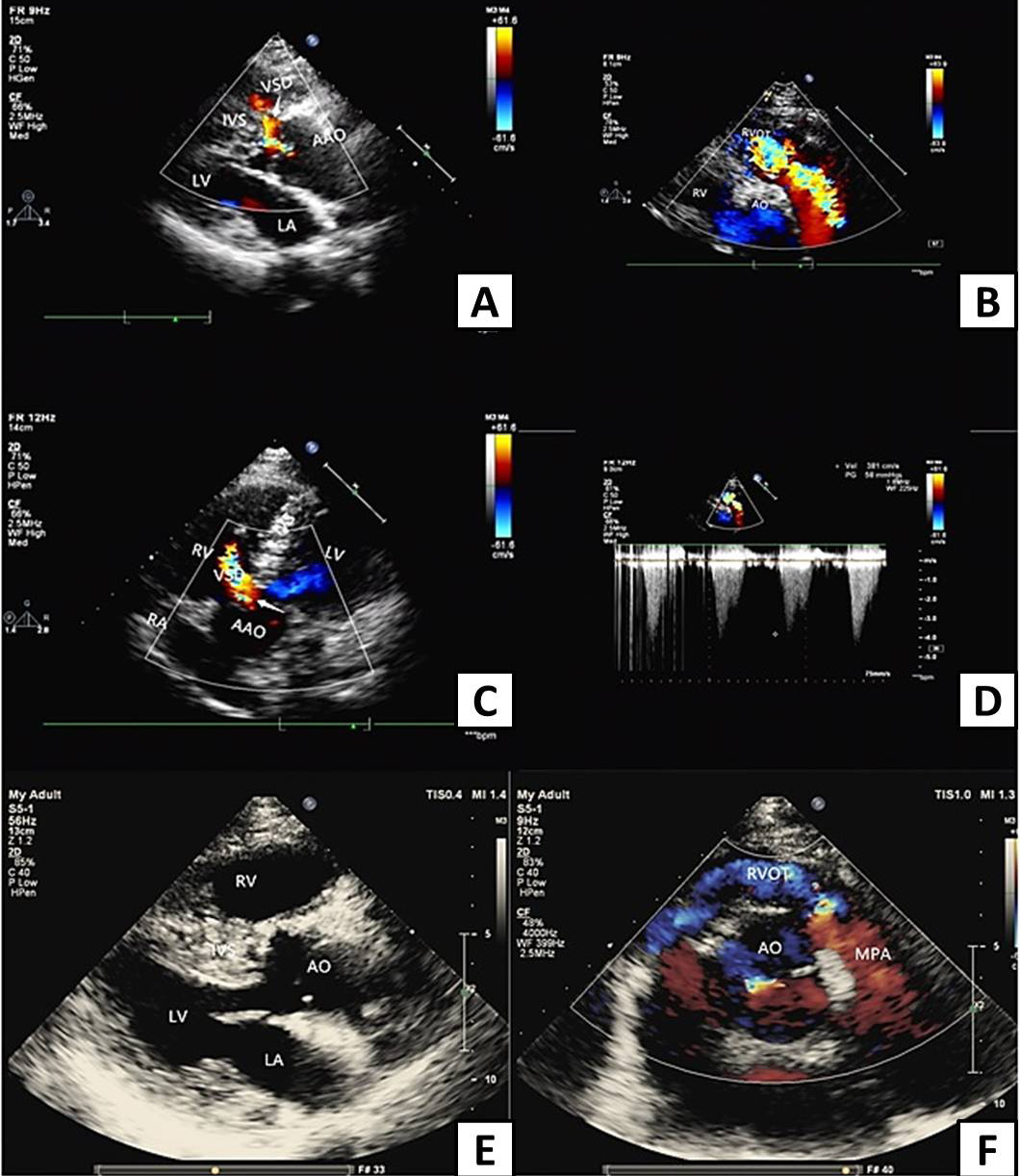
Transthoracic echocardiography before and after corrective surgery (AAO, ascending aorta. AO, aorta; RVOT, right ventricular outflow tract; VSD, ventricular septal defect; IVS, interventricular septum; RA, right atrium; LA, left atrium; RV, right ventricle; LV, left ventricle). A, Parasternal long axis view of left ventricle shows interruption of ventricular septum continuity before surgery, arrow points to left to right shunt of ventricular defect. B, Nonstandard axis view shows obvious acceleration of right ventricular outflow tract blood flow with multicolored mosaic blood flow signals before surgery. C, Apical 5-chamber view shows interruption of ventricular septum continuity before surgery, arrow points to left to right shunt of ventricular defect. D, Color doppler flow diagram of right ventricular outflow tract before surgery, the blood flow of right ventricular outflow tract was obviously accelerated, the peak systolic velocity was about 3.8 m/s, and the pressure difference was 54 mmHg. E, Long axis view of the left ventricle shows RVH, closure of the VSD and RVOT augmentation after corrective surgery. F, The short axis view of the aorta shows the blood flow of right ventricular outflow tract was nearly normal after corrective surgery, no blood flow acceleration was found.
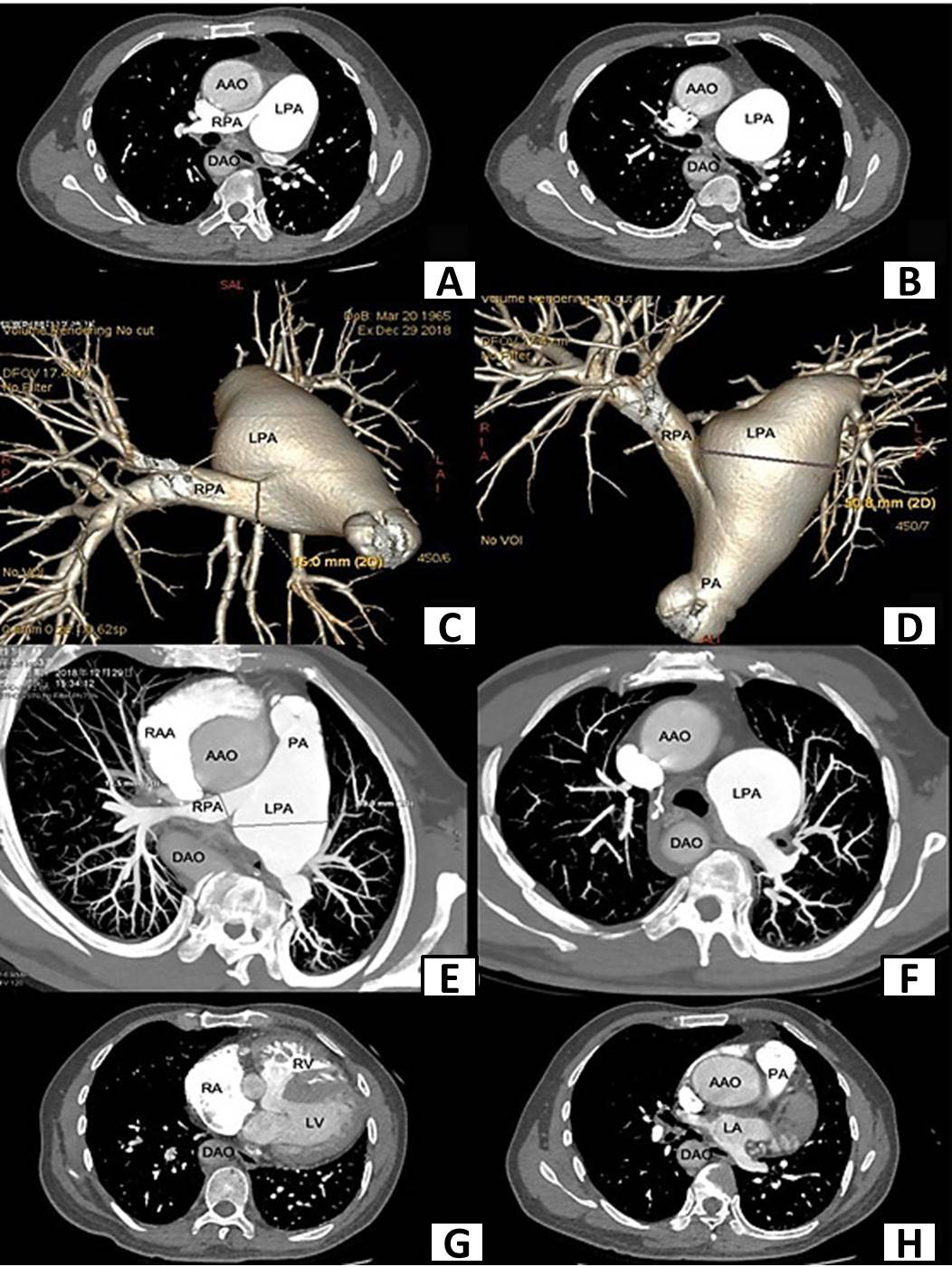
Enhanced cardiac computed tomography(CT) scan before surgery shows aortic overriding, ventricular septal defect, RVH and obstruction of right ventricular outflow tract (AAO, ascending aorta; DAO, descending aorta; LPA, left pulmonary artery; RPA, right pulmonary artery; PA, pulmonary artery; RAA, right atrial appendage; RA, right atrium; LA, left atrium; RV, right ventricle; LV, left ventricle). A, CT shows aortic override on the right pulmonary artery trunk and the dilated left pulmonary artery. B, CT shows dilated left pulmonary artery. C, CT shows stenosis of right pulmonary artery trunk and dilated left pulmonary. D, CT shows stenosis of right pulmonary artery trunk and dilated left pulmonary. E, CT shows aortic overriding, stenosis of right pulmonary artery trunk and dilated left pulmonary artery. F, CT shows dilated left pulmonary artery and thickened pulmonary branches. G, CT shows right ventricle hypertrophy and ventricular septal defect. H, CT shows aortic overriding and obstruction of right ventricular outflow tract.
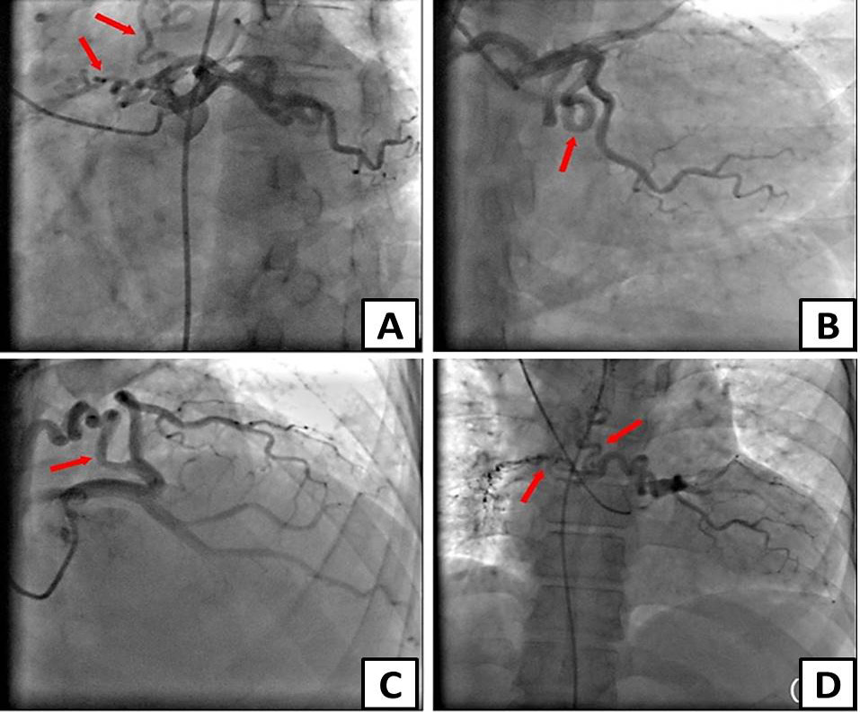
Selective coronary angiography shows a fistulous communication arising from the proximal left circum flex coronary artery to bronchial artery by different project positions. A, Coronary angiography in LAO45+CAU30 projection position. B, Coronary angiography in CAU20 projection position. C, Coronary angiography in RAO30+CRA30 projection position. D, Coronary angiography in AP projection position (red arrows show the fistulas).
References
- Al-Umairi, R.S., Al-Kindi, F. and Al-Tai, S., 2019. Prevalence and spectrum of coronary anomalies detected on coronary computed tomography angiography: A single centre experience in Oman. Sultan Qaboos Univ. Med. J., 19: p.e108. https://doi.org/10.18295/squmj.2019.19.02.005[CrossRef]
- Angeli, E., Napoleone, C.P., Oppido, G., Martìn-Suàrez, S. and Gargiulo, G., 2008. Anomalous origin of additional coronary artery arising from the pulmonary artery in complex congenital heart disease. Cardiovasc. Pathol., 17: 190-191. https://doi.org/10.1016/j.carpath.2007.03.007[CrossRef]
- Butt, K., Agha, A., Parente, R., Limback, J. and Burt, J.R., 2019. Anomalous coronary anatomy with fistula diagnosed on coronary computed tomography angiography. Cureus, 11: e4403. https://doi.org/10.7759/cureus.4403[CrossRef]
- Chowdhury, U.K., Patel, K., Gupta, S.K., Jagia, P. and Singh, S.P., 2015. Right coronary artery to pulmonary arterial fistula associated with tetralogy of Fallot: A case report and review of literature. World J. Pediat. Congenital Heart Surg., 6: 654-657. https://doi.org/10.1177/2150135115581385[CrossRef]
- Czekajska-Chehab, E., Skoczyński, M., Przybylski, P., Staśkiewicz, G., Tomaszewski, A., Siek, E., Kurzepa, J. and Drop, A., 2020. Coronary artery fistulas morphology in coronary computed tomography angiography. Folia Morphol., 79: 777-785. https://doi.org/10.5603/FM.a2019.0132[CrossRef]
- Dabizzi, R.P., Caprioli, G.I.U.S.E.P.P.E., Aiazzi, L.U.I.G.I., Castelli, C.A.R.L.O., Baldrighi, G.I.O.R.G.I.O., Parenzan, L.U.C.I.O. and Baldrighi, V.I.N.C.E.N.Z.O., 1980. Distribution and anomalies of coronary arteries in tetralogy of fallot. Circulation, 61: 95-102. https://doi.org/10.1161/01.CIR.61.1.95[CrossRef]
- Haworth, S.G. and Macartney, F.J., 1980. Growth and development of pulmonary circulation in pulmonary atresia with ventricular septal defect and major aortopulmonary collateral arteries. Br. Heart J., 44: 14-24. https://doi.org/10.1136/hrt.44.1.14[CrossRef]
- Hussain, F.T.N., Grogan, M. and Dearani, J.A., 2012. Surgical repair of tetralogy of Fallot at age 83. World J. Pediat. Congen. Heart Surg., 3: 518-520. https://doi.org/10.1177/2150135112451031[CrossRef]
- Kajihara, N., Asou, T., Takeda, Y., Kosaka, Y., Matsuhama, M., Onakatomi, Y., Yanagi, S. and Yasui, S., 2009. A symptomatic neonate with tetralogy of Fallot, an absent pulmonary valve, and a single coronary artery with a major fistula. Annls. Thor. Surg., 88: 1680-1683. https://doi.org/10.1016/j.athoracsur.2009.04.020[CrossRef]
- Kim, S.Y., Kim, E.R., Bang, J.H. and Kim, W.H., 2017. Development of a pulmonary arteriovenous fistula after a modified Glenn shunt in tetralogy of Fallot and its resolution after shunt takedown in a 57-year-old patient. Korean J. Thor. Cardiovasc. Surg., 50: 215. https://doi.org/10.5090/kjtcs.2017.50.3.215[CrossRef]
- Lee, J.J., Cho, J.Y., Lee, Y.H., Lee, B.H., Park, C.G., Seo, H.S., Oh, D.J. and Na, J.O., 2014. An adult case of tetralogy of Fallot accompanied by multiple anomalies including multidirectional coronary artery fistulas. Korean Circulat. J., 44: 196. https://doi.org/10.4070/kcj.2014.44.3.196[CrossRef]
- Li, J.L., Huang, L., Zhu, W., Ye, W.T., Yan, L.F., Zhong, X.M., Luo, H.Y., Saboo, S., Huang, M.P. and Liang, C.H., 2019. The evaluation of coronary artery-to-pulmonary artery fistula in adulthood on 256-slice CT coronary angiography: Comparison with coronary catheter angiography and transthoracic echocardiography. J. Cardiovasc. Comput. Tomogr., 13: 75-80. https://doi.org/10.1016/j.jcct.2018.10.013[CrossRef]
- McMahon, C.J., Prendiville, T. and Nolke, L., 2014. Pre-operative identification of an anomalous left coronary artery from the pulmonary artery in association with tetralogy of Fallot and an aortopulmonary window. Cardiol. Young, 24: 170-171. https://doi.org/10.1017/S1047951113000255[CrossRef]
- Punzo, B., Forte, E., Salvatore, M., Cavaliere, C. and Cademartiri, F., 2019. Case report of coronary artery fistula: Added value of multimodal advanced imaging with cardiac computed tomography and cardiac magnetic resonance. Medicine, 98: e18255. https://doi.org/10.1097/MD.0000000000018255[CrossRef]
- Stanescu, C.M. and Branidou, K., 2008. A case of 75-year-old survivor of unrepaired tetralogy of Fallot and quadricuspid aortic valve. Eur. J. Echocardiogr., 9: 167-170. https://doi.org/10.1016/j.euje.2007.06.009[CrossRef]
- Talwar, S., Sharma, P., Gulati, G.S., Kothari, S.S. and Choudhary, S.K., 2009. Tetralogy of Fallot with coronary artery to pulmonary artery fistula and unusual coronary pattern: missed diagnosis. J. Cardiac Surg., 24: 752-755. https://doi.org/10.1111/j.1540-8191.2009.00917.x[CrossRef]
- Talwar, S., Anderson, R.H., Keshri, V.K., Choudhary, S.K., Gulati, G.S. and Airan, B., 2014. Coronary-pulmonary artery fistula in tetralogy of Fallot with pulmonary atresia. Asian Cardiovasc. Thor. Annls., 22: 1003-1009. https://doi.org/10.1177/0218492313505101[CrossRef]
- Trehan, V., Mukhopadhyay, S., Yusuf, J., Rangasetty, U.C. and Gupta, M.D., 2004. Transcatheter closure of coronary-to-pulmonary fistula by nonconventional coils in a patient with tetralogy of Fallot. Pediat. Cardiol., 25: 681-683. https://doi.org/10.1007/s00246-003-0619-8[CrossRef]
- Witters, I., De Groot, R., Van Loo, K., Willekens, C., Coumans, A., Frints, S., Fryns, J.P. and Baldewijns, M., 2014. Tetralogy of Fallot with coronary artery to pulmonary artery fistula. Prenatal Diagn., 34: 1345-1346. https://doi.org/10.1002/pd.4481[CrossRef]
Services
Article usage statistics
Loading…