The decrease in lying time of high lead group could be explained by the engage of rats in other behaviours as ingestive and investigatory behaviour to cope with changes present.
The histological examination of the liver tissue for lead acetate and aluminum chloride-treated rats showed mild to moderate acute cellular swelling with hydropic degeneration and multifocal hepatic cell necrosis accompanied by lymphocytic infiltration.
Kidney histological investigation revealed that lead acetate and aluminum chloride exposure caused progressive glomerular and tubular alterations.
Interstitial, tubular and glomerular damage are also characteristic renal lesions due to lead acetate toxicity.
Selected from the article's own text.
Abstract
Amira Abd El-Moneum Goma, Hossam Gaafar Tohamy
1Department of Animal Husbandry and Animal Wealth Development, Faculty of Veterinary Medicine, Alexandria University, Edfina, Behira, Egypt; 2Department of pathology Faculty of Veterinary Medicine, Alexandria University, Edfina, Behira, Egypt.
Abstract | Animal welfare is a matter of societal concern and debate, its improvement should consider those aspects actually affecting animal’s health, through determining their impact on some welfare indicators, so, this study was conducted to investigate the effect of heavy metals toxicity on behavioural, biochemical and histopathological changes in adult rats. Twenty-five Sprague-Dawley adult rats were allotted into five groups, each of 5, two groups (GA & GB) treated with lead acetate (low 1g/l & high 2g/l) and another two (GC & GD) treated with anhydrous aluminium chloride (low 2 g/l & high 3.5 g/l) in drinking water daily for 5 weeks, while, GE (control) received drinking water without any treatment. The findings obtained indicated that feeding time, drinking, cage and total exploration frequencies increased in GB than other groups, although, lying time decreased. However, the movement activities increased in GA than others. On the other hand, glucose level decreased significantly in GB than GD and control. Furthermore, triglycerides and total lipid levels were significantly decreased in all treated groups than control. It was also revealed that the serum ALT increased significantly in GB than other groups while creatinine decreased significantly in both groups (GA & GB) treated with lead acetate than other groups with most decrement in high dose group of lead acetate. However, the urea level decreased and uric acid increased significantly in all treated groups than control. Microscopically, degenerated and necrotic hepatocytes, tubular and glomerulus’s necrosis were observed in all toxicated rats beside eosinophilic intranuclear inclusions in lining epithelium of some renal tubules of lead toxicated rats. Therefore, it can be concluded that different toxic levels of lead acetate and aluminum chloride can affect animal’s welfare by behavioural, biochemical and histopathological alterations in a dose dependent manner.
Animal welfare is an issue not only for veterinary science and animal psychology but also for biochemical, agriculture, zoology and companion animal fields because animal welfare is a socially constructed idea and it is not a permanently fixed idea . On the other hand, animals are very good indicator of environmental pollution as they inhabit the same space as human and exposed to action of the same pollutants, for that reason it is appropriate and advantageous to evaluate the negative impact of the polluted environment by heavy metals on human health by parallel evaluation of their load on animals .
Lead is a one of the major heavy metals known to be toxic to mammals. It had many sources as metallic lead present in storage batteries, paints and rubbish dumps in areas near lead industrial establishments and also from the industrial applications especially the addition of vast quantities of lead as an anti-knock agent in gasoline . Poisoning with lead is associated with behavioural and physiological problems as it has deleterious effects on neurological, haematological, gastrointestinal, reproductive, circulatory and immunological systems .
Aluminum also is the third most prevalent element and approximately 8% of total mineral components in the earth’s crust . It is broadly distributed in the environment and widely used in daily life causing its easy exposure to animals and human. It gets access through their body via pulmonary and oral route . Although only a small portion of aluminum is absorbed by gastrointestinal tract, oral intake represents the route with greatest toxicological implications , especially, it is added to drinking water for purification purposes and found in medicines as antacids . Furthermore, it was reported that aluminum exposure can lead to impairments in glucose utilization, free-radical-mediated cytotoxicity and lipid peroxidation .
The liver is the responsible organ for maintaining the body metabolic homeostasis and has been considered as the target organ for the toxic effect of lead and aluminum . Moreover, Jarrar (2003), Taib et al. (2004), Shelley et al. (2012) and Al-Qayim and Saadoon (2013) stated that it is the largest repository softy tissue for both toxins followed by kidney where a small amount was excreted in urine and that both metals affect many biological activities in these organs at molecular, cellular and intercellular levels which may result in morphological alterations.
The objective of this study was to investigate the effect of lead acetate and aluminum chloride toxicities on welfare aspects through measuring its effect on some welfare indicators as behaviour, biochemical parameters as well as histopathological changes in adult rats.
Materials and Methods
Animals and Experimental Design.
All experimental procedures were conducted in accordance with the Alexandria University Animal Ethics Committee guidelines. This research was conducted with strict rules to preserve and safeguard the animal welfare without subjecting them to any degree of suffering or stress..
Twenty-five Sprague-Dawley adult rats (three months old, 110-130g) were obtained from the Laboratory Animal Breeding Colony of the Animal health research institute, Alexandria University. They were housed in individual plastic cages (37x30x14cm) and fed on ration containing 16.3% crude protein, 6.8% fat and 3.5% crude fiber, adlibitum with water. Natural light-dark cycle was used without any alteration in lighting program. Identification were carried out by temporary marking on tail using different colour paints and remarking was employed at intervals according to diminish of paint.
After an acclimatization period of one week, the rats were randomly assigned into five groups (five rats each), two groups treated with lead acetate which were, GA (low lead acetate), that was given 1g/l lead acetate and GB (high lead acetate), which was treated with 2 g/l lead acetate in drinking water . However, the other two groups were treated with anhydrous aluminium chloride, that were GC (low aluminium chloride), which admistered 2 g/l anhydrous aluminum chloride and GD (high aluminium chloride) that was given 3.5 g/l anhydrous aluminium chloride in drinking water . The last group was GE (Control) which was given drinking water without any treatment. The application of treatment was continued for five weeks on daily bases.
Behavioural Observations
Focal sample observations were carried out three times weekly, four hours daily within periods of late morning and early afternoon to obtain a twelve hours daylight observation per week. The observed patterns were ingestive, resting, body care, investigatory behaviour and movement activities .
Biochemical Parameters
A sample of blood was collected from orbital venous plexus of ether anaesthetized rats at the end of study in a tube without anticoagulant that was left for 3 hours at 4oC to clot then centrifuged at 3000 r.p.m for 10 minutes to obtain serum which was kept at -20oC for biochemical analysis, that includes cholesterol , triglycerides , total lipid , total protein , albumin , globulin calculated by subtraction of albumin from total protein level, albumin/ globulin ratio, glucose , corticosterone , ALT (Alanine aminotransferase) and AST (Aspartate aminotransferase) according to , γ-GT (Gamma glutamyl transaminase) activity , Creatinine , urea and uric acid .
Histopathological Studies
Specimens from liver and kidney were collected directly after scarification of examined animals. The samples were fixed in 10% neutral buffered formalin, dehydrated in alcohol, cleared in xylene and embedded in paraffin, 4-6 micron thick sections were prepared and stained with haematoxylin and eosin .
Statistical Analysis
All data were analyzed by one-way analysis of variance, proc GLM, using SAS (Statistical Analysis system, version 6, 4th Edition, SAS Institute, Cary, NC. USA.). Data are expressed as means±S.E.M. and P values<0.05 were considered significant in all tests, unless stated otherwise. Analysis of significant main effects of experimental treatment was performed using multiple range comparisons with Duncan multiple range test.
Results
Behavioural Patterns
The obtained data deducted that feeding time increased significantly in GB & GD than others with the highest increment in GB. Moreover, drinking frequency showed significant increase in GB than other groups. However, lying time decreased significantly in GC and GB than other treated groups and control. On the other hand, movement activities increased significantly in GA than other treated groups and control. However, frequencies of cage and total exploration increased significantly in GB than other treated groups. Moreover, standing time, licking, scratching and trough exploration frequencies showed a non-significant effect for all treatments (Table 1).
Biochemical Parameters.
The biochemical parameters shown in Table 2 revealed that, lipid profile indicated a significant decrease in triglycerides and total lipid levels in all treated groups than control ones except GA in total lipid level. Moreover, it can be deducted that lowest level for cholesterol, triglycerides and total lipid were in GC while the highest were for GA. However, protein profile indicated that albumin level increased significantly in GA and GD than control (3.67+0.14 & 3.10+0.10 vs. 2.69+0.14 g/dl) with the highest in GA, in addition, all treated groups showed increased albumin level than control, while all decreased significantly than GA. Furthermore, it was indicated that the highest level of albumin, globulin and total protein were in GA and lowest level in GB treated rats. Moreover, glucose level decreased significantly in GB when compared to control and GD (79.43+11.45 vs. 120.33+15.59 & 128.17+5.91 mg/dl). However, there was a non-significant effect of treatment on cholesterol, total protein, globulin, albumin/ globulin ratio and corticosterone.
The liver functions enzymes indicated that ALT increased significantly in GB than other groups. Moreover, there was a significant increment in the two treated groups of aluminium chloride than control, in addition to a significant dose dependent increase in lead acetate treated groups. However, AST showed a significant increment in GB than GD. Furthermore, GGT in GC decreased significantly than control. On the other hand, the kidney functions enzymes showed that Creatinine level decreased significantly
Table 1: Least square means and their standard error to the effect of lead acetate and aluminium chloride (Alcl3) toxicities on behavioural patterns of adult rats
Item
Treatment
GA
GB
GC
GD
GE
Ingestive behaviour
Feeding (min/hr)
7.08+0.49c
16.70+2.49a
8.95+0.87bc
10.75+1.34b
6.05+0.50c
Drinking (freq/hr)
0.74+0.08b
1.53+0.18a
0.65+0.10b
0.55+0.07b
0.71+0.07b
Resting behaviour
Standing (min/hr)
0.48+0.09a
0.68+0.17a
0.75+0.15a
0.83+0.17a
0.68+0.21a
Lying (min/hr)
37.37+3.22a
28.10+4.13b
28.67+4.25b
37.78+3.16a
36.53+3.40a
Body care behaviour
Licking (freq/hr)
2.75+0.29a
3.04+0.15a
2.56+0.21a
2.00+0.12a
3.08+0.70a
Scratching (freq/hr)
2.10+0.18a
2.58+0.17a
3.05+0.20a
2.31+0.17a
3.12+0.75a
Movement activities (freq/hr)
2.26+0.38a
1.78+0.14ab
1.51+0.06b
2.05+0.24ab
1.58+0.20b
Investigatory behaviour
Cage (freq/hr)
1.00+0.18b
1.69+0.13a
1.08+0.11b
1.18+0.12ab
1.49+0.29ab
Trough (freq/hr)
0.13+0.02a
0.13+0.02a
0.11+0.02a
0.13+0.01a
0.09+0.02a
Total (freq/hr)
1.14+0.18b
1.81+0.13a
1.18+0.11b
1.31+0.13ab
1.58+0.29ab
Means within the same raw for the same category carry different superscripts are significantly different. GA: low lead acetate treated group; GB: high lead acetate treated group; GC: low aluminium chloride treated group; GD: high aluminium chloride treated group; GE: control group
Table 2: Least square means and their standard error to the effect of lead acetate and aluminium chloride (Alcl3) toxicities on the biochemical parameters of adult rats
Item
Treatment
GA
GB
GC
GD
GE
Lipid profile
Cholesterol (mg/dl)
85.64+10.54a
81.16+5.70a
73.33+5.17a
81.43+2.17a
87.91+5.81a
Triglycerides (mg/dl)
134.75+8.18b
113.31+3.54b
108.36+14.18b
115.33+2.03b
174.67+21.39a
Total lipid (mg/dl)
330.67+28.72ab
300.00+14.64b
279.67+22.81b
290.00+5.77b
374.67+29.91a
Protein profile
Total protein (g/dl)
7.18+0.08a
5.54+0.32a
6.36+0.58ab
6.15+0.33ab
6.00+0.58ab
Albumin (g/dl)
3.67+0.14a
2.82+0.14bc
2.95+0.01bc
3.10+0.10b
2.69+0.14c
Globulin (g/dl)
3.51+0.08a
2.72+0.19a
3.41+0.58a
3.05+0.23a
3.31+0.46a
Albumin/Globulin Ratio
1.05+0.06a
1.04+0.04a
0.93+0.18a
1.02+0.05a
0.84+0.09a
Carbohydrate profile
Glucose (mg/dl)
103.72+11.4ab
79.43+11.45b
94.28+10.14ab
128.17+5.91a
120.33+15.59a
Hormone
Corticosterone (ng/ml)
129.00+3.00a
127.00+0.50a
127.00+0.50a
127.00+0.50a
120.00+11.46a
Liver functions
ALT (U/l)
67.00+2.89c
109.04+3.91a
92.00+2.00b
88.33+3.18b
67.00+5.00c
AST (U/l)
20.67+2.33ab
31.33+2.60a
26.00+5.13ab
19.33+2.03b
30.00+3.00ab
GGT (U/l)
7.72+1.67ab
9.26+0.66ab
7.34+1.39b
11.19+1.39ab
12.36+1.54a
Kidney functions
Creatinine (mg/dl)
1.76+0.06bc
1.68+0.06c
1.85+0.03ab
1.97+0.05a
2.01+0.04a
Urea (mg/dl)
43.31+1.44c
46.06+1.97bc
50.28+1.99ab
45.64+1.55bc
53.85+2.22a
Uric acid (mg/l)
127.07+12.72a
138.83+10.47a
129.40+10.47a
143.57+13.26a
62.53+4.56b
Means within the same raw for the same category carry different superscripts are significantly different. For abbreviations see Table 1
in two treated groups of lead acetate than other treated groups and control with most decrement in GB. Furthermore, urea level showed significant decrease in all treated groups than control. Moreover, there was a significant decrement in GA than GC. On the other hand, uric acid level increased significantly in all treated groups than control.
Histopathological Studies
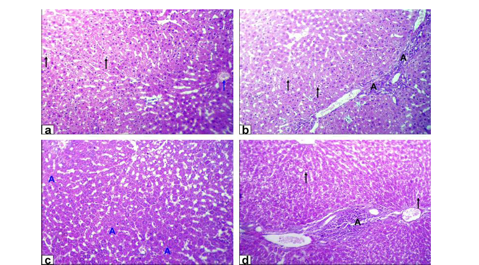
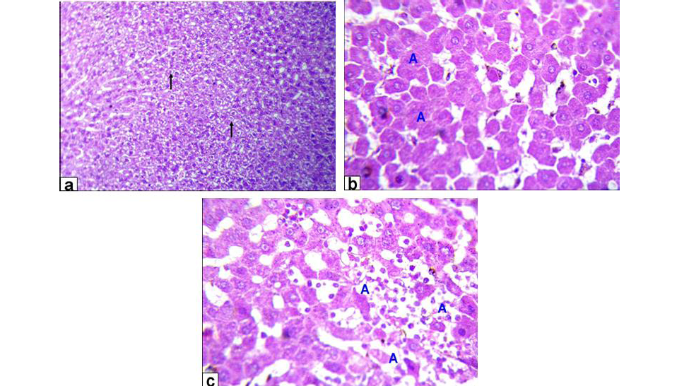
Liver of GA exhibited hydropic degeneration which characterized by the cytoplasm been replaced by clear fluids and the nucleus not affected either in shape or location beside loss of cord arrangement of hepatocytes and portal tract with few aggregations of mononuclear inflammatory cells. Moreover, that of GB showed focal necrosis of the hepatocytes beside fatty degeneration which characterized by large empty vacuoles of sharp borders, that replaced almost the cytoplasm and the portal tract with massive aggregation of mononuclear inflammatory cells which scattered in different spaces with biliary hyperplasia (Figure 1). However, the histopathological examination of the liver tissues of GC revealed remarkable degenerative changes represented by diffuse disorganization of the hepatic cords and moderate hydropic degeneration. While, that of GD exhibited multifocal to diffuse hepatocytes necrosis with fragmented nuclei, ghost or pyknotic and finally necrotic hepatocytes infiltrated with inflammatory cell (Figure 2).
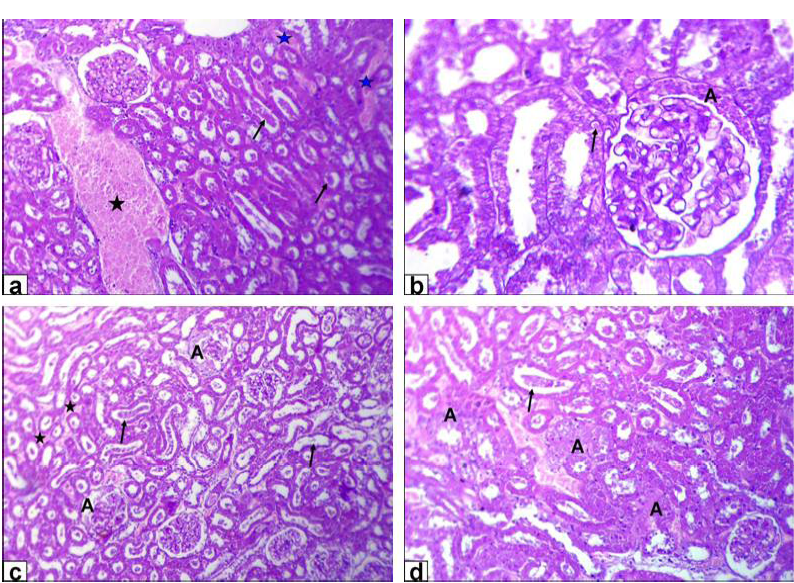
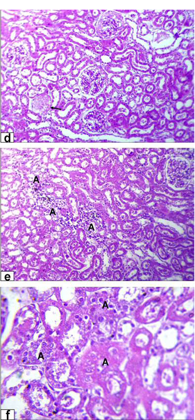
The encountered lesions of the kidney of GA rats were congestion of the blood vessel and inter-tubular blood capillaries, cellular debris in the lumen of the renal tubules beside eosinophilic intranuclear inclusions in lining epithelium of renal tubules and glomerulus’s filtrate occupied the space of bowman capsule. Moreover, tubular dilatation, haemorrhage, glomerulus’s filtrate occupied the space of bowman capsule and finally tubular necrosis were observed in rats of GB (Figure 3).
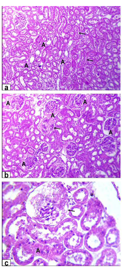
On the other hand, the noticeable lesions in that of GC was tubular dilatation with cellular debris in its lumen; glomerulus’s filtrate occupied the space of bowman capsule and
necrosis of glomerulus’s tuft of bowman capsule and necrosis of some renal tubules (Figure 4A). Moreover, the kidney of GD rats showed complete necrosis of glomerulus’s tuft of bowman capsule, interstitial nephritis with inflammatory cell infiltration and tubular necrosis (Figure 4B).
Discussion
The European Welfare Quality Project defined the criteria of animal welfare as it should include good housing, feeding, health and freedom to behave normally . Furthermore, animal behaviour and health are the most effective animal-based indicators, with researchers/advisers scoring the highest means in case of animal behaviour Moreover, good feeding and housing are from the farm level indicators which are also important for welfare improvement .
Varies neurotransmitters as monoamines, peptides and amino acids controls ingestive behaviour. Many peptides including cholecystokinin, bombesin, calcitonin, corticotrophin-releasing factor, neurotensin and somatostatin had led to a decrease in the feed intake of experimental animals, in contrast, a small number of them increases feed intake. As early as 1924 it had been reported that peripherally administered insulin stimulated feeding in under nourished diabetic children, which appeared to be secondary to the rapid decrease of glucose in blood as if glucose was maintained constant insulin decreased feed intake . Moreover, Harris et al. (1998) stated that several cerebral structures and neurotransmitters intervene in the regulation of feed intake processes as serotonin and dopamine that could play primordial role in the control of satisfaction.
Thus the increment in feeding time of high lead acetate group might be under insulin effect which led to drop in glucose level which was also clear in this study, so the in
crease in feeding time thus correlated by drinking frequency increase. Furthermore, this increase in drinking could be explained by that brain controls many aspects related to the homeostatic processes so the fluctuation in these aspects led the brain to trigger regulatory mechanisms
that include renal water and electrolyte excretion and sodium appetite, the vicinity of the third ventricle regions are especially involved in these homeostatic procedures .
Similarly, Minnema and Hammond (1994) stated that although exposure to lead initially resulted in decreased
nocturnal meal size and duration towards the end of the study, the lead exposed rats had compensated to this by increasing these two parameters during the light phase. Furthermore, the water intake of aluminium treated rats was not different from controls as demonstrated by Mahieu et al. (2009). On contrary, Benlahcen et al. (2009), Kahloula et al. (2009) and Kowalczyk et al. (2004) found a decrease in feed and water intake due to lead acetate and aluminium chloride administration when compared to control group.
The hyperactivity observed in low lead acetate group could be contributed to the hypothesis of dual aminergic and cholinergic neurotransmitters alterations, as the higher aminergic activity, the lower cholinergic activity inducing hyperactivity . Moreover, Luthamn et al. (1994) suggested that changes in motor skills and exploratory behaviour could be induced by lead, which might be in relation to altered dopamine neurotransmitter. These was in consistent with Tawari-Fufeyin et al. (2008) who indicated that guinea pigs intoxicated with lead showed periods of hyperactivity, with restless movements within the cages and in efforts to escape through the mesh holes of their individual cages could have been as a consequence of neurotoxicity of the metal. However, Roig et al. (2006) reported that exposing rats to aluminium in drinking water did not alter significantly motor activity in the open field test. On contrary, Kaoud et al. (2008) showed that lead acetate caused hypoactivity. However, Amit et al. (2012) reported that aluminum exposed mice and rats were more active in the locomotor activity.
The increment of investigatory behaviour in the high lead acetate group are in consistency with Donald et al. (1987) who reported that pups born for pregnant mice administered lead acetate had increased exploratory and social investigation at age of 3-4 weeks than controls. However, Seddik et al. (2010) found that lead acetate administration to rats caused decrease in exploratory activity when compared to control group. Furthermore, Amira and Mahrous (2013b) reported that there was non-significant effect of either metal on investigatory behaviour.
The decrease in lying time of high lead group could be explained by the engage of rats in other behaviours as ingestive and investigatory behaviour to cope with changes present. Similarly, Amira and Mahrous (2013a) and (2013b) reported that in case of lead treated rats there was a decrease in lying time in a dose dependent manner, while, in that of aluminium chloride the decrease was in low dose group only.
Lead and aluminum aggregation in liver and kidney produces toxic remnants by altering various biochemical which including a number of enzymes and cytostructural elements. Its deleterious effects are thought to be mediated via damage to the cell membranes, lipid peroxidation resulting in the loss of membrane fluidity, change membrane potential and/or increase membrane permeability .
In the present work the lipid profile indicated a significant decrease in triglycerides and total lipid levels in all treated groups. These findings were in parallel with those mentioned by Yousef (2004) and El-Nekeety et al. (2009). On contrary, Shrivastava (2011), Bhadauria (2012) showed that triglycerides levels were higher in the lead and aluminum treated group compared to the control group. Moreover, serum glucose level showed a significant decrease in lead treated groups in a dose dependent manner and not affected by aluminum treatment that agrees with Karamala et al. (2011) and Hassan et al. (2008). This hypoglycemia might be due to the dysfunction of liver as the decrease in glucose-6-phosphatase activity indicates decrease degradation of glycogen, which may result in a subsequent decrease in blood glucose level . On contrary, Krishna and Ramachandran (2009) and Bhadauria (2012) showed increase in glucose level of lead and aluminum treated rats.
Hepatic cells share in many of metabolic activities and contain a lot of enzymes. Among these, AST, ALT and ALP are commonly employed biological markers for hepatic injury and efficacy of hepatoprotective interventions . ALT and AST activity are sensitive indicators of hepatic necrosis . Moreover, one of the main indicators of hepatotoxicity is the activity of ALT .
These results may indicate degenerative changes and hypo function of liver and might also be attributed to the increased cell membrane permeability or cell membrane damage of hepatocytes caused by lead acetate . Aluminum toxicity was associated with various biochemical abnormalities and these could usually be contributed to the release of intracellular constituents into the circulation, following the loss of cell membrane integrity or interference with normal metabolism and function. Thus the alteration in these enzymatic activities may be due to infusion of them from liver cytosol into blood stream and/or liver dysfunction . On contrary to this findings, Niu et al. (2009) and Yousef (2004) found lower level of ALT and AST activities with lead acetate and aluminium chloride toxicity, in addition to, an increase in GGT level of the aluminium chloride treated mice .
Urea and uric acid are the principal waste products of protein catabolism. They are synthesized in liver from ammonia which produced as a result of amino acids de-amination , while, creatinine is the major waste product of creatine metabolism by muscle. In the kidney, creatinine is distilled by the glomerulus and excreted by the tubules and only free Creatinine appears in the blood serum . Creatinine and urea could be considered as suitable prognostic indicators of renal dysfunction in case of lead and aluminum exposure.
Rumana et al. (2002) found significant decrease in Creatinine and blood urea nitrogen in lead acetate treated rats. Moreover, Kant et al. (2009) cited that blood urea nitrogen level was decreased after exposure to aluminium sulphate. Furthermore, El-Nahal (2010) and Bhadauria (2012) showed that serum uric acid was significantly increased in lead and aluminum treated rats. This increase in uric acid may be contributed to the impairment in kidney functions . Furthermore, Sadhana et al. (2008) showed that aluminum generates oxidative stress in kidney which caused significantly rise in the activity of uric acid. On contrary to this, Karamala et al. (2011), Shrivastava (2011) and Sadhana et al. (2008) reported that lead acetate and aluminum administration resulted in an increase in serum Creatinine and urea.
The histological examination of the liver tissue for lead acetate and aluminum chloride-treated rats showed mild to moderate acute cellular swelling with hydropic degeneration and multifocal hepatic cell necrosis accompanied by lymphocytic infiltration. The severity of these lesions was dose-dependent. Similar lesions were reported by El-Sokkary et al. (2005) and Agarwal and Jain (2011). Lead-induced portal mononuclear inflammatory cells infiltration, cholestasis and biliary hyperplasia which was well-documented by Hamir and Sullivan (1983). However, fatty changes were sporadic and appeared in a few aluminums treated rat’s hepatocytes.
Kidney histological investigation revealed that lead acetate and aluminum chloride exposure caused progressive glomerular and tubular alterations. Those are in agreement with previous investigations done by Lin et al. (1993) and Abo El-Magd et al. (2015). Interstitial, tubular and glomerular damage are also characteristic renal lesions due to lead acetate toxicity. However, Schraishuhn et al. (1992), Jarrar (2003) and Mcgavin and Zachary (2007) stated that tubular changes occur earlier than glomerular and interstitial changes, including development of pathognomonic eosinophilic intranuclear inclusions in the renal tubular epithelium. These findings showed clear renal lesions associated with chronic lead poisoning and characterized by varying degree of damaged glomeruli, and degenerated proximal tubules in all intoxicated rats in the form of intranuclear inclusion formation, glomerulus’s filtrate occupied the space of bowman capsule, tubular dilatation, hemorrhage and tubular necrosis. Regarding to aluminum induced renal damage, there were tubular dilatation with presence of cellular debris within the lumina of the renal tubules, in addition to necrosis of glomerulus’s tuft of bowman capsule, interstitial nephritis with inflammatory cell infiltration and tubular necrosis was also seen.
CONCLUSION
It can be concluded that both metals affect the welfare of animals at the different stages starting from the behavioural alterations as a first trial of animal to cope with these changes reaching to the pathological affection of the main organs in the body so it is important to record this levels of changes for detection of the welfare alteration of animals in response to these environmental pollutions which indeed will be reflected also on the health of human beings.
ACKNOWLEDGEMENTS
The authors would like to acknowledge the workers who participate to accomplish this work.
CONFLICT OF INTEREST
The authors declare that they have no conflict of interest.
Authors’ contribution
Amira A Goma conceived the idea, participated in the design of the study and also performed the oral dosing of animals, behavioural observations, biochemical measures, analyze the data and written the paper. Hossam G. Tohamy conceived the idea, participated in the design of the study and performed the histopathogical studies and written the paper. All the auhtors read and approved the manuscript.
References
Abdel-Wahhab MA, Omara EA, Abdel-Galil MM, Hassan NS, Nada SA, Saeed A, El-Sayed MM (2007). Zizyphus spina-christi extract protects against aflatoxin B1-intitiated hepatic carcinogenicity. Afr. J. Trad. CAM. 4(3): 248–256.
Abo El-Magd NF, El-Karef A, El-Shishtawy MM, Eissa LA (2015). Hepatoprotective effects of glycyrrhizin and Omega3 fatty acids on Nuclear Factor-kappa B pathway in thioacetamide-induced fibrosis in rats. Egypt. J. Basic Appl. Sci. 2: 65–74.
Agarwal DR, Jain S (2011). Significant liver toxicity in albino rats upon oral aluminum administration: A serious public health implication. J. Anat. Soc. India. 60(1): 46–49. http://dx.doi.org/10.1016/S0003-2778(11)80010-9[CrossRef]
Al-Qayim MAJ, Saadoon D (2013). Assessment of the ameliorative role of proplis and malic acid in intestinal and liver functions of aluminium exposed male rats. Int. J. Sci. Nature. 4(3): 552–558.
Amira AG, Mahrous UE (2013b). Changes in behaviour and learning ability of rats Intoxicated with lead. Int. J. Biol. Sci. Eng. 7(12): 767-773.
Amira AG, Mahrous UE (2013a). Ethological problems and learning disability due to aluminium toxicity in rats. Beni-Suef. Vet. Med. J. 22: 154-159.
Amit P, Manisha C, Kumar JD, Sandeep T, Raj MD (2012). Alteration in thyroid hormones and vitamins as early markers of aluminum induced neurodegeneration in rats. Int. J. Res. Pharm. Sci. 2 (2): 137-150.
Averós X, Aparicio MA, Ferrari P, Guy JH, Hubbard C, Schmid O, Ilieski V ,Spoolder HAM (2013). The Effect of Steps to Promote Higher Levels of Farm Animal Welfare across the EU. Societal versus Animal Scientists Perceptions of Animal Welfare. Animals. 3: 786-807. http://dx.doi.org/10.3390/ani3030786[CrossRef]
Baker HJ, Lindsey JR, Weisbroth SH (1979). Housing to control research variables. In: the laboratory rats. Biological and Diseases. Academic Press, New York. http://dx.doi.org/10.1016/b978-0-12-074901-0.50015-0[CrossRef]
Bancroft JD, Stevens A, Turner DR (1996). Theory and practice of histological techniques. Newyork: Churchill livingstone, Edinburgh, London.
Benlahcen K, Sansar W, Belhabri L, Slimani M (2009). Lead in water: Neurotoxicity and stressful effect on wistar rat. Global J. Environ. Res. 3(1): 52-60.
Bhadauria M (2012). Combined treatment of HEDTA and propolis prevents aluminum induced toxicity in rats. Food Chem. Toxicol. 50: 2487–2495. http://dx.doi.org/10.1016/j.fct.2011.12.040[CrossRef]
Chinoy NJ (2001). Beneficial effects of some vitamins and calcium on fluoride and aluminium toxicity on gastrocnemius muscle and liver of male mice. Fluoride. 34: 21–33.
Coulombe JJ, Favreau L (1963). Anew simple semimicro-method for colorimetric determination of urea. Clin. Chem. 9: 102-108.
Domingo JL, Gomez M, Sanchez DJ, Llobet JM, Corbella J (1993). Effect of various dietary constituents on gastrointestinal absorption of aluminum from drinking water and diet. Res. Commun. Chem. Pathol. Pharmacol. 79(3): 377–380.
Donald JM, Cutler MG, Moore MR (1987). Effects of lead in the laboratory mouse. Development and social behaviour after lifelong exposure to 12 microM lead in drinking fluid. Neuropharmacology. 26(4): 391-399. http://dx.doi.org/10.1016/0028-3908(87)90194-8[CrossRef]
El-Demerdash FM (2004). Antioxidant effect of vitamin E and selenium on lipid peroxidation, enzyme activities and biochemical parameters in rats exposed to aluminium. J. Trace Elem. Med. Biol. 18: 113–121. http://dx.doi.org/10.1016/j.jtemb.2004.04.001[CrossRef]
El-Nahal DM (2010). Effect of using pectin on lead toxicity. J. Am. Sci. 6(12): 541-554.
El-Nekeety AA, El-Kady AA, Soliman MS, Hassan NS, Abdel-Wahhab MA (2009). Protective effect of Aquilegia vulgaris (L) against lead acetate induced oxidative stress in rats. Food Chem. Toxicol. 47: 2209-2215. http://dx.doi.org/10.1016/j.fct.2009.06.019[CrossRef]
El-Sokkary GH, Abdel-Rahman GH, Kamel ES (2005). Melatonin protects against lead- induced hepatic and renal toxicity in male rats. Toxicology. 213: 25–33. http://dx.doi.org/10.1016/j.tox.2005.05.003[CrossRef]
Goldberg AM, Hanin I (1976). Biology of cholinergic function. Raven press, New York.
Gouda IM, Abdel-Aziz SA, Ahmed AA, Lotfic MM, Soliman MM (1985). Changes in some Liver function in experimentally lead poisoned goats. Archiv fur Experimentelle Veterinarimedizin. 39: 257-267.
Gromysz-Kalkowska K, Kanoniuk D, Szubartowska E, Unkiewicz-Winiarczyk A (2004). Influence of drinking water-administered aluminum on morphology and respiratory function of blood in rats. Polish J. Environ. Stud. 13(5): 515-519.
Gryzunov YA, Koplik EV, Smolina NV, Kopaeva LB, Dobretsov GE, Sudakov KV (2006). Conformational properties of serum albumin binding sites in rats with different behaviour in the open field test. Stress. 9(1): 53-60. http://dx.doi.org/10.1080/10253890600677790[CrossRef]
Hamir AN, Sullivan ND (1983). Extra-neural lesions in experimental lead toxicosis of dogs. J Small Anim. Pract. 24: 437–444. http://dx.doi.org/10.1111/j.1748-5827.1983.tb00383.x[CrossRef]
Harris RBS, Zhou J, Youngblood BD, Rybkin II, Smagin GN, Ryan DH (1998). Effect of repeated stress on body weight and body composition of rats fed low and high fat diets. Am. J. Physiol. (Regul. Integr. Comp. Physiol). 275(44): R1928-R1938.
Hassan AA, Jassim HM (2010). Effect of treating lactating rats with lead acetate and its interaction with vitamin E or C on neuro-behaviour, development and some biochemical parameters in their pups. Iraqi J. Vet. Sci. 24(1): 45-52.
Hassan MF, Sadek MA, Abd-El-Razik FH, Kamel EA (2008). Risk of aluminum toxicity and its relation to some biochemical changes in healthy, diabetic and hyperlepidemic rats. Egypt. J. Natural Toxins. 5: 100-120.
Jarrar BM (2003). Histological and histochemical alterations in the kidney induced by lead. Ann. Saudi. Med. 23 (1-2): 10–15.
Johnson AK, Thunhorst RL (1997). The neuroendocrinology of thirst and salt appetite: Visceral sensory signals and mechanisms of central integration. Front. Neuroendocrinol. 18: 292-353. http://dx.doi.org/10.1006/frne.1997.0153[CrossRef]
Josephson B, Gyllensward A (1975). Colorimetric determination of total protein. Scand. J. Clin. Lab. Invest. 9: 29. http://dx.doi.org/10.3109/00365515709088110[CrossRef]
Kahloula K, Slimani M, Abdelkader A (2009). Behavioural and neurochemical studies of perinatal lead exposed in rat wistar. Eur. J. Scient. Res. 35 (4): 603-614.
Kant V, Srivastava AK, Verma PK, Raina R (2009). Alterations in biochemical parameters during subacute toxicity of fluoride alone and in conjunction with aluminum sulphate in goats. Biol. Trace. Elem. Res. 130: 20-30. http://dx.doi.org/10.1007/s12011-008-8311-8[CrossRef]
Kaoud HA, Kamel MM, Abd-El-Razek AH (2008). Effect of neurotoxic metals on neurobehavioural and learning ability in rat pups. J. Egypt. Vet. Med. Assoc. 68: 1-14.
Kaplan MM (1987). Laboratory tests. In: Diseases of the liver. J.B. Lippincott Co., Philadelphia.
Karamala SK, Ch SL, Y A, Rao CS, Vasulu S, Pidugu AP (2011). Hematobiochemical changes of lead poisoning and amelioration with Ocimum sanctum in wistar Albino rats. Vet. World. 4(6): 260-263. http://dx.doi.org/10.5455/vetworld.4.260[CrossRef]
Kashyap SR, Diab DL, Baker AR, Yerian L, Bajaj H, Gray-McGuire C, Schauer PR, Gupta M, Feldstein AE, Hazen SL, Stein CM (2009). Triglyceride levels and not adipokine concentrations are closely related to severity of nonalcoholic fatty liver disease in an obesity surgery cohort. Obesity. 17: 1696-1701. http://dx.doi.org/10.1038/oby.2009.89[CrossRef]
Kowalczyk E, Kopff A, Kedziora J, Blaszczyk J, Kopff M, Niedworok J, Fijalkowski P (2004). Effect of long-term aluminum chloride intoxication on selected biochemical parameters and oxidative –antioxidative balance in experimental animals. Polish J. Environ. Stud. 13(1): 41-43.
Kozak M, Kralova E, Sviatko P, Bilek J ,Bugarsky A (2002). Study of the content of heavy metals related to environmental load in urban areas in Slovakia. Bratsil Lek Listy. 103(7-8): 231-237.
Krishna H, Ramachandran AV (2009). Biochemical alterations induced by the acute exposure to combination of chlorpyrifos and lead in Wistar rats. Biol. Med. 1(2): 1-6.
Landrigan PJ, Boffetta P, Apostoli P (2000). The reproductive toxicity and carcinogenicity of lead: a critical review. Am. J. Ind. Med. 38: 231–243. http://dx.doi.org/10.1002/1097-0274(200009)38:3<231::AID-AJIM2>3.0.CO;2-O[CrossRef]
Larsen K (1972). Creatinine assay by a reaction-kinetic principle. Clin. Chim. Acta. 41: 209-217. http://dx.doi.org/10.1016/0009-8981(72)90513-X[CrossRef]
Lavicoli I, Carelli G, Stanek EJ, Castellino N, Calabrese EJ (2003). Effects of low doses of dietary lead on red blood cell production in male and female mice. Toxicol. Lett. 137: 193-199. http://dx.doi.org/10.1016/S0378-4274(02)00404-6[CrossRef]
Levine AS, Morley JE, Gosnell BA, Billington CJ, Krahn DD (1986). Neuropeptides as regulators of consummatory behaviours (Review). J. Nutr. 116: 2067-2077.
Lin JL, Yeh KH, Tseng HC, Chen WY, Lai HH, Lin YC (1993). Urinary N-acetylglucosaminidase excretion and environmental lead exposure. Am. J. Nephrol. 13: 442–447. http://dx.doi.org/10.1159/000168661[CrossRef]
Luthamn J, Lindqvist, E, Gerhardt GA, Olson L, Hoffer BH (1994). Alterations in central monoamine systems after postnatal lead acetate treatment in rats. Environ. Res. 65: 100-118. http://dx.doi.org/10.1006/enrs.1994.1024[CrossRef]
Mahieu S, Contini MDC, Gonzalez M, Millen N (2009). Melatonin reduces oxidative damage induced by aluminum in rat kidney. Toxico. Lett. 190: 9-15. http://dx.doi.org/10.1016/j.toxlet.2009.06.852[CrossRef]
Martin P, Bateson P (1993). Measuring Behaviour: An Introductory Guide. Cambridge University Press, Cambridge, London. http://dx.doi.org/10.1017/cbo9781139168342[CrossRef]
McCann BS, Magee MS, Broyles FC, Vaughan M, Albers JJ, Knopp RH (1995). Acute psychological stress and epinephrine infusion in normolipidemic and hyperlipidemic men: Effects on plasma lipid and apoprotein concentrations. Psychosomatic Med. 57: 165-176. http://dx.doi.org/10.1097/00006842-199503000-00009[CrossRef]
McGavin MD, Zachary JF (2007). Pathologic basis of veterinary disease. Mosby-Elsevier, St. louis, Missouri. 4: 1476.
Minnema DJ, Hammond PB (1994). Effect of lead exposure on patterns of food intake in weanling rats. Neurotoxicol. Teratol. 16 (6): 623-629. http://dx.doi.org/10.1016/0892-0362(94)90040-X[CrossRef]
Misawa T, Shigeta S (1992). Behavioural effects of repeated aluminum administration in the rat. Takai. J. Exp. Clin. Med. 17 (5): 155-159.
Niu R, Sun Z, Cheng Z, Li Z, Wang J (2009). Decreased learning ability and low hippocampus glutamate in offspring rats exposed to fluoride and lead. Environ. Toxicol. Pharmacol. 28: 254-258. http://dx.doi.org/10.1016/j.etap.2009.04.012[CrossRef]
Ochmanski W, Barabasz W (2000). Aluminum-occurrence and toxicity for organisms. Przegl Lek. 57: 665–668.
Patra RC, Swarup D, Dwivedi SK (2001). Antioxidant effects of alpha-tocopherol, ascorbic acid and Lmethionine on lead induced oxidative stress to the liver, kidney and brain in rats. Toxicology. 162: 81–88. http://dx.doi.org/10.1016/S0300-483X(01)00345-6[CrossRef]
Patrick L (2006). Lead Toxicity Part II: the role of free radical damage and the use of antioxidants in the pathology and treatment of lead toxicity. Altern. Med. Rev. 11(2): 114–127.
Pfister HP, Ivinskis A (1983). Prenatal psychological stress effects on offspring behaviour in rats. Austr. J. Psychol. 35(1): 21-36. http://dx.doi.org/10.1080/00049538308255300[CrossRef]
Reitman S, Frankel S (1957). A Colorimetric method for the determination of serum glutamic oxalacetic and glutamic pyruvic transaminases. Am. J. Clin. Pathol. 28: 56-63. http://dx.doi.org/10.1093/ajcp/28.1.56[CrossRef]
Roig JL, Fuentes S, Colomina MT, Vicens P, Domingo JL (2006). Aluminum, restraint stress and aging: Behavioural effects in rats after 1 and 2 years of aluminum exposure. Toxicology. 218: 112-124. http://dx.doi.org/10.1016/j.tox.2005.10.006[CrossRef]
Rumana S, Mishra GV, Vohora SB (2002). Effect of therapeutic doses of calcined arsenic and lead preparations in rats. Ind. J. Vet. Pathol. 26: 81- 82.
Sadhana S, Sangeeta S, Ramesh M (2008). Co-therapy of chelating agents and Ca/Fe against aluminum induced nephrotoxicity. Ind. J. Pharmacol. 40: 161.
Sanders T, Liu Y, Buchner V, Tchounwou PB (2009). Neurotoxic effects and biomarkers of lead exposure: A review. Rev. Environ. Health. 24 (1): 15–45. http://dx.doi.org/10.1515/REVEH.2009.24.1.15[CrossRef]
Sarov GM, Vlaykova TI (2005). Changes in blood glucose, triglycerides and lipid peroxidation products in rabbits after hanging fixation. Bulgarian J. Vet. Med. 8 (3): 157-161.
Schraishuhn J, Kaufer-Weiss I, Weiss E (1992). Light and electron-microscopic studies of calf kidneys after exposure to subtoxic lead levels. Berl Munch Tierarztl Wochenschr. 105: 290–293
Seddik L, Bah TM, Aoues M, Benderdour M, Slimani M (2010). Dried leaf extract protects against lead induced neurotoxicity in wistar rats. Eur. J. Scient. Res. 42(1): 139-151.
Shati AA, Alamri SA (2010). Role of saffron (Crocus sativas L.) and honey syrup on aluminum induced hepatotoxicity. Saudi. Med. J. 31(10): 1106-1113.
Shelley R, Kim NS, Parsons PJ, Lee BK, Jaar B, Fadrowski J, Agnew J, Matanoski GM, Schwartz BS, Steuerwald AJ, Todd AC, Simon D, Weaver VM (2012). Associations of multiple metals with kidney outcomes in lead workers. Occup. Environ. Med. 69(10): 727–735. http://dx.doi.org/10.1136/oemed-2012-100765[CrossRef]
Shrivastava S (2011). S-Allyl-Cysteines reduce amelioration of aluminum induced toxicity in rats. Am. J. Biochem. Biotechnol. 7(2): 74-83. http://dx.doi.org/10.3844/ajbbsp.2011.74.83[CrossRef]
Stafford K (2014). Sheep veterinarians and the welfare of sheep: No simple matter. Small Ruminant Res. 118: 106-109. http://dx.doi.org/10.1016/j.smallrumres.2013.12.008[CrossRef]
Suradkar SG, Ghodasara DJ, Vihol P, Patel J, Jaiswal V, Prajapati KS (2009). Haemato-biochemical alterations induced by lead acetate toxicity in Wistar rats. Vet. World. 2(11): 429-431.
Taib NT, Jarrar BM, Mubarak M (2004). Ultrastructural alterations in hepatic tissues of white rats (Rattus norvegicus) induced by lead experimental toxicity. Saudi. J. Biol. Sci. 11: 11–20.
Tawari-Fufeyin P, Ogie-Odia EA, Asemota OC, Balogun AF (2008). Use of Amaranthus hybridus to reduce toxic effects of some heavy metals in guinea pig (Cavia porcellus) tissues. Biosci. Res. Commun. 20: 277-281.
Testolin G, Erba D, Ciappellano D, Bermano G (1996). Influence of organic acids on aluminum absorption and storage in rat tissues. Food Additives Contaminants. 13 (1): 21–27. http://dx.doi.org/10.1080/02652039609374378[CrossRef]
Thomas L (1992). Enzymatic colorimetric determination of cholesterol. In: Labour and Diagnoze. Volume 4.
Tietz NW (1994). Textbook of clinical chemistry, Volume 2, W.B. Saunders Co., Philadelphia, P.A. Pp. 851.
Trinder P (1969). Rapid colorimetric method for determination of blood glucose. Annu. Clin. Biochem. 6: 24-27. http://dx.doi.org/10.1177/000456326900600108[CrossRef]
Turkez H, Yousef MI, Geyikoglu F (2010). Propolis prevents aluminium-induced genetic and hepatic damages in rat liver. Food Chem. Toxicol. 48: 2741–2746. http://dx.doi.org/10.1016/j.fct.2010.06.049[CrossRef]
Verstraeten SV, Aimo L, Oteiza PI (2008). Aluminium and lead: molecular mechanisms of brain toxicity. Arch. Toxicol. 82: 789–802. http://dx.doi.org/10.1007/s00204-008-0345-3[CrossRef]
Watanabe S (2007). How animal psychology contributes to animal welfare. Appl. Anim. Behav. Sci. 106: 193-202. http://dx.doi.org/10.1016/j.applanim.2007.01.003[CrossRef]
Whitehead TP, Bevan EA, Miano L, Leonardi A (1991). Defects in diagnostic kits for determination of urate in serum. Clin. Chem. 37(6): 879-881.
Wilson MA, Johnston MV, Goldstein CW, Blue ME (2000). Neonatal lead exposure impairs development of rodent barrel field cortex. Proc. Nat. Acad. Sci. 97(10): 5540-5545. http://dx.doi.org/10.1073/pnas.97.10.5540[CrossRef]
Yang RZ, Park S, Reagan WJ, Goldstein R, Zhong S, Lawton M, Rajamohan F, Qian K, Liu L, Gong DW (2009). Alanine aminotransferase isoenzymes: molecular cloning and quantitative analysis of tissue expression in rats and serum elevation in liver toxicity. Hepatology. 49 (2): 598-607. http://dx.doi.org/10.1002/hep.22657[CrossRef]
Young DS, Thomas DW, Friedman RB, Pestaner LC (1972). Effects of drugs on clinical laboratory tests. Clin. Chem. 18 (10): 1041-1303.
Yousef MI (2004). Aluminum induced changes in hemato-biochemical parameter, lipid peroxidation and enzyme activities of male rabbits: Protective role of ascorbic acid. Toxicology. 199: 47-57. http://dx.doi.org/10.1016/j.tox.2004.02.014[CrossRef]