
The purpose of this study was to investigate the impact of toll like receptor 9 (TLR9) signaling pathway and its immunoregulatory roles in neonatal acute lung injury (NALI) , laying a theoretical basis for the clinical therapy of NALI. Forty peripheral blood mononuclear cells (PBMCs) of newborns with acute lung injury (ALI) were recruited as experimental group (EG), and another 40 PBMCs of normal newborns were selected as normal group (NG). PCR was used to detect TLR9 mRNA expression. The PBMCs were cultured by non-CpG ODN (control group, CG), CpG ODN (experimental group, EG), and blank culture (BG) for 72 h. Proliferation of PBMCs was measured with a β liquid scintillator, and the concentration of interferon (IFN)-α was determinded. It was revealed that the intensity of TLR9 mRNA expression in the PBMCs of patients with ALI was 0.76±0.06, which differed slightly versus NG (0.74±0.04) (P>0.05). The number of proliferating cells was markedly different from the CG and the BG after using TLR9 activator (P<0.05); TLR9 agonist could increase IFN-α production obviously versus CG and BG (P<0.05). It was concluded that the intensity of TLR9 mRNA expression in PBMCs of patients with ALI differed inconsiderably from that of healthy newborns. TLR9 signaling pathway was involved in immune regulation in newborns with ALI, including enhancing the proliferation ability of PBMC and increasing the production of IFN-α.
Short Communication · Volume 56, 25 March 2025 · Open Access
Immunoregulatory Effect of Toll Like Receptor 9 Signaling Pathway in Neonatal Acute Lung Injury
Affiliations & Notes ⌄
- 1Department of Pediatrics, Shanghai East Hospital Affiliated to Tongji University, Shanghai, 200120, China. * Corresponding author: yujiao35937346@163.com
Article Info ⌄
- Journal:
- Pakistan Journal of Zoology
- Published:
- 25 March 2025

From the article
- The results revealed that TLR9 mRNA was expressed in PMBC of all lung injury patients and all healthy participants.
- The average expression intensity of TLR9 mRNA in PBMC of ALI patients was 0.76±0.06, and the average expression intensity of TLR9 mRNA in PBMC of NG was 0.74±0.04.
- The effect of TLR9 signaling pathway in NALI is achieved by participating in immune regulation, and its specific effects are to enhance the proliferation ability of PBMC and increase the production of IFN-α.
- On this basis, it could be considered that TLR9 was involved in the immune regulation of patients with acute pneumonia, and its regulation included promoting the increase of PMBC proliferation intensity and increasing the output of IFN-α.
Selected from the article's own text.
Abstract
Materials and methods
Peripheral blood mononuclear cells (PBMCs) of 40 newborns with ALI were recruited as experimental group (EG) and PBMCs of normal newborns was recruited as normal group (NG) for this study. 0-3 years old patients with ALI having received no drugs and with no other lung diseases were included. Patients with psychiatric diseases or other systemic illnesses, concurrent with additional lung diseases were excluded.
The expression intensity of TLR9 mRNA in the EG and the NG were analyzed by PCR. For this, the PBMCs were cultured by non-CpG ODN (control group, CG), CpG ODN (EG), and blank culture (blank group, BG). After 72-h culture, the proliferation changes of PBMCs were measured by β liquid scintillator, and the concentration of interferon (IFN)-α was measured by ELISA.
For isolation of PBMCs, the peripheral venous blood was injected into the heparin anticoagulant tube, diluted with equal volume of phosphate buffer saline (PBS) and centrifuged at 2000 rpm for 30 min at 18-20°C. PBMC has antigen-recognizing receptors and mitogen receptors on its surface, and the corresponding cell clones can proliferate under the stimulation of specific antigens. The concentration of IFN-α in samples was detected by double-antibody sandwich ELISA method.
The experiment’s data underwent analysis using SPSS 19.0. Measurement data were presented as means ± standard deviation, while count data were represented in percentages and concentrations. Statistical analysis revealed significant differences when P<0.05.
Results
From the newborns recruited, the gender distribution was 18 female and 21 male patients with ALI aged ≤3 years, with a M (SD) age of 1.5 (0.94) years. The results showed that there are no significant differences between the study groups so it can be concluded that they are homogenous groups.
Table I shows the descriptive data for the expression of TLR9 mRNA intensity at different ages and genders in the EG and the NG. Figure 1 displayed the data of TLR9 mRNA intensity expressed at different ages in the patient group and NG. The results revealed that TLR9 mRNA was expressed in PMBC of all lung injury patients and all healthy participants. The average expression intensity of TLR9 mRNA in PBMC of ALI patients was 0.76±0.06, and the average expression intensity of TLR9 mRNA in PBMC of NG was 0.74±0.04. Therefore, the EG and the NG demonstrated neglect able differences (P>0.05).
Table I. Intensity map of TLR9 mRNA expression in study groups.
| Age (year) | Normal group | Patient group | ||
| Male | Female | Male | Female | |
| Age≤1 | 0.76 | 0.73 | 0.73 | 0.77 |
| 1<Age≤2 | 0.77 | 0.75 | 0.79 | 0.78 |
| 2<Age≤3 | 0.72 | 0.71 | 0.75 | 0.74 |
Figure 2 shows the value-added data of PBMC under the action of TLR9 inducer at different ages. The PBMC of patients with ALI were activated by CpG ODN for three days, which was compared with CG (ODN) and BG after three days of culture. What’s more, the cell suspension was tested with a β liquid scintillator. The experimental data indicated that the concentration of PBMCs in patients with ALI ((1.72±0.13) ×10 cells/mL) was markedly superior to that of PBMCs in CG ((0.35±0.11) ×10 cells/mL) (P<0.05) and the cell concentration of PBMCs in BG ((0.26±0.13) ×10 cells/mL) (P<0.05). Thus, the above disclosed that EG had statistically marked difference versus CG and BG (P<0.05).
The concentration data of IFN-α in research objects at different ages under the action of agonists (Fig. 3). After three consecutive days of CpG ODN stimulation, the data disclosed that the concentration of IFN-α in the PBMC supernatant of patients with ALI (193.7±3.8ng/L) increased notably versus CG (25.7±1.6ng/L) (P<0.05) and the concentration of BG (18.4±1.3ng/L) (P<0.05). Therefore, the results demonstrated substantial difference between EG and CG (P<0.05).
Discussion
There are many factors that lead to NALI . Its pathological and physiological characteristics are mainly reduced lung volume, decreased lung compliance, ventilation, or blood flow proportional imbalance , and the development to the severe stage is called acute respiratory distress syndrome. The physiological anatomy of the respiratory tract of the newborn shows that the nasopharynx, trachea, and bronchi are narrowed, and mucus secretion is small , and the immune function of the newborn is not fully developed, so it is more likely to suffer from ALI. The treatment of ALI is mainly to improve the oxygenation of the patient, and then adjust the body function. TLRs play a crucial role in natural immunity as a class of protein molecules . These single transmembrane non-catalytic proteins are adept at identifying conserved structures from microorganisms . Upon breaching physical barriers like skin or mucous membranes, TLRs recognize microorganisms, prompting the body to trigger immune cell responses. There are currently 11 members of the human TLRs family that have been found in mammals and humans . TLR9 is the main receptor in the innate immune system that recognizes the unmethylated CpG DNA of bacteria and viruses. The effect of TLR9 signaling pathway in NALI is achieved by participating in immune regulation, and its specific effects are to enhance the proliferation ability of PBMC and increase the production of IFN-α.
In the experiment of detecting the expression of TLR9 mRNA, the PMBC of all lung injury patients and healthy participants expressed TLR9 mRNA, and the average expression intensity of TLR9 mRNA expression in the PBMC of ALI patients was 0.76±0.06, which was compared with the average of NG (0.74±0.04). Therefore, the experimental results revealed neglect able difference between the patient group and NG (P>0.05). PBMC of EG of ALI patients were cultured for 3 days, and they were also cultivated in CG and BG, which were detected by the β liquid scintillator. PBMC concentration of ALI patients was (1.72±0.13)×10, which was higher dramatically than the concentration of CG ((0.35±0.11)×10) (P<0.05) and BG ((0.26±0.13)×10) (P<0.05). Three days after CpG ODN stimulation, IFN-α (193.7 ± 3.8 ng/L) in the supernatant of ALI patients rose substantially versus CG (25.7±1.6 ng/L) (P<0.05) and BG (18.4±1.3 ng/L) (P<0.05). The experimental results were similar to the current research status, and suggested that the intensity of expression of TLR9 mRNA in the PBMC of patients with ALI differed slightly from that of healthy newborns. The TLR9 signaling pathway was involved in immune regulation in NALI, and its effects included enhancing the proliferation ability of PBMC and increasing the production of IFN-α.
Conclusion
In this study, the patient group and NG were first selected, and the two groups were detected by PCR. The expression intensity of TLR9 mRNA was observed between groups, and no marked difference was found. On this basis, it could be considered that TLR9 was involved in the immune regulation of patients with acute pneumonia, and its regulation included promoting the increase of PMBC proliferation intensity and increasing the output of IFN-α. Consequently, future experiments will involve larger sample sizes to delve deeper into analyzing the TLR9 signaling pathway’s impact on NALI. Ultimately, this study’s findings aim to offer scientific data support and a theoretical framework for understanding the TLR9 signaling pathway’s mechanism in NALI.
Acknowledgment
Thanks to the members from Shanghai East Hospital Affiliated to Tongji University, the group collected samples, obtained data, and theoretical guidance.
Funding
The study received no external funding.
IRB approval
This study was approved by the Advanced Studies Research Board of Shanghai East Hospital Affiliated to Tongji University, Shanghai, China.
Ethical approval
The study was carried out in compliance with guidelines issued by Ethical Review Board Committee of Shanghai East Hospital Affiliated to Tongji University, China. The official letter would be available on fair request to corresponding author.
Statement of conflict of interest
The author has declared no conflict of interest.
Figures
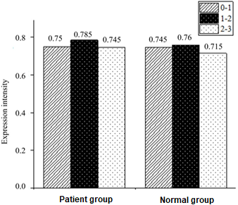
Comparison on expression intensity between NG and patient group.
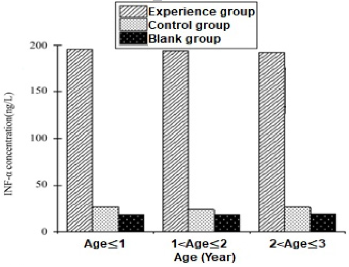
Proliferation of peripheral blood mononuclear cells.
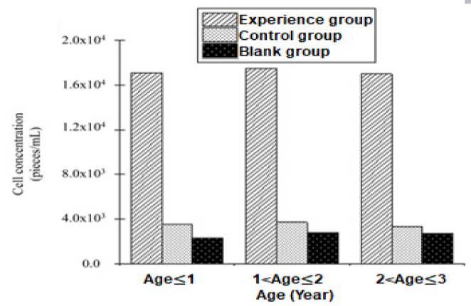
Concentrations of IFN-α.
References
- Abed, M., Elhariri, M., El-Helw, R., Khattab, M.S., Setta, A. and Abed, M., 2020. J. World’s Poult. Res., 10: 60-66. https://doi.org/10.36380/scil.2020.wvj8[CrossRef]
- Blagev, D.P., Harris, D., Dunn, A.C., Guidry, D.W., Grissom, C.K. and Lanspa, M.J., 2019. Lancet, 394: 2073-2083. https://doi.org/10.1016/S0140-6736(19)32679-0[CrossRef]
- Cao, C., Yin, C.F., Shou, S.T., Wang, J., Yu, L.C., Li, X.N. and Chai, Y.F., 2018. Shock, 50: 595-605. https://doi.org/10.1097/SHK.0000000000001104[CrossRef]
- De Dios, R., Nguyen, L., Ghosh, S., McKenna, S. and Wright, C.J., 2020. Immunology, 160: 64-77. https://doi.org/10.1111/imm.13182[CrossRef]
- Goonewardene, K.B., Popowich, S., Gunawardana, T., Gupta, A., Kurukulasuriya, S., Karunarathna, R., Chow-Lockerbie, B., Ahmed, K.A., Tikoo, S.K., Foldvari, M., Willson, P. and Gomis, S., 2017. Avian Dis., 61: 503-511. https://doi.org/10.1637/11684-060617-Reg.1[CrossRef]
- Huang, R.Q., Qin, C.J., Wang, J.M., Hu, Y.Q., Zheng, G.P., Qiu, G.G., Ge, M.H., Tao, H.K., Shu, Q., Xu, J.G., 2019. Aging, 11: 7996-8014. https://doi.org/10.18632/aging.102314[CrossRef]
- Kellner, M., Noonepalle, S., Lu, Q., Srivastava, A., Zemskov, E. and Black, S.M., 2017. Adv. exp. Med. Biol., 967: 105-137. https://doi.org/10.1007/978-3-319-63245-2_8[CrossRef]
- Makita, Y., Suzuki, H., Kano, T., Takahata, A., Julian, B.A., Novak, J. and Suzuki, Y., 2020. Kidney Int., 97: 340-349. https://doi.org/10.1016/j.kint.2019.08.022[CrossRef]
- Naik, E., Chi, Y., Milley, R., Calacsan, C., Chipman, S., Tüting, T., Coffman, R. and Guiducci, C., 2017. Cancer Res., 77: 5596-5596. https://doi.org/10.1158/1538-7445.AM2017-5596[CrossRef]
- Schaedler, E., Remy-Ziller, C., Hortelano, J., Kehrer, N., Claudepierre, M.C., Gatard, T., Jakobs, C., Préville, X., Carpentier, A.F. and Rittner, K., 2017. Vaccine, 35: 577-585. https://doi.org/10.1016/j.vaccine.2016.12.020[CrossRef]
- Schmitt, H., Ulmschneider, J., Billmeier, U., Vieth, M., Scarozza, P., Sonnewald, S., Reid, S., Atreya, I., Rath, T., Zundler, S., Langheinrich, M., Schüttler, J., Hartmann, A., Winkler, T., Admyre, C., Knittel, T., Dieterich Johansson, C., Zargari, A., Neurath, M.F. and Atreya, R., 2020. J. Crohns. Colitis., 14: 508-524. https://doi.org/10.1093/ecco-jcc/jjz170[CrossRef]
- Sekheri, M., El-Kebir, D., Edner, N. and Filep, J.G., 2020. Proc. natl. Acad. Sci. USA, 117: 7971-7980. https://doi.org/10.1073/pnas.1920193117[CrossRef]
- Sommariva, M., Le Noci, V., Storti, C., Bianchi, F., Tagliabue, E., Balsari, A. and Sfondrini, L., 2017. Cell. Immunol., 313: 52-58. https://doi.org/10.1016/j.cellimm.2017.01.004[CrossRef]
- Wu, G.N., Zhu, Q.Q., Zeng, J.L., Gu, X.L., Miao, Y.Y., Xu, W.J., Lv, T.F. and Song, Y., 2019. J. Thorac. Dis., 11: 4816-4828. https://doi.org/10.21037/jtd.2019.10.26[CrossRef]
- Xie, L., Liu, S.Y., Cheng, J.H., Wang, L.J., Liu, J. and Gong, J.B., 2017. Regul. Toxicol. Pharmacol., 89: 148-154. https://doi.org/10.1016/j.yrtph.2017.07.028[CrossRef]
- Xu, W.J., Wang, X.X., Jin, J.J., Zou, Q., Wu, L., Lv, T.F., Wan, B., Zhan, P., Zhu, S.H., Liu, H.B., Zhao, N.W., Li, C.J. and Song, Y., 2019. Am. J. Physiol. Lung. Cell. Mol. Physiol., 316: L567-L577. https://doi.org/10.1152/ajplung.00190.2018[CrossRef]
- Yan, J., Li, J.J., Zhang, L., Sun, Y., Jiang, J., Huang, Y., Xu, H., Jiang, H. and Hu, R., 2018. Free Radic. Biol. Med., 121: 78-85. https://doi.org/10.1016/j.freeradbiomed.2018.04.557[CrossRef]
- Ye, C., Li, H.T., Bao, M.W., Zhuo, R., Jiang, G.N. and Wang, W.X., 2020. Aging, 12: 6120-6128. https://doi.org/10.18632/aging.103010[CrossRef]
- Zhou, B.H., Yan, J.L., Guo, L., Zhang, B., Liu, S., Yu, M.C., Chen, Z., Zhang, K.W., Zhang, W.T., Li, X.Q., Xu, Y.F., Xiao, Y.S., Zhou, J., Fan, J., Hung, M.C., Li, H. and Ye, Q.H., 2020. Theranostics, 10: 6530-6543. https://doi.org/10.7150/thno.44417[CrossRef]
Services
Article usage statistics
Loading…