Our results showed that there was a negative relationship between the poultry farms’ hygiene and the prevalence of Enterobacteriaceae.
The duck farms were the most contaminated farms with Salmonella and E. coli; in contrast, the breeder chicken farms recorded the lowest prevalence.
Our result demonstrated that Salmonella species showed 100% resistance against doxycycline, ampicillin, and amoxicillin.
The results revealed that multidrug resistance was observed in all Salmonella isolates against four antibiotics or more and in all E. coli isolates against three antibiotics or more (except in O44).
Selected from the article's own text.
Abstract
Enterobacteriaceae threaten the success of the poultry industry, as they cause great economic losses due to the high mortalities and the high treatment cost. They cause serious public health hazards, as they are the major cause of food-borne infections. The study aimed to investigate the prevalence of Enterobacteriaceae in twelve poultry farms located in Qalubia Governorate, Egypt, and their antimicrobial resistance. A total number of 2160 samples included litter, pen litter, stored feed, feed from the feeders, water, drinkers, droppings, dust, swabs from walls, birds’ cloaca, worker’s hands, and wheels of vehicles. The results showed that there was a negative relationship between the prevalence of Enterobacteriaceae and the hygienic measures enforced in the poultry farms under study. The highest prevalence of Salmonella and E. coli was recorded in duck farms (36.2% and 54.3% respectively) and the lowest prevalence was recorded in breeder chicken farms (10.2% and 29.3% respectively). The isolated Salmonella species showed high resistance (100%) against doxycycline, ampicillin, and amoxicillin, while E. coli species showed resistance (90%) against oxytetracycline, doxycycline, ampicillin, and amoxicillin. Both Salmonella and E. coli species were highly susceptible to gentamicin. Gene tetA and blaSHV were detected in Gene tetA and blaSHV were detected in 28.5% & 57.1% of Salmonella, respectively, and 70% & 60% of E. coli serotypes, respectively. The application of good biosecurity programs including strict measures in poultry farms is the preferable method to reduce the risk of pathogenic bacteria and reduce the use of antibiotics.
Poultry is an important source of protein due to its cheaper prices and high nutritional value. Therefore, poultry farming has become one of the most important industries worldwide and has significantly developed and increased in recent years . Poultry hygiene is a critical point, especially in intensive farms with closed housing that facilitates the spreading of bacterial contamination such as Enterobacteriaceae . Enterobacteriaceae are widely distributed in nature and can be found in soil, feed, and water. They normally inhabit the gastrointestinal tract of birds, humans, and animals and might cause serious infections in poultry under certain circumstances.
Salmonella, Escherichia coli (E. coli), and Shigella are the major cause of food-borne infection all around the world. Poultry and its products are considered the main vehicle for these pathogens causing serious economic losses and huge public health hazards, in addition to huge numbers of annual mortalities . Salmonella causes serious clinical symptoms and high mortality at young ages less than 6 weeks, older chicks may show stunting and uneven growth . E. coli causes economic loss in broilers due to high mortality and weight loss and a decrease in egg production and profitability in layer and breeder farms . Shigella can infect chickens (shigellosis) causing gastroenteritis, the development of serious complications, and death and it is known for its zoonotic capabilities . Antimicrobials are widely used in poultry farms as one of the methods to control the bacterial diseases that result from improper hygiene; they aren’t only used to treat and prevent infectious diseases but also as prophylaxis, and growth promotors. On the other side, bacteria had built up great resistance to antibacterial substances. This complex situation increases the development of multi-drug resistant bacteria (MAR) in poultry .
The present study aimed to investigate the prevalence of some Enterobacteriaceae species such as Salmonella, E. coli, and Shigella in different poultry farms and monitor the antimicrobial resistance of the isolated Enterobacteriaceae species with the detection of some resistance genes such as tetA and blaSHV.
MATERIAL AND METHODS
Sampling
A total number of 2160 samples and swabs were collected from twelve poultry farms, 180 samples, and swabs from each farm in three visits per farm, and five samples were collected per visit from each type of samples and swabs. The samples included litter, pen litter, stored feed, feed from the feeders, water, drinkers, droppings, dust, swabs from walls, birds’ cloaca, worker’s hands, and wheels of vehicles, 180 samples per each. The collection of samples was approved with Institutional Approval Number (BUFVTM 04-07-22).
Poultry farms
The present study was carried out on twelve poultry farms. The selection of the farms was based on their geographical location, hygienic level, housing system, and type of production. All farms are located in Qalyubia Governorate, Egypt, as well as all farms use public chlorinated tap water as a water source. The most used antibiotics in studied poultry farms were oxytetracycline, doxycycline, amoxicillin, and flumequine. The basic information on poultry farms under study and the applied hygienic measures were listed in (Table 1).
Preparation of samples
One gram of each sample was put in nine ml of buffer peptone water (BPW) to be ready for bacteriological examination .
Isolation and identification of Enterobacteriaceae
Isolation of Salmonella: The previously prepared samples were incubated aerobically in BPW at 37ºC for 24 h. From the pre-enrichment tubes, one ml was inoculated into nine ml Rappaport Vassiliadis (RV) broth and incubated aerobically at 42ºC for 24 h. A loop full of selectively enriched broth was streaked separately onto Xylose Lysine Desoxycholate (XLD) agar and Hektoen enteric (HE) agar and incubated at 37ºC for 24 h. The suspected colonies were pink with or without black centers colonies on XLD and clear colonies with or without black centers on HE agar. One colony from the presumptive Salmonella colonies was subcultured onto XLD agar until the pure homogenous colonies were obtained. The pure suspected colonies were subcultured onto nutrient agar plates for further identification. These procedures were carried out after .
Isolation of E. coli: The previously prepared samples were incubated aerobically in BPW at 37ºC for 24 h. A loop full of the non-selective enriched broth was streaked onto Eosin Methylene Blue (EMB) agar and incubated at 37°C for 24 h. The suspected colonies were metallic green in reflected light with a blue-black center in transmitted light. The typical colonies were subcultured onto EMB agar until the pure cultures with homogenous colonies were obtained. The pure colonies were subcultured onto nutrient agar plates for further identification. These procedures were carried out after .
Isolation of Shigella: The samples were pre-enriched in peptone water and incubated anaerobically at 41.5ºC for 20 h. A loop full of the non-selective enriched samples was inoculated onto McConkey agar (low selectivity), XLD agar (moderate selectivity), and HE agar (high selectivity) and incubated at 37ºC for 24 h, the suspected colonies were smooth colorless colonies on McConkey agar, pale red on XLD agar, and ranged from clear to pale green colonies on HE agar. The pure suspected colonies were subcultured onto McConkey agar plates for further identification. These procedures were carried out after (ISO, 2004).
Biochemical identification of Enterobacteriaceae: The purified suspected colonies of Salmonella, E. coli, and Shigella were identified based on biochemical tests panel following the standard test protocol described in FDA’s Bacteriological Analytical Manual (FDA, 2012), , and , respectively.
Table 1: The basic information and hygienic measures of poultry farms under study.
Farms
Type of the flock
Housing system
Ventilation
Feed
and watering system
Fence
Foot bath
Worker hygiene
visit
ors
con
trol
hygienic disposal of wastes
Cleaning and disinfecti
on 4program
Farm 1
Broiler chicken
Open deep litter
Mechanical and
natural
Manual
-
-
Bad
Bad
Not applied
Weak
Farm 2
Broiler chicken
Open deep litter
Mechanical and
natural
Manual
-
-
Fair
Fair
Not applied
Fair
Farm 3
Broiler chicken
Open deep litter
Mechanical and
natural
Auto
mated
+
+
Fair
Fair
applied
Fair
Farm 4
Layer chicken
Open deep litter
Natural
Manual
-
+
Fair
Fair
applied
Good
Farm 5
Layer chicken
Open deep litter
Natural
Manual
-
_
Bad
Bad
Not applied
Weak
Farm 6
Layer chicken
Battery, open system
Mechanical and natural
Auto
mated
+
+
Fair
Fair
applied
Good
Farm 7
Broiler breeder chicken
Closed deep litter
Mechanical
Auto
mated
+
+
Good
Good
applied
Good
Farm 8
Broiler breeder chicken
Closed deep litter
Mechanical
Auto
mated
_
+
Good
Good
applied
Good
Farm 9
Broiler breeder chicken
Closed deep litter
Mechanical
Auto
mated
+
+
Good
Good
applied
Good
Farm 10
Breeder ducks
Open deep litter
Natural
Manual
_
_
Bad
Bad
Not
applied
Weak
Farm 11
Breeder ducks
Open deep litter
Natural
Manual
_
_
Bad
Bad
Not
applied
Weak
Farm 12
Breeder ducks
Open deep litter
Natural
Manual
_
_
Bad
Bad
Not
applied
Weak
(+) Positive, (-) Negative
Table 2: Antimicrobial discs, concentration, and interpretation of their action on the isolated Enterobacteriaceae.
Antimicrobial agent
Disc content (ug)
Resistant (mm)
Intermediate (mm)
Susceptible (mm)
Oxytetracycline (O30)
30
≤ 11
12-14
≥15
Doxycycline (DO30)
30
≤ 10
11-13
≥14
Enrofloxacin (EX5)
5
≤ 12
13-16
≥17
Norfloxacin (NX10)
10
≤ 12
13-16
≥17
Flumequine (UB30)
30
≤ 10
11-13
≥14
Ciprofloxacin(CIP5)
5
≤ 15
16-20
≥21
Ampicillin (AMP10)
10
≤ 13
14-16
≥17
Amoxicillin (AMX10)
10
≤ 13
14-16
≥17
Cefotaxime (CTX30)
30
≤ 13
14-20
≥21
Ceftriaxone (CTR30)
30
≤ 13
14-20
≥21
Gentamicin (GEN10)
10
≤ 12
13-14
≥15
(≤) equal or less, (≥) equal or more
Table 3: The prevalence of Salmonella and E. coli in collected samples and swabs from different poultry farms (mean ± SE).
Parameters
Salmonella
E. coli
Broiler chicken
Farms
Breeder chicken farms
Layer chicken farms
Duck farms
Total
Broiler
chicken Farms
Breeder chicken farms
Layer
chicken farms
Duck
farms
Total
Wall
6.7±
3.33eB
0±
0eC
6.7±
6.67dB
17.8±
4.01fA
7.8
17.8±
7.78fgB
17.8±
5.21dB
33.3±
10efA
17.7±
7.03hB
21.7
Stored feed
0±
0fB
0±
0eB
0±
0eB
6.7±
4.71gA
1.7
2.2±
2.22iD
4.4±
2.94eC
13.6±
6.61hB
20±
4.71gA
10
Feeder
26.7±
8.16cB
6.7±
3.33deC
0±
0eD
62.2±
7.78bcA
23.9
48.9±
7.54cC
35.6±
5.56cD
53.3±
6.67cB
93.3±
4.71abA
54.5
Water source
0±
0fA
0±
0eA
0±
0eA
0±
0hA
0
4.4±
2.94hiB
0±
0eC
6.7±
4.71hB
33.3±
7.45deA
14.5
Drinker
35.6±
6.48bB
6.7±
4.71deD
15.6±
9.3cC
75.6±
7.29aA
33.3
20±
8.82efD
33.3±
7.45cC
40±
8.82deB
86.7±
5.77bcA
41.7
Stored litter
6.7±
4.71eB
0±0eD
4.4±
4.44deC
15.6±
5.56fA
6.7
33.3±
8.16dB
0±
0eD
46.7±
6.67dA
24.4±
6.48fgC
26.1
Pen litter
51.1±
9.49aB
35.6±
6.48aD
44.4±
9.88aC
66.7±
6.67bA
49.5
68.9±
8.89bC
68.9±
4.84abC
84.4±
5.56aB
97.8±
2.22aA
80
Dust
13.3±
5.77dB
13.3±
4.71cB
8.9±
4.84dC
33.3±
5.77dA
17.2
28.9±
5.88dB
17.8±
7.03dC
33.3±
5.77efA
31.1±
4.84efA
27.8
Cloaca
53.3±
6.67aB
22.2±
6.19bD
28.89±
6.76bC
64.4±
8.01bcA
42.2
75.6±
7.29abB
64.4±
4.44bC
75.6±
8.68bB
84.4±
5.6cA
75
Droppings
48.9±
6.76aB
31.1±
8.24aD
44.4±
9.30aC
60±
8.82cA
46.1
80±
6.67aC
75.6±
5.56aD
88.9±
7.54aB
95.6±
2.94aA
85
Hand
0±0fB
2.2±
2.22deAB
0±0eB
4.4±
2.94ghA
1.7
11.1±
5.88ghD
17.8±
5.21dC
24.4±
8.68gB
40±
9.43dA
23.3
Wheel
11.1±
5.88deB
6.7
±4.71deC
0±0eD
26.7±
11.06eA
11.1
26.7±
10.00deB
17.8±
5.21dC
31.1±
9.49fgA
26.7±
8.16efgB
25.6
Total
21.1
10.2
12.8
36.2
20
33.7
29.3
44.4
54.3
40.4
a, b & c: There is no significant difference (P>0.05) between any two means for each farm separately, within the same column have the same superscript letter.
Table 4: Different Salmonella and E. coli serotypes were isolated from different poultry farms.
Salmonella strains
Broiler chicken farms
Breeder chicken farms
Layer chicken farms
Duck farms
Percentage of total serotypes (%)
S. Agona
+
+
+
+
39.76
S. Kentucky
+
+
-
+
24.1
S.Derby
+
+
-
+
16.87
S. Typhimurium
+
+
+
+
8.43
S. Enteritidis
+
-
+
+
7.22
S. Molade
-
-
-
+
2.4
S.Virchow
-
-
-
+
1.2
O26
-
+
+
+
22.9
O44
+
+
+
+
20.83
O119
-
+
-
+
14.58
O86
-
+
-
+
11.11
O124
-
+
+
+
6.94
O114
+
+
-
+
6.94
O55
+
-
-
+
5.56
O112
+
-
-
-
4.17
O164
+
-
-
-
1.39
O157
+
-
-
+
1.39
O91
-
-
+
-
1.39
O152
-
+
-
+
.69
O128
-
-
+
-
.69
O153
+
-
-
+
.69
O127
-
+
-
+
.69
(+) Positive, (-) Negative
Table 5: Antibiotic susceptibility of isolated Salmonella and E. coli serotypes.
Antimicrobial agent
Salmonella
E. coli
S
I
R
S
I
R
NO
%
NO
%
NO
%
NO
%
NO
%
NO
%
Oxytetracycline (O30)
1
14.3
1
14.3
5
71.4
1
10
0
0
9
90
Doxycycline (DO30)
0
0
0
0
7
100
1
10
0
0
9
90
Enrofloxacin (EX5)
4
57.1
0
0
3
42.9
5
50
2
20
3
30
Norfloxacin (NX10)
4
57.1
2
28.6
1
14.3
6
60
2
20
2
20
Flumequine (UB30)
2
28.6
2
28.6
3
85.7
4
40
2
20
4
40
Ciprofloxacin(CIP5)
0
0
5
71.4
2
28.6
5
50
2
20
3
30
Ampicillin (AMP10)
0
0
0
0
7
100
0
0
1
10
9
90
Amoxicillin(AMX10)
0
0
0
0
7
100
1
10
0
0
9
90
Cefotaxime (CTX30)
3
42.9
3
42.9
1
14.3
2
20
8
80
0
0
Ceftriaxone (CTR30)
4
57.1
1
14.3
2
28.6
7
70
1
10
2
20
Gentamicin (GEN10)
7
100
0
0
0
0
8
80
2
20
0
0
Serological identification of Enterobacteriaceae: The positive isolates of Salmonella were serologically identified according to Kauffman’s white scheme , by using Salmonella antiserum according to for the detection of Somatic (O) and flagellar (H) antigens. Meanwhile, the positive E. coli were serologically identified according to by using rapid diagnostic E. coli antisera sets for detection of somatic (O) and capsular (K) antigens.
Antibiotic Resistance of Enterobacteriaceae: The disk diffusion method was done according to to test the sensitivity of isolated Enterobacteriaceae by using eleven antibiotics. The concentrations of antimicrobial discs and the diameters of the inhibition zone of the tested strains were demonstrated in (Table 2). MAR index for each strain was determined according to the following formula, isolates classified as intermediate were considered sensitive to MAR index .
MAR index = No. of resistance / total No. of tested antibiotics.
Conventional Polymerase Chain Reaction (cPCR)
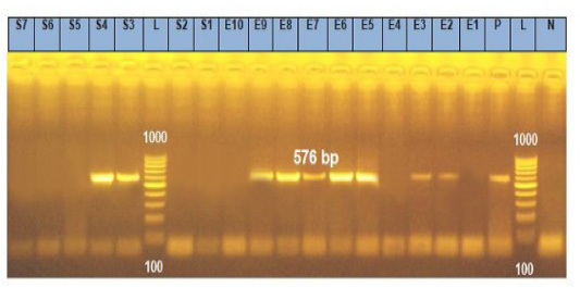
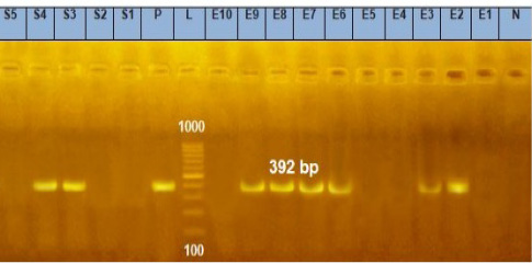
Molecular detection of antibiotic resistance genes by using cPCR was carried out after (Momtaz et al., 2012). The isolated Salmonella and E. coli serotypes were subjected to cPCR for detection of two resistant genes, these genes were the blaSHVresistance gene for β lactams (amoxicillin and ampicillin) and tetA resistance genes for tetracyclines (oxytetracycline and doxycycline). The genomic DNA extraction was carried out using a QIAamp DNA mini kit (Catalogue No.51304). The master mix was carried out according to the Emerald Amp GT PCR master mix (Takara, Code No. RR310A kit). The Oligonucleotide primers for tetA gene F, 5′GGTTCACTCGAACGACGTCA′3 and R, 5′CTGTCCGACAAGTTGCATGA′3 with 576 bp according to (Dipineto et al., 2006). While primers ofthe blaSHVgene were F, 5′AGGATTGACTGCCTTTTTG′3 and R, 5′ATTTGCTGATTTCGCTCG′3, with 392 bp according to (Bisi-Johnson et al., 2011). The cycling conditions of the primers during cPCR were carried out according to a specific Emerald Amp GT PCR master mix (Takara kit).
Statistical Analysis
The statistical analyses were carried out following (Mahmoud and Abd Abd El-Hamed, 2018) to analyze the prevalence of Salmonella and E. coli in collected samples and swabs from different poultry farms by using General Linear Models (GLM) of SPSS Statistics 25 (IBM Corp., Somers, NY, USA). Studied trials were subjected to a two-way ANOVA. Multiple comparisons were carried out by applying the Duncan test. The significance level was set at a P value < 0.05.
RESULTS
Enterobacteriaceae prevalence
Salmonella and E. coli had the highest prevalence in the duck farms (36.2% and 54.3% respectively). In contrast, the lowest prevalence of Salmonella and E. coli were in the breeder chicken farms (10.19 % and 29.28 % respectively). The prevalence of Salmonella was the highest in pen litter (49.45%), while it was the lowest in stored feed and hand swabs (1.67% per each). On the other hand, the prevalence of E. coli was the highest in droppings (85%), while it was the lowest in stored feed (10%), as shown in (Table 3).
Identification of Salmonella and E. coli
Seven serotypes of Salmonella were isolated; the most isolated Salmonella serotype was S. Agona (39.76%). On the other hand, fifteen E. coli serotypes were isolated; the most isolated E. coli serotype was O26 (22.9%), as shown in (Table 4).
Antibiotic Susceptibility of Salmonella and E. coli Serotypes
The different Salmonella serotypes showed 100% resistance against doxycycline, ampicillin, and amoxicillin. In contrast, the serotypes of Salmonella showed 100% sensitivity to gentamicin. Moreover, the different serotypes of E. coli showed the highest resistance (90%) against oxytetracycline, doxycycline, ampicillin, and amoxicillin. In contrast, all serotypes of E. coli were susceptible to gentamicin and cefotaxime, as shown in (Table 5). The highest MAR index (.727) was shown in S. Enteritidis followed by S. Kentucky (.545) and E. coli O164 had the highest MAR index (.909), followed by O114 (.727), as shown in (Table 6).
Molecular characterization of Salmonella and E. coli isolates using conventional PCR (cPCR)
The resistant gene tetA was detected in S. Typhimurium and S. Enteritidis serotypes, in addition to O26, O119, O124, O114, O55, O164, and O157 serotypes. While the resistant gene blaSHV was detected in S. Agona, S. Kentucky S. Typhimurium, and S. Enteritidis, in addition, O26, O119, O124, O114, O164, and O157 serotypes of E.coli as shown in (Tables 7 and 8) and (Figures 1 and 2).
Table 6: Antimicrobial resistance profile of isolated Salmonella and E. coli serotypes.
Strains
Antimicrobial resistance profile
MAR index
S. Agona
O, DO, EX, AMP, AMX
.454
S. Kentucky
O, DO, EX, UB,AMP, AMX,
.545
S. Derby
O, DO, AMP, AMX
.364
S.Typhimurium
O, DO, AMP, AMX,
.364
S. Enteritidis
O, DO, EX, CIP, AMP, AMX, CTX,CTR
.727
S. Molade
O,DO, AMP, AMX,
.364
S. Virchow
O, DO, AMP, AMX
.364
O26
O, DO, EX , UB, AMP, AMX
.454
O44
AMP
.090
O119
O, DO, AMP, AMX
.364
O86
O, DO, AMP, AMX
.364
O124
O, DO, UB, AMP, AMX
.454
O114
O,DO, EX, NX, UB,CIP, AMP, AMX
.727
O55
O, DO,CIP, AMP, AMX
.454
O112
O, DO, AMP, AMX
.364
O164
O,DO, EX, NX, UB,CIP, AMP, AMX, CTX,CTR
.909
O157
O, DO, AMP
.273
Table 7: The tetA and blaSHV resistant genes in isolated Salmonella from different poultry farms.
Salmonella isolate
tetA
blaSHV
S. Agona
-
+
S. Kentucky
-
+
S. Derby
-
-
S. Typhimurium
+
+
S. Enteritidis
+
+
S. Molade
-
-
S. Virchow
-
-
Total
28.5%
57.1%
(+) Positive, (-) Negative
Table 8: The tetA and blaSHV resistant genes in isolated E. coli from different poultry farms.
E. coli sample
tetA
blaSHV
O26
+
+
O44
-
-
O119
+
+
O86
-
-
O124
+
+
O114
+
+
O55
+
-
O112
-
-
O164
+
+
O157
+
+
Total
70%
60%
(+) Positive, (-) Negative
DISCUSSION
Enterobacteriaceae are serious contamination facing poultry industries as they are one of the most important groups of bacteria which can infect the poultry and cause dangerous diseases. Improper biosecurity measures and poor hygiene in poultry farms are the main cause of the introduction and spreading of Enterobacteriaceae .
Our results showed that there was a negative relationship between the poultry farms’ hygiene and the prevalence of Enterobacteriaceae. The duck farms were the most contaminated farms with Salmonella and E. coli; in contrast, the breeder chicken farms recorded the lowest prevalence. This is might be due to the variation in the level of hygienic measures of each farm as breeder chicken farms were the highest, unlike the duck farms . Moreover, the variation in the survival capabilities of Salmonella and E. coli in poultry farms is affected by many factors such as poultry husbandry systems, antibiotic use, environmental temperature, stress factors, in addition, age, type, immune status, and physiological status of birds .
Salmonella and E. coli were isolated from litter, droppings, birds’ cloaca, drinkers, feed, dust, vehicles’ wheels, farms’ walls, and workers’ hands, but Salmonella prevalence was the highest in pen litter (49.45%). The main source of Salmonella spreading in poultry farms was the contaminated litter that could be contaminated by the surrounding environment, dust, insects, free-living animals, and rodents . On the other hand, the contamination of E. coli was the highest in birds’ droppings (85%), birds’ droppings were the main source of E. coli contamination in different poultry farms .
The isolated Salmonella serotypes were S. Agona, S. Kentucky, S. Derby, S Typhimurium, S. Enteritidis, S. Molade, and S. Virchow. While, the isolated E. coli serotypes were O26, O44, O119, O86, O124, O114, O55, O112, O164, O157, O91, O152, O128, O153, and O127. The appropriate management practices, proper hygiene, and biosecurity measures in poultry farms are very essential for the control of Enterobacteriaceae .
Our results indicated that all examined samples were free from Shigella species and this result may be due to the high sensitivity of Shigella species to unfavorable macroclimatic environmental conditions. It is destroyed by dryness and direct sunlight and is sensitive to different antibiotics .
The routine prophylactic use of antibiotics in different poultry farms leads to an increase in the prevalence of an antibiotic-resistance against many bacterial species which is considered one of the most important public health hazards . Our result demonstrated that Salmonella species showed 100% resistance against doxycycline, ampicillin, and amoxicillin. Moreover, Salmonella species showed 85.7% resistance against flumequine. In previous studies, this high Salmonella resistance was reported against the same antibiotics in Egypt , India , Pakistan , Bangladesh , western Algeria and Côte d’Ivoire .
While, the E. coli species showed 90% resistance against oxytetracycline, doxycycline, ampicillin, and amoxicillin . This resistance may be attributed to the prolonged use of these antibiotics . On the other hand, E. coli isolates showed relatively low resistance against norfloxacin and cefotaxime (20% each). A previous incompatible study in Pakistan reported that E. coli isolates showed high resistance against flumequine, enrofloxacin, ciprofloxacin, and norfloxacin (76.7%, 79.6%, 82.5%, and 73.7% respectively), it demonstrated that this high resistance was due to extensive use of these antibiotics as feed additives in tested poultry farms for diseases prevention .
Gentamicin and cefotaxime antibiotics had the highest effect against E. coli species . In addition, Gentamicin had the highest effect on Salmonella species. This result was previously reported in, Egypt . A previous study in Egypt reported that the isolated Salmonella showed complete resistance against gentamicin and ceftriaxone (100 %). It demonstrated that these antibiotics were commonly used by poultry producers as a preventive tool . In contrast, these antibiotics weren’t commonly used in the tested poultry farms in our study and this proved the strong relationship between prophylactic use of the antibiotics and the development of antimicrobial resistance in poultry farms.
The results revealed that multidrug resistance was observed in all Salmonella isolates against four antibiotics or more and in all E. coli isolates against three antibiotics or more (except in O44). This high prevalence of multidrug resistance in Salmonella and E. coli has been previously reported in Egypt , Bangladesh , Pakistan , western Algeria and in Côte d’Ivoire .
The tetA gene was detected in (28.5%) of Salmonella and (70%) of E. coli serotypes, while the blaSHV gene was detected in (57.1%) of Salmonella and (60%) of E. coli serotypes. These genes were previously detected in Salmonella and E. coli isolates from poultry farms by .
Our results showed that the serotypes of Salmonella and E. coli which had the highest MAR index also had the two resistant genes (tetA and blaSHV). In contrast, the serotypes which had the lowest MAR index didn’t have the two resistant genes (tetA and blaSHV). The results of the disk diffusion test were confirmed by the results of cPCR .
CONCLUSION
Improving the hygiene practices and enforced application of the maximum biosecurity measures in different poultry farms can reduce the prophylactic use of antibiotics and as a result, the drug residues can be minimized in eggs and poultry meat. In addition; minimize the anti-microbial resistance and reduce the cost of using antibiotics.
ACKNOWLEDGEMENTS
The authors especially thank the poultry producers who participated and helped to complete this study. The authors also extend their thanks and appreciation to all members of the Department of Hygiene and Veterinary Care, Faculty of Veterinary Medicine, Benha University for their assistance in completing this study.
CONFLICT OF INTEREST
The authors declared that they had no competing interests.
AUTHORS CONTRIBUTION
All authors contributed equally to this study.
NOVELTY STATEMENT
This study was the first study in Qalubia governorate, Egypt aimed to detect resistance genes of the field Enterobacteriaceae strains already present in the poultry farm environment and isolated from the environmental samples as feeders, drinkers, dust, walls, and wheels of vehicles.
Figures
Figure 1. Figure 1. Figure 2.
References
Al-baqir A, Hussein A, Ghanem I, Megahed M (2019). Characterization of paratyphoid Salmonellae isolated from broiler chickens at Sharkia governorate, Egypt. Zagazig Vet. J. 47(2): 183-192. https://doi.org/10.21608/zvjz.2019.10544.1028[CrossRef]
Amer MM, Mekky HM, Amer AM, Fedawy HS (2018). Antimicrobial resistance genes in pathogenic escherichia coli isolated from diseased broiler chickens in Egypt and their relationship with the phenotypic resistance characteristics. Vet. World. 11(8): 1082. https://doi.org/10.14202/vetworld.2018.1082-1088[CrossRef]
Assoumy MA, Bedekelabou AP, Teko-Agbo A, Ossebi W, Akoda K, Nimbona F, Bada-Alambédji R (2021). Antibiotic resistance of Escherichia coli and Salmonella spp. strains isolated from healthy poultry farms in the districts of Abidjan and Agnibilékrou (Côte d’Ivoire). Vet. World. 14(4): 1020. https://doi.org/10.14202/vetworld.2021.1020-1027[CrossRef]
Ballal M, Devadas SM, Shetty V, Bangera SR, Ramamurthy T, Sarkar A (2016). Emergence and serovar profiling of non-typhoidal Salmonellae (NTS) isolated from gastroenteritis cases–a study from south India. Infect. Dis. 48(11-12): 847-851. https://doi.org/10.3109/23744235.2016.1169553[CrossRef]
Bisi-Johnson MA, Obi CL, Vasaikar SD, Baba KA, Hattori T (2011). Molecular basis of virulence in clinical isolates of escherichia coli and Salmonella species from a tertiary hospital in the eastern cape, south Africa. Gut pathogens. 3(1): 1-8. https://doi.org/10.1186/1757-4749-3-9[CrossRef]
Blaak H, van Hoek AH, Hamidjaja RA, van der Plaats RQ, Kerkhof-de Heer L, de Roda Husman AM, Schets FM (2015). Distribution, numbers, and diversity of esbl-producing E. coli in the poultry farm environment. PloS one. 10(8): e0135402. https://doi.org/10.1371/journal.pone.0135402[CrossRef]
CLSI (2015). Performance standards for antimicrobial susceptibility testing; twenty-fifth informational supplement. CLSI document M100-S25. Wayne, PA: Clinical and Laboratory Standards Institute.
Cruickshank R, Duguid J, Marmion B, Swain RH (1975). Medical microbiology. 12th ed.; Edinburg, London and new york. Prev. Ed. In fao library.
Cusack T, Ashley E, Ling C, Rattanavong S, Roberts T, Turner P, Wangrangsimakul T, Dance D (2019). Impact of clsi and eucast breakpoint discrepancies on reporting of antimicrobial susceptibility and AMR surveillance. Clin. Microbiol. Infect. 25(7): 910-911. https://doi.org/10.1016/j.cmi.2019.03.007[CrossRef]
Diab MS, Zaki RS, Ibrahim NA, Abd El Hafez MS (2019). Prevalence of multidrug resistance non-typhoidal Salmonellae isolated from layer farms and humans in Egypt. World Vet. J. 9(4): 280-288. https://doi.org/10.36380/scil.2019.wvj35[CrossRef]
Dipineto L, Santaniello A, Fontanella M, Lagos K, Fioretti A, Menna L (2006). Presence of Shiga toxin‐producing escherichia coli o157: H7 in living layer hens. Lett. Appl. Microbiol. 43(3): 293-295. https://doi.org/10.1111/j.1472-765X.2006.01954.x[CrossRef]
El-Sharkawy H, Tahoun A, El-Gohary AE-GA, El-Abasy M, El-Khayat F, Gillespie T, Kitade Y, Hafez HM, Neubauer H, El-Adawy H (2017). Epidemiological, molecular characterization and antibiotic resistance of Salmonella enterica serovars isolated from chicken farms in Egypt. Gut Pathogens. 9(1): 1-12. https://doi.org/10.1186/s13099-017-0157-1[CrossRef]
FDA (2012). Fda’s bacteriological analytical manual (bam).Chapter 5Salmonella, Washington, dc.
Gupta MD, Islam M, Sen A, Sarker MS , Das A (2017). Prevalence and antibiotic susceptibility pattern of escherichia coli in cattle on Bathan and intensive rearing system. Microb. Health. 6(1): 1-4. https://doi.org/10.3329/mh.v6i1.34062[CrossRef]
Gwida M, Hotzel H, Geue L, Tomaso H (2014). Occurrence of Enterobacteriaceae in raw meat and in human samples from Egyptian retail sellers. Int. Scholar. Res. Notices 2014. https://doi.org/10.1155/2014/565671[CrossRef]
Harakeh S, Yassine H, Gharios M, Barbour E, Hajjar S, El-Fadel M, Toufeili I, Tannous R (2005). Isolation, molecular characterization and antimicrobial resistance patterns of Salmonella and escherichia coli isolates from meat-based fast food in Lebanon. Sci. Total Environ. 341(1-3): 33-44. https://doi.org/10.1016/j.scitotenv.2004.09.025[CrossRef]
Hassan A, Osama M (2021). Incidence of Salmonellae and E. coli in meals served in Egyptian hotels. Benha Vet. Med. J. 41(1): 120-123. https://doi.org/10.21608/bvmj.2021.86837.1452[CrossRef]
Hussain J, Rabbani I, Aslam S, Ahmad H (2015). An overview of poultry industry in Pakistan. World’s Poult. Sci. J. 71(4): 689-700. https://doi.org/10.1017/S0043933915002366[CrossRef]
Ibrahim HA, Abo El-Makarem HS (2021). Bacterial food poisoning in chicken and some chicken product. Alexandria J. Vet. Sci. 69(1). https://doi.org/10.5455/ajvs.117676[CrossRef]
ISO E (2004). 7937. Microbiology of food and animal feeding stuffs—horizontal method for the enumeration of clostridium perfringens—colony-count technique. International Organization for Standardization: Geneva, Switzerland.
Jozić S, Lušić DV, Ordulj M, Frlan E, Cenov A, Diković S, Kauzlarić V, Đurković LF, Totić JS, Ivšinović D (2018). Performance characteristics of the temperature-modified iso 9308-1 method for the enumeration of Escherichia coli in marine and inland bathing waters. Marine Pollut. Bullet. 135: 150-158. https://doi.org/10.1016/j.marpolbul.2018.07.002[CrossRef]
Kamboh AA, Shoaib M, Abro SH, Khan MA, Malhi KK, Yu S (2018). Antimicrobial resistance in Enterobacteriaceae isolated from liver of commercial broilers and backyard chickens. J. Appl. Poult. Res. 27(4): 627-634. https://doi.org/10.3382/japr/pfy045[CrossRef]
Kauffman G (1974). Kauffmann white scheme. J. Acta. Path. Microbiol. Sci. 61: 385.
Khan M A, Göpel Y, Milewski S, Görke B (2016). Two small RNAs conserved in Enterobacteriaceae provide intrinsic resistance to antibiotics targeting the cell wall biosynthesis enzyme glucosamine-6-phosphate synthase. Front. Microbiol. 7: 908. https://doi.org/10.3389/fmicb.2016.00908[CrossRef]
Kim A, Lee YJ, Kang MS, Kwag SI, Cho JK (2007). Dissemination and tracking of Salmonella spp. in integrated broiler operation. J. Vet. Sci. 8(2): 155-161. https://doi.org/10.4142/jvs.2007.8.2.155[CrossRef]
Kok T, Worswich D, Gowans E (1996). Some serological techniques for microbial and viral infections. Practical Medical Microbiology (Collee, J.; Fraser, A.; Marmion, B. and Simmons, A., eds.), 14th ed., Edinburgh, Churchill Livingstone, UK: 179-204.
Matin MA, Islam MA, Khatun MM (2017). Prevalence of colibacillosis in chickens in greater Mymensingh district of Bangladesh. Vet. World. 10(1): 29. https://doi.org/10.14202/vetworld.2017.29-33[CrossRef]
Messens W, Herman L, De Zutter L, Heyndrickx M (2009). Multiple typing for the epidemiological study of contamination of broilers with thermotolerant campylobacter. Vet. Microbiol. 138(1-2): 120-131. https://doi.org/10.1016/j.vetmic.2009.02.012[CrossRef]
Mahmoud RD, Abd Abd El-Hamed R (2018). Organic acids as potential alternate for antibiotic as growth promoter in Japanese quail. Egyptian Poult. Sci. J. 38(2): 359-373.
Momtaz H, Rahimi E, Moshkelani S (2012). Molecular detection of antimicrobial resistance genes in E. coli isolated from slaughtered commercial chickens in Iran. Vet. Med. 57(4): 193-197. https://doi.org/10.17221/5916-VETMED[CrossRef]
Nechyporenko O, Berezovskyy A, Fotina T, Petrov R, Fotin A (2018). Efficiency of complex disinfecting measures in the conditions of poultry farming. Науковий вісник Львівського національного університету ветеринарної медицини та біотехнологій імені СЗ Ґжицького. 20(92). https://doi.org/10.32718/nvlvet9234[CrossRef]
Noha MA, Halla EKB (2019). Effect of different hygienic levels on Salmonella and antimicrobial resistance in layer cages system. . American-Eurasian J. Agric. Environ. Sci. 19(5): 350-356.
Omara ST, Zawrah MF, Samy A (2017). Minimum bactericidal concentration of chemically synthesized silver nanoparticles against pathogenic Salmonella and shigella strains isolated from layer poultry farms. J. App. Pharm. Sci. 7(8): 214-221.
Park S, Woodward C, Kubena L, Nisbet D, Birkhold S, Ricke S (2008). Environmental dissemination of foodborne Salmonella in preharvest poultry production: Reservoirs, critical factors, and research strategies. Crit. Rev. Environ. Sci. Technol. 38(2): 73-111. https://doi.org/10.1080/10643380701598227[CrossRef]
Parvin MS, Hasan MM, Ali MY, Chowdhury EH, Rahman MT, Islam MT (2020). Prevalence and multidrug resistance pattern of Salmonella carrying extended-spectrum β-lactamase in frozen chicken meat in Bangladesh. J. Food Protect. 83(12): 2107-2121. https://doi.org/10.4315/JFP-20-172[CrossRef]
Pashazadeh P, Mokhtarzadeh A, Hasanzadeh M, Hejazi M, Hashemi M, de la Guardia M (2017). Nano-materials for use in sensing of Salmonella infections: Recent Adv. Biosens. Bioelectron. 87: 1050-1064. https://doi.org/10.1016/j.bios.2016.08.012[CrossRef]
Phagoo L, Neetoo H (2015). Antibiotic resistance of Salmonella in poultry farms of Mauritius. J. Worlds Poult. Res. 5(3): 42-47.
Rukambile E, Sintchenko V, Muscatello G, Kock R, Alders R (2019). Infection, colonization and shedding of campylobacter and Salmonella in animals and their contribution to human disease: A review. Zoon. Pub. Health. 66(6): 562-578. https://doi.org/10.1111/zph.12611[CrossRef]
Saliu E-M, Vahjen W, Zentek J (2017). Types and prevalence of extended–spectrum beta–lactamase producing Enterobacteriaceae in poultry. Anim. Health Res. Rev. 18(1): 46-57. https://doi.org/10.1017/S1466252317000020[CrossRef]
Soliman E, Hassan R (2017). Evaluation of superphosphate and meta-bisulfide efficiency in litter treatment on productive performance and immunity of broilers exposed to ammonia stress. Adv. Anim. Vet. Sci. 5(6): 253-259.
Vandekerchove D, De Herdt P, Laevens H, Pasmans F (2004). Risk factors associated with colibacillosis outbreaks in caged layer flocks. Avian Pathol. 33(3): 337-342. https://doi.org/10.1080/0307945042000220679[CrossRef]
Waghamare R, Paturkar A, Vaidya V, Zende R, Dubal Z, Dwivedi A, Gaikwad R (2018). Phenotypic and genotypic drug resistance profile of Salmonella serovars isolated from poultry farm and processing units located in and around Mumbai city, India. Vet. World. 11(12): 1682. https://doi.org/10.14202/vetworld.2018.1682-1688[CrossRef]
Yahya M, Hammadi K, Faiza FF (2021). The antibiotic resistance study of Enterobacteriaceae, Yersiniaceae and Morganellaceae bacteria isolated from broilers (outside veterinary control) in western Algeria. European J. Biolog. Res. 11(2): 217-23.