
Leptospirosis is a zoonotic bacterial disease affecting both humans and animals. This is the first study aiming to detect the Leptospira spp. in bovine kidneys collected from selected wet markets in Kota Bharu, Kelantan and subsequently determine the predominant Leptospira species. Additionally, the pathological alterations to the bovine kidneys as a result of leptospirosis were also analyzed. A total of fifty bovine kidney samples (n=50) were collected from four wet markets within Kota Bharu town and the samples were tested using a polymerase chain reaction (PCR) assay. From the results, thirteen samples (n=13; 26%) were found to be positive for the 16S rRNA gene. DNA sequencing of the positive samples revealed the presence of Leptospira borgpetersenii (n=11) and Leptospira interrogans (n=1), which are known to be pathogenic Leptospira species in cattle and humans. One sample cannot be sequenced due to poor yield and quality. Furthermore, the kidneys that were PCR-positive showed significant histopathological lesions of bovine leptospirosis, consisting of interstitial nephritis, glomerular atrophy and tubular necrosis. In conclusion, the present study demonstrated the presence of L. borgpetersenii and L. interrogans in local cattle and this is an indisputable public health risk to the public considering that the samples were obtained from the local wet markets.
Research Article · Volume 57, Issue 2, 25 February 2025 · Open Access
Histopathological and Molecular Analyses of Leptospira borgpetersenii and Leptospira interrogans in Bovine Kidneys in Kota Bharu, Kelantan, Malaysia
· · · · · · · · · · ·
Affiliations & Notes ⌄
- 1Department of Veterinary Preclinical Sciences, Faculty of Veterinary Medicine, Universiti Malaysia Kelantan, Jalan Padang Tembak, 16100 Pengkalan Chepa, Kota Bharu, Kelantan, Malaysia 2Department of Veterinary Paraclinical Studies, Faculty of Veterinary Medicine, Universiti Malaysia Kelantan, Jalan Padang Tembak, 16100 Pengkalan Chepa, Kota Bharu, Kelantan, Malaysia 3Department of Veterinary Diagnostics, Faculty of Veterinary Medicine, Universiti Malaysia Kelantan, Jalan Padang Tembak, 16100 Pen
Article Info ⌄
- Journal:
- Pakistan Journal of Zoology
- Published:
- 25 February 2025

From the article
- The present study described the presence of leptospiral DNA in fresh bovine kidneys sold at the wet markets and examined the degree of histopathological lesions correlated to the infected kidneys.
- In conclusion, L. borgpetersenii and L. interrogans were detected in the bovine kidney samples collected from wet markets in Kota Bharu, Kelantan.
- To date, this was the first study in Malaysia that successfully detected Leptospira spp. in bovine kidney samples.
- The presence of pathogenic Leptospira spp. in cattle sold as beef, presents a zoonotic risk to the people working with the animals as well as visitors to the wet markets.
Selected from the article's own text.
Abstract
INTRODUCTION
Leptospirosis is a zoonotic disease that can affect a wide range of mammalian hosts including humans ; . Despite being much underreported and neglected in many countries, the disease was estimated to cause more than a million human cases globally, with approximately 60,000 annual deaths . Leptospirosis can produce mild to severe clinical manifestations depending on the susceptibility of the host, inoculum dose and the pathogenicity or the virulence factor of the infecting strain . Studies of leptospirosis are extensive in humans over the years but studies on animal leptospirosis are relatively lacking. In Malaysia, leptospirosis is an endemic disease due to its tropical climate. The warm and humid weather in Malaysia allow this organism to thrive longer in the environment and increase the risk of exposure. This contributes to the dramatic increase in the number of reported cases in Malaysia. According to the Ministry of Health Malaysia, there was a marked increase of leptospirosis cases from 12.5 per 100,000 population in 2012 to 15.0 per 100,000 population in 2013 .
Cattle are one of the major leptospires reservoir, and the disease; bovine leptospirosis (BL), is one of the leading causes of reproductive disorder in cattle which is characterized by failure to conceive, embryonic loss and abortion . The worldwide prevalence of BL is not known, possibly due to the silent clinical signs exhibited by the animals that often go undiagnosed by veterinarians and farmers . In Malaysia, BL is listed as a notifiable disease by the Department of Veterinary Services, but it has received marginal attention in research despite its potential impact on both animal and human health. Cattle can carry leptospires within their renal system, and can transmit pathogenic leptospires to human via direct contact with contaminated urine and the environment ; . The human-cattle interface is one of the important factors for leptospirosis transmission especially among agricultural workers and people living in the vicinity of livestock farms . One cross-sectional study in Malaysia on BL showed that nearly 90% of cattle have been exposed to Leptospira spp. and this would potentially increase the risk of transmission to humans tending the animals . In another study involving 120 cattle farmers in northeastern Malaysia, it was found that most farmers are highly exposed to Leptospira spp. showing 72.5% seropositivity, reflecting the high occupational risk associated to cattle farming .
Despite the importance of BL in cattle populations and its potential implications for human health, there are several gaps of knowledge in the literature. One is the lack of comprehensive data on the prevalence and distribution of Leptospira species in cattle herds in Malaysia, including the specific serovars that are prevalent in different states/areas ; . BL is typically associated with Leptospira borgpetersenii serovar Hardjo (Hardjobovis type) and Leptospira interrogans serovar Hardjo (Hardjoprajitno type) . In the tropics of South America, BL can also be caused by Leptospira santarosai and Leptospira noguchii . Therefore, understanding the diversity of Leptospira species circulating in cattle populations is crucial for developing effective control measures.
Furthermore, there is a lack of data on the genetic characteristics of Leptospira strains isolated from cattle in Malaysia. Molecular detection tools such as the polymerase chain reaction (PCR) assays have been widely used to detect Leptospira spp. in various species worldwide ; and has been regarded as the most convenient method employed for the surveillance of BL. Molecular techniques can provide valuable information on the genetic diversity, population structure, and transmission dynamics of Leptospira strains. Besides that, there is also a need for more studies on the pathological lesions associated with leptospirosis in bovine kidneys. Using histopathological evidence is important to visualize changes to the tissue lesions relating to the infection and to demonstrate the carrier state of the animals. Additionally, understanding the pathological changes caused by BL together with complementary histopathology evidence can provide insights into the disease’s pathogenesis and help in the diagnosis and management of BL.
The state of Kelantan, which is located at the northeast of Peninsular Malaysia, has one of the highest gross domestic products in agriculture production (Department of Statistics Malaysia, 2023). Cattle herding is one of the substantial farming activities in the state, producing approximately between 83,000 and 85,000 cattle per year; the third highest after Johor and Pahang states (Department of Veterinary Services Malaysia, 2023). Despite one of the top states with the highest cattle population, there are limited BL studies being carried out here ; . Moreover, Kelantan is one of the states in Malaysia with the highest human leptospirosis cases due to yearly flooding that occurs every monsoon season that would eventually lead to leptospirosis outbreaks due to the spreading of Leptospira spp. by the flood water . The spread of pathogenic Leptospira could have been magnified during the flooding season as other carrier animals such as rats and dogs could also shed the bacteria into flood waters, thus transmitting the bacteria to different animal hosts, including to healthy cattle and humans. Hence, this study was conducted to detect the presence of Leptospira spp. in bovine kidneys and to determine the predominant species in Kelantan cattle. Moreover, this study also aimed to investigate the possible pathological lesions in the bovine kidneys as a result of leptospirosis.
MATERIALS AND METHODS
Sample collection and preparation
Convenience sampling was carried out by collecting a total of 50 bovine kidney samples (about 50 g from each animal) obtained from butchers in four wet markets in Kota Bharu, Kelantan, Malaysia. The study objectives were explained, and verbal consent was obtained from all the butchers before any samples were obtained. Since we do not want to interfere with the butchers when they are busy attending to customers, we approached them after they were done for the day to explain our study and to obtain their consent. Subsequently, we obtained several suitable dates when they will be available to provide us with the kidney samples. Sampling was performed from December 2021 until February 2022. Knives, other butchering tools and the hands of the butchers were washed with detergent before cutting open the carcasses. Additionally, we provided gloves, protective goggles and aprons to the butchers as a biosafety precaution.
Kidney samples were obtained directly from the carcasses and directly placed into a sterile sealed bag. Each kidney sample was kept individually in a sterile sealed bag and stored in a polystyrene box with ice packs for transportation. To ensure that the samples were fresh for molecular detection, the samples were processed immediately upon arriving at the laboratory, and then stored at -80°C until further analysis. For histopathological examination, selected kidney samples were fixed in 10% (v/v) neutral buffer formalin for 24 h before staining procedure. Ethical approval application was submitted to the Institutional Animal Care and Use Committee, Universiti Malaysia Kelantan, however we were informed that this was not required since the study samples were obtained from third parties (e.g., butchers) and culturing of Leptospira was not performed.
Molecular detection of Leptospira spp.
All kidney samples were subjected to DNA extraction prior to molecular detection and purification. Approximately 25 g of fresh bovine kidney tissues, directly excised from the renal lobe were sectioned, weighed, and homogenized using a commercial DNA extraction kit (Geneaid, Taiwan), following the manufacturer’s instructions.
Leptospiral DNA was amplified by PCR using a set of primers targeting the 16S rRNA (Table I), and the PCR protocol and product visualization were carried out as described by Kamaruzaman et al. (2022). The PCR reaction mixture was prepared by mixing 12.5 μl of the PCR mastermix (Promega, USA), 1.0 μl of 10 mM of both forward and reverse primers, 0.5 μl of nuclease free water and lastly 5.0 μl of the extracted DNA, for a final 20.0 μl reaction per sample. The conditions of the PCR cycle was set using the following steps; denaturation at 95°C for 5 min, followed by denaturation at 95°C for 30 sec, annealing at 60°C for 1 min, extension at 72°C for 30 sec for 35 cycles, and the final extension was set at 72°C for 5 min. Similar to a previous study , L. interrogans serovar Canicola was used as a positive control for this study. The PCR was repeated twice to confirm the presence of PCR-positive products on the gel. PCR-positive samples were then purified using the NucleoSpin Gel and PCR Clean-up (Macherey-Nagel, Germany) according to the manufacturer’s instructions and their concentrations were quantified using the NanoPhotometer P-360 (Implen, Germany). Samples below 20 ng/μl were discarded. All products were submitted for sequencing analysis by a third-party service provider (Apical Scientific, Malaysia). The resulting DNA sequences were then analyzed by using ChromasPro software version 10.0 for DNA assembly and editing. Next, a phylogenetic tree was constructed using the Molecular Evolutionary Genetics Analysis version 11.0 (MEGA 11) (PSU, USA) to determine the genetic relationships amongst the Leptospira spp. obtained from this study using 15 reference Leptospira spp. across three clades representing the pathogenic, intermediate and non-pathogenic groups, and one outlier strain of a similar spirochete (Leptonema illini) as an outgroup.
Histopathological examination
Six kidney samples representing both positive and negative results upon molecular detection were trimmed and fixed in 10% (v/v) formaldehyde for 24 h. The fixed tissues were then dehydrated using 70% (v/v) ethanol for 15 min to remove the water before adding xylene to the tissue for 20 min to clear the remaining alcohol in the specimen. Next, the tissues were then embedded using molten wax to form a block. The block was then sectioned using a rotary microtome to a thickness of about 3-4 μm. Finally, the tissue sections were stained with Haematoxylin and Eosin (H and E) and were observed under a compound microscope to evaluate the lesions. The histopathology lesions were classified using an established scoring system (Table II).
Table I. The primers used in this study.
| Primer name | Target gene | Product size (bp) | Sequences | Reference |
| Lep F Lep R | 16S rRNA | 330 | 5’- GGC GGC GCG TCT TAA ACA TG- 3’ 5’- TCC CCC CAT TGA GCA AGA TT- 3’ |
Table II. Histopathology scoring system used for kidney evaluation adapted from Prakoso et al. (2020).
| Score | Severity | Duration | Distribution | Exudate |
| 0 | NHC | NHC | NHC | NHC |
| 1 | Minimal | Acute | Focal | Suppurative |
| 2 | Mild | Chronic | Multifocal | Fibrinous |
| 3 | Moderate | Chronic active | Locally extensive | Necrotizing |
| 4 | Severe | - | Diffuse | Fibrinopurulent |
| 5 | - | - | - | Granulomatous |
NHC, No histopathological changes.
Results
Molecular detection of Leptospira spp.
Table III. PCR detection results of Leptospira spp. in kidney samples in this study.
| Wet market | No. of samples | No of positive by PCR |
| A | 23 | 10 |
| B | 16 | 3 |
| C | 7 | 0 |
| D | 4 | 0 |
| Total | 50 | Total positive: 13 (26%) |
Phylogenetic analysis
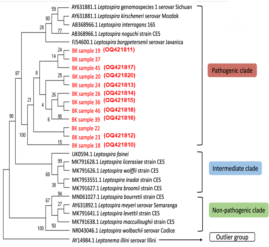
All PCR-positive samples (n=12) were purified and submitted for sequencing except for one sample due to poor yield and quality. Phylogenetic analysis against representatives Leptospira genomospecies pathogenicity class (pathogenic, intermediate, non-pathogenic group), revealed that all samples from the present study were clustered within the pathogenic Leptospira subclade, together with other pathogenic Leptospira spp. (L. borgpetersenii, L. interrogans, Leptospira kirschneri and L. noguchii) (Fig. 2). Consequently, comparison of the amplified 16S rRNA sequences against the Basic Local Alignment Search Tool (BLAST) database, revealed that all samples have nearly 90% similarities and identities with L. borgpetersenii (n=11; Samples no. 18, 19, 22, 23, 24, 26, 36, 37, 39, 45 and 46) and L. interrogans (n=1; Sample no. 20) strains (Fig. 2).
Histopathological evaluation
Representatives of three PCR-positive and three PCR-negative bovine kidney samples were selected for histopathology examination. Grossly, all samples collected from four wet markets did not have evidence of macroscopic or obvious gross lesions indicating the presence of leptospires. However, morphological changes and microscopic lesions were seen in all samples upon evaluation. The H and E scores indicate the severity of the histopathological lesions, for which the higher score indicates an increase in the severity of the lesions (Table IV). All of the samples showed severe diffuse necrotizing nephritis, regardless of whether they were PCR-positive or negative. Sample no. 22 has the highest score which showed severe chronic active diffuse necrotizing tubulointerstitial nephritis characterized by interstitial infiltration of inflammatory cells, perivascular cuffing hyaline degeneration and tubular necrosis (Table IV, Fig. 3A-D). The remaining samples showed mixed histopathological lesions consisting of severe chronic diffuse necrotizing tubulointerstitial nephritis (Sample no. 32) (Table IV, Fig. 3E-F) and severe diffuse necrotizing glomerulonephritis (Samples no. 3, 11, 18 and 45), respectively (Figure not shown).
Table IV. Histopathology scoring of bovine kidney samples.
| Sample no.* | PCR results | Morphological diagnosis | H and E score |
| 3 | Negative | Severe diffuse necrotizing glomerulonephritis | 11 |
| 11 | Negative | Severe diffuse necrotizing glomerulonephritis | 11 |
| 32 | Negative | Severe chronic diffuse necrotizing tubulointerstitial nephritis | 13 |
| 18 | Positive | Severe diffuse necrotizing glomerulonephritis | 11 |
| 22 | Positive | Severe chronic active diffuse necrotizing tubulointerstitial nephritis | 14 |
| 45 | Positive | Severe diffuse necrotizing glomerulonephritis | 11 |
DISCUSSION
The state of Kelantan is one of the most affected states in Malaysia with high human leptospirosis cases due to the post-monsoon season that results in high rainfall and floods that facilitate the transmission of leptospirosis . Despite the high awareness of leptospirosis amongst the locals due to its endemic nature in the country, leptospirosis is considered a neglected disease as there is a lack of knowledge pertaining to the transmission of the disease from cattle to human . In Malaysia, most cattle farmers do not practice leptospirosis vaccination as it not considered compulsory from the veterinary authority as compared to other top priority diseases such as foot and mouth disease (FMD), haemorrhagic septicaemia (HS) and most recently, lumpy skin disease (LSD). Additionally, Leptospira vaccines in cattle lack cross-reactivity among serovars and provide only a short-term protection (Grassmann et al., 2017), both of which are the major pitfalls for controlling the disease at the farm level.
Investigations on BL in Kelantan have only began to be described in recent years, though most of the studies involved serology ; and only one study utilizing molecular techniques by Sabri et al. (2019). The present study described the presence of leptospiral DNA in fresh bovine kidneys sold at the wet markets and examined the degree of histopathological lesions correlated to the infected kidneys.
This is the first study in Kelantan that successfully detected Leptospira spp. in bovine kidney samples. Bovine kidneys are often sold as offal, which are readily available at markets for local consumption and can be easily collected to screen for BL. Molecular detection, using PCR provides rapid results in contrast to other time-consuming methods and it is also used as the confirmatory diagnostic test due to the higher sensitivity and specificity . PCR has been extensively used to detect the presence of leptospiral DNA by targeting certain genes such as the universal 16S rRNA and the other surface proteins of the species, such as OmpL1, LipL32, LipL36 and LipL41 ; . From this study, wet market A has the highest number of positive samples compared to the other markets (Table III). This could be due to the butchers obtaining beef from the same infected farms/sources. From our observation, the PCR-positive samples were traced to several individual butchers in the wet markets, as we collected the cattle kidney samples every alternate day (data not shown). It is important to note that we collected several samples from the same butcher at the same time and Leptospira spp. were not detected in all samples from the same butcher. Cross contamination between samples is possible since slaughtering of the cattle was done at the abattoir and the fresh carcasses were directly brought to the market for sale. However, if cross contamination occurred at the abattoir, we would expect all the kidney samples to be PCR-positive. This was not the case in the present study as there was clear demarcation of PCR-positive and negative samples. In this case, the histopathology images could complement the PCR results as Leptospira PCR-positive kidneys have relatively more pronounced tissue damage as compared to Leptospira PCR-negative kidneys.
From the phylogenetic analysis, all samples showed nearly 90% similarities with L. borgpetersenii (n=11) and L. interrogans (n=1), which are amongst the highly pathogenic Leptospira species. Cattle are known as a natural reservoir for L. borgpetersenii that have a global distribution and infection in cattle may result in abortion and reproductive failures ; . Due to its smaller genetic content compared to other closely-related pathogenic Leptospira species, L. borgpetersenii is considered a host-restricted pathogen which may not be able to survive outside the host . Cattle can be infected with L. borgpetersenii and L. interrogans (with or without clinical signs) via various possibilities, such as direct contact with other cattle in the herd , venereal transmission , maternal transmission during pregnancy or suckling . The detection of both L. borgpetersenii and L. interrogans in this study may also be suggestive of concurrent presence of different Leptospira spp. as a result of the post-flood effect during the monsoon season, considering that the samples were obtained during the monsoon period of November until March with continuous rainfalls and floods at the northeast part of Malaysia, including the Kelantan state . During the monsoon season, the movement of cattle to higher grounds by the farmers may facilitate the transmission of Leptospira via cattle urination to the environment. The risk of Leptospira exposure to other animals and humans sharing the same contaminated environment is increased during this period. As some pathogenic Leptospira species can survive in the water, the distribution of the bacteria is enhanced through common drinking water during floods which can potentially cause an outbreak . However, in this study we could not determine the type of serovars or serogroup under L. borgpetersenii and L. interrogans using tissue samples as the identification is only possible via Microscopic Agglutination Test (MAT), which is the current gold standard for diagnosis of leptospirosis approved by the World Organisation for Animal Health (WOAH) (World Organisation for Animal Health, 2023).
Following the systemic infection, Leptospira are usually cleared out from the circulation by the antibodies at about 10 days after infection. However, the bacteria may evade the immune response, colonizing the renal tubules, which are antibody free sites. Leptospiral renal colonization occurred in the infected hosts at the leptospiruria phase, in which the bacteria are shed in the urine. The presence of leptospiral bacteria can cause direct or indirect damage to the kidney due to the leptospiral antigen initiated immune response, resulting in certain histopathological alterations . These leptospiral antigens can be found in renal structures such in the proximal tubule cells and renal interstitium in the form of clumps consisting of mainly macrophages. Grossly, infected bovine kidneys with leptospirosis should appear as whitish spots on the surface however, this was not observed in this study. The white-spotted appearance is suggestive of interstitial nephritis, which is common in BL. However, this is not considered a pathognomonic lesion and other diseases may also cause a similar appearance . The finding should be supported by other diagnostic approaches, such as molecular detection and histopathological evaluation for confirmation.
Generally, microscopic renal lesions related to BL are characterized by chronic disease, with the infiltration of inflammatory cells composing of predominantly lymphoplasmocytic cells . These findings are consistent for chronic infection in cattle, possibly at the carrier state. Taken together, the presence of Leptospira whether in the kidney or urine samples, and in the absence of clinical signs are indicative for chronic infection . Similarly, other animal species such as dogs and rodents also showed major renal changes indicating chronic progressive lesions such as glomerulonephritis and interstitial nephritis ; . Evidently, the lesions seen in this area are caused by the toxin component from leptospiral outer membrane proteins (OMPs) that is able to trigger a series of proinflammatory effects upon stimulation of nitric oxide production by the proximal tubules cells causing cellular changes and inflammation . Unlike rodents, canine leptospirosis presents either hyperacute, acute, subacute, or chronic cases, which will depend on the infecting Leptospira serovars versus the native serovars or naturally acquired serovar . Acute canine leptospirosis often presents with kidney failure whereas in the case of bovine leptospirosis, acute manifestation is not common in adult cattle compared to the subclinical or chronic infection, of which they remain a life-long carrier, able to transmit the disease throughout their lifetime. Interestingly, the immunization aim for each species is different; vaccination in dogs is aimed at conferring a long-lasting immunity and reducing the impact of bacterial load toward the target organs , whilst in cattle, vaccination aims to mainly prevent reproductive failure associated with pregnancy losses and reduced conception rate .
Furthermore, other mixed lesions were observed in this study; such as inflammatory cells infiltration, tubular necrosis, glomerular atrophy and hyaline degeneration. All of these are indicative for leptospirosis, including those samples that presented negative results upon molecular detection (Fig. 3 E-F). However, these findings could also be suggestive for mixed or concurrent infections from other pathogens such as bacterial pyelonephritis that can cause renal damage, resulting in similar histopathological changes as leptospirosis. Thus, in our opinion, the H and E method would not be considered as an ideal stain to demonstrate renal carriage and lesion associated to leptospirosis due to its non-specificity that may cause misinterpretation with other diseases. We recommend the use of additional histopathological assays such as silver staining or Warthin-Starry techniques to support the detection and visualize the alterations specific for BL ; .
CONCLUSIONS
In conclusion, L. borgpetersenii and L. interrogans were detected in the bovine kidney samples collected from wet markets in Kota Bharu, Kelantan. Tissue damage was evident in the kidneys via histopathology analysis. To date, this was the first study in Malaysia that successfully detected Leptospira spp. in bovine kidney samples. This study also adds to the current body of knowledge on the epidemiology of BL in Malaysia. The presence of pathogenic Leptospira spp. in cattle sold as beef, presents a zoonotic risk to the people working with the animals as well as visitors to the wet markets. Further investigation such as tracing the source of infection to where the cattle originated as well as screening the herd via MAT for the presence of anti-Leptospira antibodies, including individual farmers or butchers at the wet markets in Kelantan can be very useful in determining the exposure level, as well as identifying the common leptospiral serovars within the locality. Moreover, leptospiral isolation from the kidney and urine samples from live animals can be used to determine circulating leptospiral serovars. A proper characterization of leptospiral isolates is critical for the provision of evidence-based knowledge to support the development and commercialization of multivalent vaccines containing serovars that are circulating among local cattle populations. Using One Health approaches; proper surveillance program and campaigns to increase the public awareness should be carried out to address the situation, with the aim of preventing the disease from spreading in the population.
Funding
This study was supported by the Faculty of Veterinary Medicine, Universiti Malaysia Kelantan, and the Ministry of Higher Education, Malaysia in parts through the Fundamental Research Grant Scheme (FRGS) (Grant no: FRGS/1/2021/SKK06/UMK/02/1 awarded to INAK) and the Higher Institution Centre of Excellence program (TIDREC-2023 and MO002-2019 awarded to SA).
IRB approval
Ethical approval application was submitted to the Institutional Animal Care and Use Committee, Universiti Malaysia Kelantan, however we were informed that this was not required since the study samples were obtained from third parties (e.g., butchers) and culturing of Leptospira was not performed.
Ethical statement
All experimental procedures were conducted in accordance with the Biosafety & Biosecurity Committee and the Institutional Animal Care and Use Committee, Universiti Malaysia Kelantan.
Statement of conflict of interest
The authors have declared no conflict of interest.
Figures
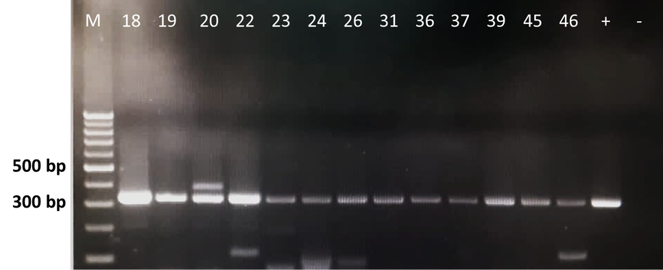
Leptospira 16S rRNA PCR amplification of bovine kidney samples. M: 1 kb marker, +/− (positive/negative) controls and the Leptospira PCR-positive samples were placed in between M and the controls (Samples no. 18, 19, 20, 22, 23, 24, 26, 31, 36, 37, 39, 45 and 46). L. interrogans serovar Canicola was used as positive control in this study.
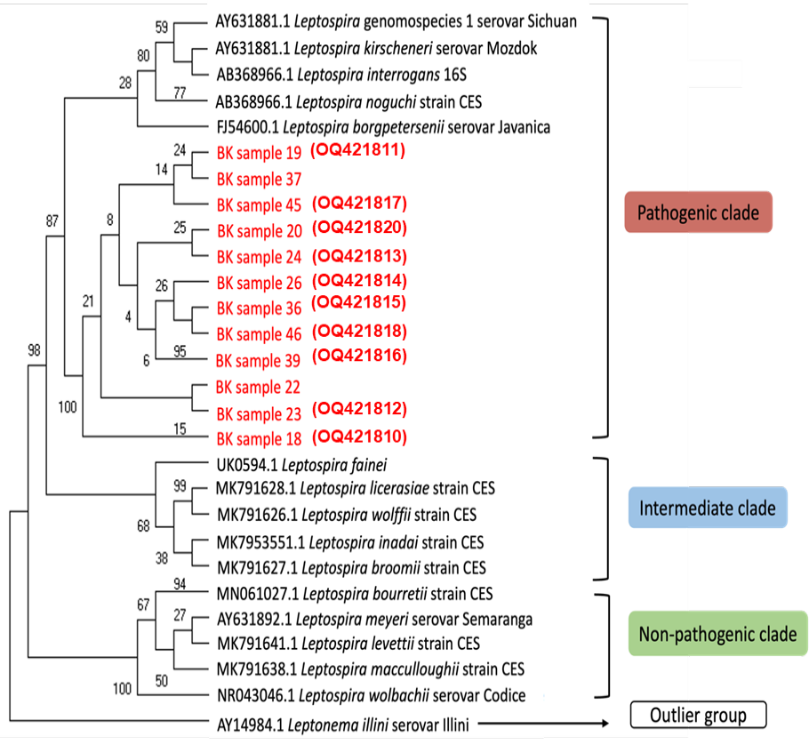
The phylogenetic analysis of 16S rRNA nucleotide sequences across various Leptospira group using several genomospecies as reference and one outlier group by maximum likelihood method constructed using MEGA11 [15]. Bootstrapping was performed 1,000 times, and all positions containing gaps and missing data were removed. Sequences obtained as part of this study are highlighted in red and the accession numbers generated from this study are in red parentheses.
Histopathological changes in the bovine kidney tissue samples; A, glomerular atrophy with slight deposition of homogenous hyaline droplets (tailed-arrow). B, deposition of pinkish homogenous hyaline cast within the glomerulus (arrow). C, infiltration of inflammatory cells (neutrophils and lymphocytes) in the renal interstitial (tailed-arrow). D, eosinophilic homogenous cytoplasmic hyaline droplets in the tubular epithelial cells (tailed-arrow), renal tubular necrosis (thin arrow) and margination of inflammatory cells in the blood vessel lumen (thick arrow). E, renal tubular necrosis (thin arrow) was prominent with infiltration of inflammatory cells in the renal interstitial (tailed-arrow). Eosinophilic homogenous cytoplasmic hyaline droplets were observed in the tubular epithelial cells (thick arrow). F, deposition of pinkish homogenous hyaline cast within the glomerulus (thick arrow), renal tubular necrosis (thin arrow) with inflammatory cells in the renal interstitial (tailed-arrow). Images A-D were PCR-positive (Sample no. 22), whilst images E and F were PCR-negative (Sample no. 32). (H and E stain; magnification of 400x).
References
- Abdul Rahman, M.S., Khairani, B.S., Zakaria, Z., Hassan, L. and Azri, R.M., 2020. Seroprevalence and distribution of leptospiral serovars in livestock (cattle, goats, and sheep) in flood-prone Kelantan, Malaysia. J. Vet. Res., 65: 53–58. https://doi.org/10.2478/jvetres-2021-0003[CrossRef]
- Agudelo-Flórez, P., Murillo, V.E., Londoño, A.F. and Rodas, J.D., 2013. Histopathological kidney alterations in rats naturally infected with Leptospira. Biomedica, 33 (Suppl 1): 82-88. https://doi.org/10.7705/biomedica.v33i0.686[CrossRef]
- Allan, K.J., Halliday, J.E., Moseley, M., Carter, R.W., Ahmed, A., Goris, M.G., Hartskeerl, R.A., Keyyu, J., Kibona, T., Maro, V.P., Maze, M.J., Mmbaga, B.T., Tarimo, R., Crump, J.A. and Cleaveland, S., 2018. Assessment of animal hosts of pathogenic Leptospira in northern Tanzania. PLoS Negl. Trop. Dis., 12: e0006444. https://doi.org/10.1371/journal.pntd.0006444[CrossRef]
- Aono, F.H., Cooke, R.F., Alfieri, A.A. and Vasconcelos, J.L., 2013. Effects of vaccination against reproductive diseases on reproductive performance of beef cows submitted to fixed-timed AI in Brazilian cow-calf operations. Theriogenology, 79: 242-248. https://doi.org/10.1016/j.theriogenology.2012.08.008[CrossRef]
- Aymée, L., Gregg, W.R., Loureiro, A.P., Di Azevedo, M.I.N., Pedrosa, J.d.S., de Melo, J.D.S.L., Carvalho-Costa, F.A., de Souza, G.N. and Lilenbaum, W., 2021. Bovine genital leptospirosis and reproductive disorders of live subfertile cows under field conditions. Vet. Microbiol., 261: 109213. https://doi.org/10.1016/j.vetmic.2021.109213[CrossRef]
- Azizi, S., Kheirandish, R. and Rahimi, E., 2014. Comparison of polymerase chain reaction and warthin-starry techniques to detect Leptospira spp. in kidneys of slaughtered cattle. Onderstepoort J. Vet. Res., 81: e1-e6. https://doi.org/10.4102/ojvr.v81i1.821[CrossRef]
- Benacer, D., Thong, K.L., Min, N.C., Verasahib, K., Galloway, R.L., Hartskeerl, R.A., Souris, M. and Mohd Zain, S.N., 2016. Epidemiology of human leptospirosis in Malaysia, 2004-2012. Acta Trop., 157: 162-168. https://doi.org/10.1016/j.actatropica.2016.01.031[CrossRef]
- Biscornet, L., de Comarmond, J., Bibi, J., Mavingui, P., Dellagi, K., Tortosa, P. and Pagès, F., 2020. An observational study of human leptospirosis in Seychelles. Am. J. Trop. Med. Hyg., 103: 999–1008. https://doi.org/10.4269/ajtmh.19-0228[CrossRef]
- Bulach, D.M., Zuerner, R.L., Wilson, P., Seemann, T., McGrath, A., Cullen, P.A., Davis, J., Johnson, M., Kuczek, E., Alt, D.P., Peterson-Burch, B., Coppel, R.L., Rood, J.I., Davies, J.K. and Adler, B., 2006. Genome reduction in Leptospira borgpetersenii reflects limited transmission potential. Proc. natl. Acad. Sci., 103: 14560–14565. https://doi.org/10.1073/pnas.0603979103[CrossRef]
- Carvalho, S.M., Gonçalves, L.M., Macedo, N.A., Goto, H., Silva, S.M., Mineiro, A.L., Kanashiro, E.H. and Costa, F.A., 2011. Infecção por leptospiras em ovinos e caracterização da resposta inflamatória renal. Pesq. Vet. Bras., 31: 637–642. https://doi.org/10.1590/S0100-736X2011000800001[CrossRef]
- Chaurasia, R., Salovey, A., Guo, X., Desir, G. and Vinetz, J.M., 2022. Vaccination with Leptospira interrogans PF07598 gene family-encoded virulence modifying proteins protects mice from severe leptospirosis and reduces bacterial load in the liver and kidney. Front. Cell. Infect. Microbiol., 12: 926994. https://doi.org/10.3389/fcimb.2022.926994[CrossRef]
- Costa, F., Hagan, J.E., Calcagno, J., Kane, M., Torgerson, P., Martinez-Silveira, M.S., Stein, C., Abela-Ridder, B. and Ko, A.I., 2015. Global morbidity and mortality of leptospirosis: A systematic review. PLoS Negl. Trop. Dis., 9: e0003898. https://doi.org/10.1371/journal.pntd.0003898[CrossRef]
- Dash, B.R., Dhaygude, V.S., Gadhave, P.D., Garud, K.V. and Kadam, D.P., 2018. Molecular detection of Leptospira spp. from canine kidney tissues and its association with renal lesions. Vet. World, 11: 530-534. https://doi.org/10.14202/vetworld.2018.530-534[CrossRef]
- Daud, A., Fuzi, N.M., Arshad, M.M., Kamarudin, S., Mohammad, W.M., Amran, F. and Ismail, N., 2018a. Leptospirosis seropositivity and its serovars among cattle in northeastern Malaysia. Vet. World, 11: 840–844. https://doi.org/10.14202/vetworld.2018.840-844[CrossRef]
- Daud, A.B., Mohd Fuzi, N.M.H., Wan Mohammad, W.M.Z., Amran, F., Ismail, N., Arshad, M.M. and Kamarudin, S., 2018b. Leptospirosis and workplace environmental risk factors among cattle farmers in northeastern Malaysia. Int. J. Occup. environ. Med., 9: 88-96. https://doi.org/10.15171/ijoem.2018.1164[CrossRef]
- Department of Statistics Malaysia, 2023. Available at: https://www.dosm.gov.my/v1/uploads/files/5_Gallery/2_Media/4_Stats%40media/4-Press_Statement/2022/06.%20JUN/KDNK%20Negeri%202021.pdf/ (accessed 2 February 2023).
- Department of Veterinary Services Malaysia, 2023. Available at: https://www.dvs.gov.my/dvs/resources/user_1/2021/BPSPV/penerbitan/PERANGKAAN_24_8.pdf/ (accessed 2 February 2023).
- Ellis, W.A., 1999. Leptospirosis. In: Diseases of swine. 8th edition (eds. A.D. Leman, B.E. Straw, W.L. Mengeling, S. D’Allaire and D.J. Taylor). Iowa State University Press, USA. pp. 483-493.
- Ellis, W.A., 2015. Animal leptospirosis. Curr. Top. Microbiol. Immunol., 387: 99–137. https://doi.org/10.1007/978-3-662-45059-8_6[CrossRef]
- Gökmen, T.G., Soyal, A., Kalayci, Y., Önlen, C. and Köksal, F., 2016. Comparison of 16S rRNA-PCR-RFLP, LipL32-PCR and OMPL1-PCR methods in the diagnosis of leptospirosis. Rev. Inst. Med. Trop. Sao Paulo., 58: 64. https://doi.org/10.1590/S1678-9946201658064[CrossRef]
- Grassmann, A.A., Souza, J.D. and McBride, A.J., 2017. A universal vaccine against leptospirosis: Are we going in the right direction? Front. Immunol., 8: 256. https://doi.org/10.3389/fimmu.2017.00256[CrossRef]
- Guedes, I.B., de Souza, G.O., de Souza Rocha, K., Cavalini, M.B., Neto, M.S.D., de Paula Castro, J.F., de Souza Filho, A.F., Negrão, M.P., Cortez, A., de Moraes, C.C.G. and Heinemann, M.B., 2021. Leptospira strains isolated from cattle in the Amazon region, Brazil, evidence of a variety of species and serogroups with a high frequency of the Sejroe serogroup. Comp. Immunol. Microbiol. Infect. Dis., 74: 101579. https://doi.org/10.1016/j.cimid.2020.101579[CrossRef]
- Haake, D.A. and Levett, P.N., 2017. Leptospira species (Leptospirosis). In: Mandell, Douglas, and Bennett’s infectious disease essentials (eds. J.E. Bennett, R. Dolin and M.J. Blaser). Elsevier, USA. pp. 354-355.
- Hegazy, Y., Elmonir, W., Orieby, A.F., Eldesoukey, I.E., Fransis, M. and Al-Gaabary, M.H., 2021. Leptospirosis as a neglected burden at human-cattle interface in mid-delta of Egypt. J. Infect. Dev. Ctries., 15: 704–709. https://doi.org/10.3855/jidc.13231[CrossRef]
- Islam, A.A., Ijaz, M., Hussain, F.S., Shoaib, M., Fakhar-e-Alam, Kulyar, M. and Yasmeen, K., 2019. Leptospirosis: Rising nuisance for cattle and threat to public health. In: Bacterial cattle diseases (ed. H.A.H.E. Kaoud). Intech Open, United Kingdom.
- Kamaruzaman, I., Mokhtar, M., Ting, H., Yuan, Y., Shah, A., Loong, T., Shaharulnizim, N., Reduan, M., Hamid, F., Noralidin, N., Abdul Manaf, N.A., Che Wan Zalati, C.W.S., Loong, S.K., Clegg, S. and Abu-Bakar, L., 2022. Molecular detection of pathogenic Leptospira spp. in urban rodents from wet markets in northeast Malaysia. J. Adv. Vet. Anim. Res., 9: 275. https://doi.org/10.5455/javar.2022.i593[CrossRef]
- Ko, A.I., Goarant, C. and Picardeau, M., 2009. Leptospira: The dawn of the molecular genetics era for an emerging zoonotic pathogen. Nat. Rev. Microbiol., 7: 736–747. https://doi.org/10.1038/nrmicro2208[CrossRef]
- Loong, S.K., Abd-Majid, M.A., Teoh, B.T., Cheh, M.J., Khor, C.S., Chao, C.C., Khoo, J.J. and AbuBakar, S., 2022. Leptospirosis among dengue-negative febrile patients in Selangor, Malaysia. Am. J. trop. Med. Hyg., 107: 397-400. https://doi.org/10.4269/ajtmh.20-0656[CrossRef]
- Loureiro, A.P. and Lilenbaum, W., 2020. Genital bovine leptospirosis: A new look for an old disease. Theriogenology, 141: 41–47. https://doi.org/10.1016/j.theriogenology.2019.09.011[CrossRef]
- Loureiro, A.P., Pestana, C., Medeiros, M.A. and Lilenbaum, W., 2017. High frequency of leptospiral vaginal carriers among slaughtered cows. Anim. Reprod. Sci., 178: 50–54. https://doi.org/10.1016/j.anireprosci.2017.01.008[CrossRef]
- Mérien, F., Amouriaux, P., Perolat, P., Baranton, G. and Saint, G.I., 1992. Polymerase chain reaction for detection of Leptospira spp. in clinical samples. J. clin. Microbiol., 30: 2219–2224. https://doi.org/10.1128/jcm.30.9.2219-2224.1992[CrossRef]
- Narkkul, U., Thaipadungpanit, J., Srisawat, N., Rudge, J.W., Thongdee, M., Pawarana, R. and Pan-ngum, W., 2021. Human, animal, water source interactions and leptospirosis in Thailand. Sci. Rep., 11: 3215. https://doi.org/10.1038/s41598-021-82290-5[CrossRef]
- Orjuela, A.G., Parra-Arango, J.L. and Sarmiento-Rubiano, L.A., 2022. Bovine leptospirosis: Effects on reproduction and an approach to research in Colombia. Trop. Anim. Hlth. Prod., 54: 251. https://doi.org/10.1007/s11250-022-03235-2[CrossRef]
- Peek, S. and Divers, T.J., 2018. Rebhun’s diseases of dairy cattle: 3rd edn. Saunders, USA.
- Prakoso, Y.A., Widyarini, S. and Kurniasih, K., 2020. Histopathology of the organs from cattle with leptospirosis. Adv. Anim. Vet. Sci., 8: 1220-1224. https://doi.org/10.17582/journal.aavs/2020/8.11.1220.1224[Full text][CrossRef]
- Radi, M.F., Hashim, J.H., Jaafar, M.H., Hod, R., Ahmad, N., Mohammed Nawi, A., Baloch, G.M., Ismail, R. and Farakhin, A.N.I., 2018. Leptospirosis outbreak after the 2014 major flooding event in Kelantan, Malaysia: A spatial-temporal analysis. Am. J. trop. Med. Hyg., 98: 1281–1295. https://doi.org/10.4269/ajtmh.16-0922[CrossRef]
- Rissi, D.R. and Brown, C.A., 2014. Diagnostic features in 10 naturally occurring cases of acute fatal canine leptospirosis. J. Vet. Diagn. Invest., 26: 799-804. https://doi.org/10.1177/1040638714553293[CrossRef]
- Sabri, A.R., Khairani-Bejo, S., Zunita, Z. and Hassan L., 2019. Molecular detection of Leptospira sp. in cattle and goats in Kelantan, Malaysia after a massive flood using multiplex polymerase chain reaction. Trop. Biomed., 36: 165-171.
- Sadiq, M.B., Hamid, N.A., Yusri, U.K., Ramanoon, S.Z., Mansor, R., Affandi, S.A., Watanabe, M., Kamaludeen, J. and Syed-Hussain, S.S., 2021. Ruminant farmers’ knowledge, attitude and practices towards Zoonotic Diseases in Selangor, Malaysia. Prev. Vet. Med., 196: 105489. https://doi.org/10.1016/j.prevetmed.2021.105489[CrossRef]
- Silva, F.G., Freitas, J.C., Anzai, E.K., Hashimoto, V.Y., Giraldi, N., Delbem, Á.C., Bracarense, A.P., Reis, A.C. and Vasconcellos, S.A., 2005. Leptospires detection in kidney, liver and uterus of cows slaughtered in Paraná state, Brazil. Braz. J. Microbiol., 36: 38-42. https://doi.org/10.1590/S1517-83822005000100008[CrossRef]
- Taddei, S., Moreno, G., Cabassi, C.S., Schiano, E., Spadini, C. and Cavirani, S., 2021. Leptospira seroprevalence in Colombian dairy herds. Animals, 11, 785. https://doi.org/10.3390/ani11030785[CrossRef]
- Tamura, K., Stecher, G. and Kumar, S., 2021. MEGA11: Molecular evolutionary genetics analysis version 11. Mol. Biol. Evol., 38: 3022–3027. https://doi.org/10.1093/molbev/msab120[CrossRef]
- World Organisation for Animal Health, 2023. Available at: https://www.woah.org/fileadmin/Home/fr/Health_standards/tahm/3.01.12_LEPTO.pdf (accessed 2 February 2023).
- Yang, C.W., Wu, M.S., Pan, M.J., Hsieh, W.J., Vandewalle, A. and Huang, C.C., 2002. The Leptospira outer membrane protein LipL32 induces tubulointerstitial nephritis-mediated gene expression in mouse proximal tubule cells. J. Am. Soc. Nephrol., 13: 2037-2045. https://doi.org/10.1097/01.ASN.0000022007.91733.62[CrossRef]
- Zamir, L., Baum, M., Bardenstein, S., Blum, S.E., Moran-Gilad, J., Perry Markovich, M., King, R., Lapid, R., Hamad, F., Even-Tov, B. and Elnekave, E., 2022. The association between natural drinking water sources and the emergence of zoonotic leptospirosis among grazing beef cattle herds during a human outbreak. One Hlth., 14: 100372. https://doi.org/10.1016/j.onehlt.2022.100372[CrossRef]
Cited by 1 article
In journals published by ResearchersLinks.
Services
Article usage statistics
Loading…