Research Article · Volume 10, Issue 5, 19 April 2022 · Open Access
Histomorphology and Histochemistry of the Oviduct in Laying Turkey Hens with Emphasis on the Sperm Host Glands
· · · ·
Affiliations & Notes ⌄
1Department of Cytology and Histology, Faculty of Veterinary Medicine, Beni-Suef University, Beni-Suef 62511, Egypt
2Department of Anatomy and Embryology, Faculty of Veterinary Medicine, Beni-Suef University, Beni-Suef 62511, Egypt. *Correspondence | Usama Kamal Moawad, Department of Cytology and Histology, Faculty of Veterinary Medicine, Beni-Suef University, Beni-Suef 62511, Egypt
Anatomical, histological, histochemical, and morphometric studies of the turkey hen’s oviduct under investigation showed five portions; infundibulum, magnum, isthmus, uterus (shell gland), and vagina with a prominent sperm host gland (sperm storage tubules) at the uterovaginal junction.
Each portion of the turkey’s oviduct varied concerning the length, shape, number, height of mucosal folds, and glandular contents as well as the thickness of muscular coat which allow the turkey hens to be adaptive for egg formation and transportation along their reproductive tract during the egg-laying period.
Absence of lamina muscularis mucosa, a continuation of the lamina propria with tunica submucosa forming lamina propria-submucosa filled with tubular glands, collagen fibers, and immunocompetent cells, was apparent along the entire turkey oviduct except for the vagina.
Histochemically, the neutral mucopolysaccharides were predominant in all portions of the oviduct, including their surface epithelium and proprial glands, while acid mucopolysaccharides dominated the mucosal epithelium of the infundibulum, magnum, isthmus, and vagina.
Selected from the article's own text.
Abstract
The current study was conducted to investigate the histomorphological and histochemical structure as well as the morphometrical analysis of all parts of the adult turkey hens’ (Meleagris gallopavo) oviduct during the egg production period. For this purpose, twenty four adult turkey hens were used. Gross examination and measurements of the oviduct compartments were obtained, and four specimens for each portion were used for scanning electron microscopic examination. Histologically, pieces of different oviduct portions were subjected to routine histological processing. Macroscopically, the oviduct had five portions; infundibulum, magnum, isthmus, uterus, and vagina. Microscopically, the wall of the oviduct is composed of four concentric layers; mucosa, submucosa, muscularis, and serosa but lacks lamina muscularis mucosa. The lamina propria submucosa contained immune competent cells and tubular glands in all portions of the oviduct except the infundibular funnel and vagina that devoid of any glands. The neutral mucopolysaccharides were predominant in supra-nuclear portions of the mucosal epithelium in all portions of the oviduct. In contrast, acid mucopolysaccharides were dominated in the infundibulum, magnum, isthmus, and vagina but were absent in the proprial glands. Sperm host glands were prominent in the uterovaginal portion. The vaginal tunica muscularis was the thickest compared with other oviduct parts forming the vaginal sphincter. In conclusion, the five portions of the turkey’s oviduct varied concerning the length, shape, number, height of the mucosal folds, and glandular contents as well as the thickness of the muscular coat, which allow the turkey hens to be adaptive for egg formation and transportation along their reproductive tract during the egg-laying period.
The avian oviduct is an amazing organ; it produces all of the laid egg components (egg-white and eggshell) except the yolk. It undergoes a series of hormonal (steroid hormones), biochemical, neuronal, and cellular changes during egg formation .
Anatomically, the avian oviduct extending from ovary to cloaca is formed from five distinct segments; the infundibulum that is responsible for the formation of a strong perivitelline membrane and chalaza around the egg yolk , the magnum which is the albumin secreting region , the isthmus that responsible for the formation of the shell membranes around the egg white , the uterus which forms the egg shell and the vagina that connects the uterus to the cloaca . Thus, the egg formation and transportation, as well as the sperm storage, transportation, the site of fertilization (infundibulum), and early embryonic development are the main oviduct’s functions . By the action of the fimbriated region of the infundibulum, the librated ovum from the ovary during ovulation is gathered into the ostium then transported to the oviduct where the albumen, shell membranes, hard shell, and cuticle are formed .
Histologically, the oviduct wall is composed of four distinct concentric layers; mucosa, submucosa, muscularis, and serosa where the mucosal surface epithelium vary according to the stage of production . Sperm host glands (sperm storage tubules or spermatic tubules) are avian specialized tubular mucosal invaginations resides at the junction between the uterus and vagina as sperm reservoir in their lamina propria , and are considered the primary storage site, where the infundibulum is the secondary storage portion . In domestic birds, once the sperms enter the female reproductive tract, they can survive up to 2–15 weeks according to the species, compared to a relatively short life span (several days) of mammalian sperm . The ability of sperms to reside in the sperm storage tubules (SSTs) of the uterovaginal junction (UVJ) is the biological basis of sustained fertility in chicken and turkey hens, according to Bakst et al. (2010), and the differences in the duration of fertility between the domestic fowl (2 to 3 weeks) and turkeys (10 to 15 weeks) are partly related to their respective numbers of SSTs (the mean numbers of SSTs for chickens are 4,893 and 30,566 for turkeys). SSTs are located in the lamina propria of mucosal folds in the UVJ and infundibulum . Fertilization occurs in the infundibulum .
Information about the histomorphological structure of the female reproductive organs of turkey hens is very scarce . Therefore, the current investigation aimed to describe the gross morphology, histochemistry, histomorphometry, and surface architecture of the laying turkey hen’s oviduct with particular emphasis on sperm host glands.
MATERIALS AND METHODS
Mature large turkey hens (Meleagris gallopavo) (n = 24) in egg production (40-45 weeks old) were purchased from local poultry slaughterhouses at Beni-Suef Governorate, Egypt. The oviduct was carefully removed from the surrounding viscera and grossly examined. Mesaurements for each part were taken using a Varnier calliper and photographed using a digital camera. The nomenclature in the current study was adopted according to .
Histological examination
Immediately after slaughtering of birds (n= 20), representative tissue specimens were collected from different parts of the oviduct (infundibulum, magnum, isthmus, uterus, and vagina) in addition to the uterovaginal portions. The collected specimens were immediately fixed in Bouin’s fluid for 24h, then processed using a routine paraffin embedding technique to prepare paraffin blocks. The paraffin blocks were sectioned using a rotary microtome to obtain 4-5µm paraffin sections and stained with the following stains; Harris’ Haematoxylin and Eosin (H and E stain) for demonstration of general histological structures, PAS stain for detection of neutral mucopolysaccharides, Alcian blue (AB) stain for detection of acid mucopolysaccharides, and Crossman’s trichrome stain for demonstration of collagen and smooth muscle fibers. The above-mentioned stains and techniques were conducted as outlined by Suvarna et al. (2019).
Scanning electron microscopy
For scanning electron microscopy (SEM), four specimens for each part of the oviduct were used. The specimens were fixed in 3% Glutaraldehyde solution in phosphate buffer (pH 7.2 to 7.4), post fixed with 1% osmium tetroxide in 0.1M sodium cacodylate buffer at pH 7.2 for 1h at 4°C. After that, the specimens were dehydrated through a graded series of ethanol and critical point dried (with carbon dioxide). Then the specimens were attached to aluminium stubs facing upwards, covered with colloidal carbon tabs, and then sputtered with gold palladium. The samples were examined and photographed with a JEOL/EO-JSM-6510 LV SEM at the Faculty of Science, Beni-Suef University, Egypt. The specimens were prepared and processed for SEM as outlined by Bozzola and Russel (1998).
Histomorphometric measurements
The thickness of mucosa, submucosa, musclaris, and serosa, in addition to the mucosal fold length of all oviductal portions were measured in 20 chosen sections using the Image-J analysis software with the aid of LEICA DFC290 HD system digital camera connected to a light microscope (X10 objective lens). The obtained morphometric measuremens are presented in Table 1.
Statistical analysis
The obtained data were subjected to one-way ANOVA using IBM SPSS Statistics for Windows (IBM SPSS 22; IBM Inc.) and subsequent multiple comparison Tukey test was applied to determine the differences between means. Data is expressed as mean ± SD with dissimilar superscript letters (significantly different at P< 0.05).
RESULTS and Discussion
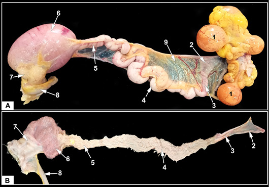
Macroscopically, the oviduct of laying turkey hens had five portions; infundibulum, magnum, isthmus, uterus, and vagina (Figure 1). The oviduct was a highly convoluted muscular duct, 85.5 ± 5.5 cm long, extended from the ovary (with follicles at different stages of maturation) to the cloaca and filled most of the dorsal and caudal parts of the left side of the abdominal cavity. The oviduct was suspended from the left side of the abdominal cavity by a thin, folded dorsal mesentery which continued around the duct to form the ventral mesentery. Histologically, the examined sections of oviductal portions, in addition to the UV portion revealed four tunics arranged concentrically from inward to outward as follows; mucosa, submucosa, muscularis, and serosa (Figures 2-7).
The infundibulum
The infundibulum was divided into a thin-walled funnel and a thick tubular region. The SEM analysis revealed spirally oriented longitudinal ridges that increased in depth toward the magnum and definite mucosal crypts appeared as deep folding of the epithelium (Figure 3A).
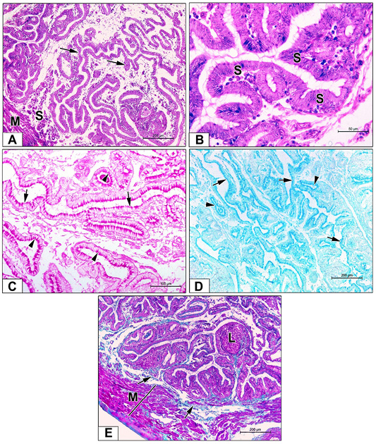
Histologically, the tunica mucosa of the infundibular funnel was composed of extensive finger-like folds with some anastomosis occupying the entire lumen (Figure 2A). These folds were long, thin, and highly branched carrying primary, secondary, and sometimes tertiary folds with shallow surface indentations running in a spiral manner. Moreover, some short primary pyramidal folds were observed. The surface epithelium was invaginated forming few mucosal glands with variable diameters. The surface epithelium was simple tall columnar ciliated and non-ciliated (secretory) cells with oval vesicular nuclei and acidophilic cytoplasm (Figure 2B). The surface epithelial cells and mucosal glands (invaginations of the surface epithelium) strongly reacted with PAS (Figure 2C) and AB (Figure 2D) stains. The propria-submucosa was thin (Figure 2A), and formed the mucosal folds’ core composed of fine collagen fibers, diffused fibroblasts, immunocompetent cells, lymphatic nodules, and devoid of tubular glands (Figure 2E). The tunica muscularis was very thin composed of inner circular and outer longitudinal smooth muscle fibers (SMFs) surrounded externally by the tunica serosa loose connective tissue covered by a mesothelial layer (Figure 2E).
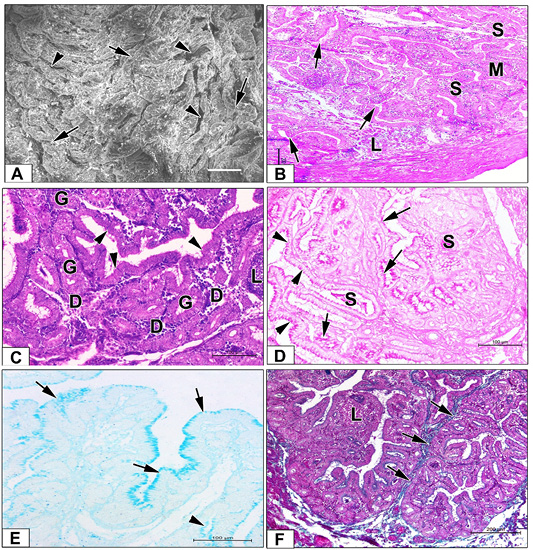
The tubulus infundibularis was thick-walled and extensively folded. Their mucosal folds were tall, highly branched, and thicker than those of the funnel part with deep surface invaginations (Figure 3B). The surface epithelium was simple columnar ciliated and non-ciliated (secretory) cells containing oval vesicular nuclei and surrounded by acidophilic cytoplasm (Figure 3C). Numerous mucosal glands with variable sizes were noticed to show the same cell lining of the surface epithelium (Figure 3C). The surface epithelial cells and the mucosal glands showed intense PAS-positive reactions (Figure 3D), whereas only surface epithelial cells showed an AB-positive reaction (Figure 3E). Numerous closely aggregated tubular glands surrounded by fine collagen fibers, connective tissue cells and solitary lymph nodules were the main components of the lamina propria (Figure 3F). These proprial glands were lined with tall columnar or even pyramidal cells with rounded basally situated nuclei and lightly acidophilic cytoplasm (Figure 3C). PAS-positive secretory materials were seen in their lumina (Figure 3D). The tunica muscularis was slightly thicker than that of the funnel part and was formed from ill-defined inner circular and outer longitudinal SMFs covered externally by the serosal layer (Figure 3B).
The magnum
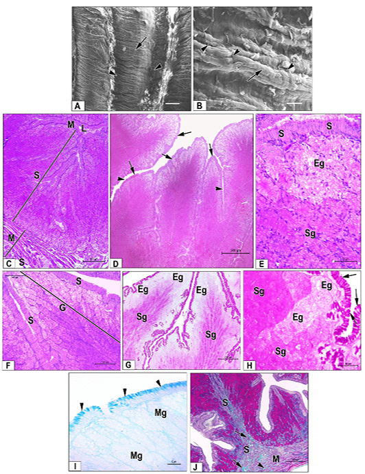
The magnum was the longest and most conspicuous part. It was easily distinguished from the infundibulum by its pronounced thicker wall (Figure 1). In SEM analysis, the mucosal folds of the magnum were more voluminous, with narrow clefts in between. The surface of the mucosal folds had transverse secondary folds with numerous pits that were presumed to be the gland’s openings (Figure 4A, B).
Histologically, the tunica mucosa of the magnum was exhibited by long, broad and thick leaf-like primary folds with deep surface invaginations that were occupied most
Table 1: Histomorphometric measurements of the oviduct portions.
UV junction
Vagina
Uterus
Ismuth
Magnum
Infundibulum
892.79±
46.256c,d
775.80±
75.674b,d
1453.5± 57.090a,b,c
578.11± 39.565 a,b
1097.3±
50.806
884.64±
71.938
Mucosal fold length
879.14 ±
80.290a,d,e
375.02±
38.626b,c
362.50± 35.735b,c
860.44± 93.805a
1025.7±
76.736a
127.08±
12.756
Mucosal fold thickness
136.62±
6.129b
161.68±
8.084
130.11±
12.229b
160.34±
8.989
209.24±
28.155a
101.15±
9.251
Submucosa thickness
1317.2±
71.081a,b,c,d,e
1633.6±
45.990 a,b,c
1329.5± 37.715a,b,c,d
451.91± 31.625
367.58±
25.610a
440.75±
14.251
Muscularis thickness
61.497±
4.962
68.823±
5.790a,b,c
69.693±
4.114a,b,c
46.538±
3.720
45.022±
5.014
47.728±
3.881
Serosa thickness
Data are expressed as means ± SD with dissimilar superscript letters (significantly differing at P < 0.05): (a) significantly different from theinfundibulum value; (b) significantly different from the magnum; (c) significantly different from the isthmus value; (d) significantly different from the uterus value and (e) significantly different from the vagina value.
of the magnum wall thickness (Figure 4C, D). The surface epithelium was composed of tall ciliated cells and secretory columnar cells. The nuclei of the later cells were rounded and basally located, while that of the former were oval and centrally located (Figure 4E, F). The supra-nuclear portions of the surface epithelium were strongly reacted with PAS (Figure 4G, H) and AB (Figure 4I) stains.
The propria-submucosa formed the core of the mucosal folds and was made up of fibro-cellular connective tissue intermingled with smooth muscle bundles (Figure 4J). Well-developed closely aggregated tubular glands occupied the lamina propria and opened to the surface epithelium (Figure 4C, D), lined by simple columnar or pyramidal cells with rounded basally situated nuclei (Figure 4E). These glands were showed three morphological phases of secretory activity; regenerating, secretory, and exhausting “resting” (Figure 4F, G). The glands underlying the surface epithelium at the apical and lateral parts of the mucosal folds showed exhausted morphological features (Figure 4G), they had prominent lumina, their epithelial cell lining exhibited vacuolated cytoplasm and negatively reacted with PAS (Figure 4G, H). The regenerating glands appeared to have a well-defined lumen and their secretory cells had acidophilic cytoplasm. On the other hand, the secretory glands (Figure 4E, F) showed ill-defined cell outlines, acidophilic secretory materials filled the lumen, and the cytoplasm of their secretory cells was deeply acidophilic and strongly reacted with PAS (Figure 4G, H). The glands at the three phases showed a negative reaction to the AB stain (Figure 4I). The muscularis tunica was formed of well-defined inner circular and outer longitudinal layers of SMFs separated by collagen fibers with numerous blood vessels and covered externally by serosal tunica (Figure 4J).
The isthmus
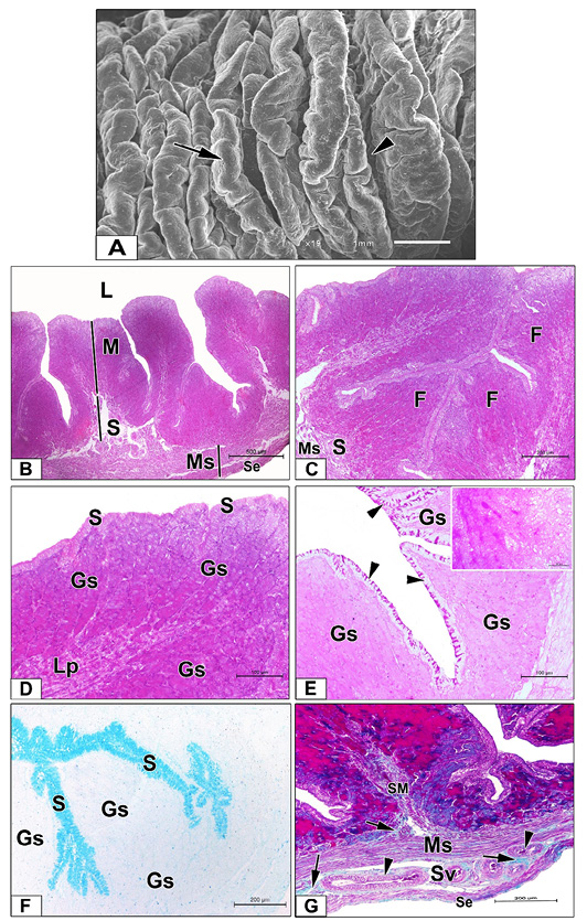
The isthmus was the relatively shortest section of the oviduct. Its diameter was less than that of the magnum (Figure 1). The SEM revealed a longitudinal orientation of its mucosal folds and parallel rows of discontinuous furrows. These folds were branched into secondary and tertiary ones (Figure 5A).
Histologically, the isthmus mucosa showed shorter, less broad primary folds than those of the magnum giving a prominent star-shaped lumen (Figure 5B, C). The mucosal surface epithelial cells were simple to pseudo-stratified ciliated columnar epithelium. The ciliated cells had apically located oval nuclei, while the secretory ones contained basally situated rounded nuclei (Figure 5D). The apical portions of the surface epithelium strongly reacted with PAS (Figure 5E) and AB (Figure 5F) stains. The propria-submucosa was made up of numerous collagenic fibers intermingled with smooth muscle bundles (Figure 5G). Numerous well-developed, closely aggregated tubular glands occupied the lamina propria and opened to the surface epithelium (Figure 5D). They were lined by simple columnar or pyramidal cells with rounded basally situated nuclei (Figure 5D). The glands showed three morphological phases of secretory activity; regenerating, secretory, and exhausting (resting) with similar morphological features of the magnum. The secretory glands reacted intensely, while the exhausted glands reacted moderately with PAS stain (insert image). On the other hand, the isthmus glands reacted negatively to the AB stain (Figure 5F). The tunica muscularis was composed of well-defined inner circular and outer longitudinal layers of SMFs. A well-developed stratum vasculare was predominant between the two layers of tunica muscularis consisting of loose connective tissue accompanied by numerous blood vessels (Figure 5G). The tunica serosa was composed of lamina subserosa (loose connective tissue) covered by mesothelial cells (Figure 5B, G).
The uterus
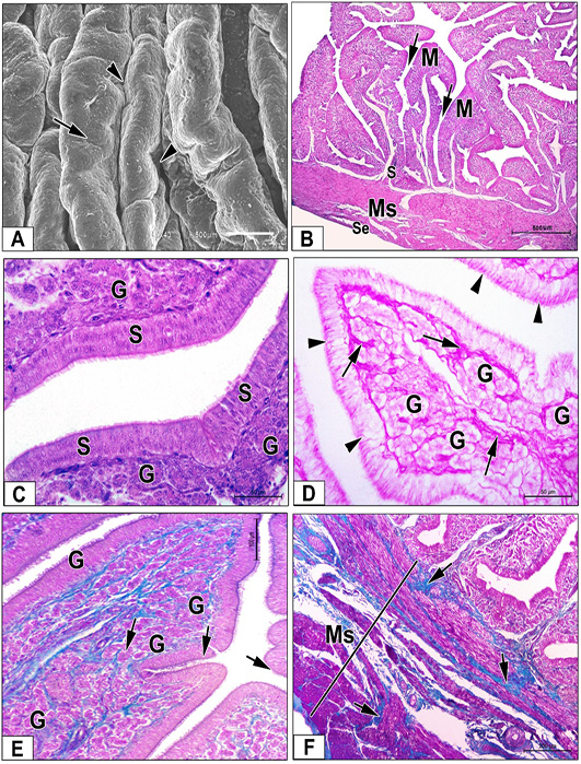
The uterus was an expanded portion of the oviduct. It was more fusiform than the isthmus (Figure 1). By SEM examination, the uterine mucosal folds appeared longer, thicker, and more complex than the isthmus. These folds were more compressed, longitudinally oriented, and obscured by secondary circular folds. Abundant voluminous interfold spaces were seen between folds (Figure 6A).
Microscopically, the uterine mucosa was packed with complex primary mucosal folds intermittent with some secondary ones. These folds were thin, long and sometimes short thick ones were recognized in-between separated by deep grooves “invaginations” (Figure 6B). The lamina epithelialis mucosa consisted of columnar ciliated and non-ciliated secretory cells (Figure 6C), containing apically located PAS-positive secretory granules (Figure 6D). Contrary, the surface epithelial cells negatively reacted with AB stain. The uterine lamina propria-submucosa was made up of vascular fibrous connective tissue that filled the core of the mucosal folds and was packed with loosely arranged simple tubular glands “uterine glands” had narrow lumina and simple cuboidal lining epithelium with rounded vesicular nuclei and acidophilic cytoplasm (Figure 6C). These glands moderately reacted with PAS stain (Figure 6D) and negatively reacted with AB stain. In addition, fine collagenic fibers surrounding the uterine glands were noticed (Figure 6E). The tunica muscularis was thick and composed of well-developed inner circular and outer longitudinal SMFs separated by highly vascularized connective tissue (Figure 6F). The outer serosal layer consisted of thick loose connective tissue covered by mesothelial cells (Figure 6B, F).
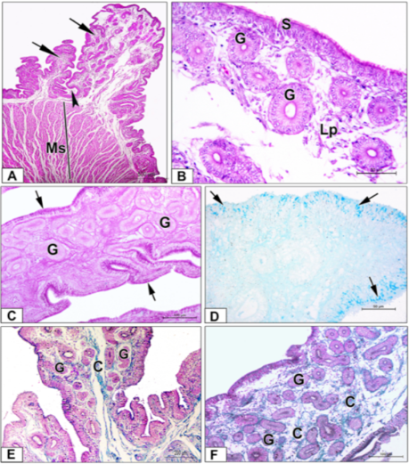
At the UV junction, the mucosal folds became shorter and thicker with broad apices surrounded by deep crypts (Figure 7A). The uterine epithelium was lined all folds at the junctional site (Figure 7B). The most characteristic feature of this portion is the presence of the sperm host glands. These glands appeared with variable sizes, prominent lumina, and were lined by a single layer of columnar cells with regularly arranged rounded nuclei close to the basement membrane and lightly stained vacuolated cytoplasm (Figure 7B). The surface epithelial cells showed intense PAS (Figure 7C) and AB (Figure 7D) reactions, while the glandular cells showed moderate apical positive PAS (Figure 7C) and negative AB (Figure 7D) reactions. The glands were opened into the mucosal crypts and surrounded by loose connective tissue of the lamina propria (Figure 7B) and fine collagen fibers (Figure 7E, F). The tunica muscularis appeared very thick and was surrounded by serosa.
The vagina
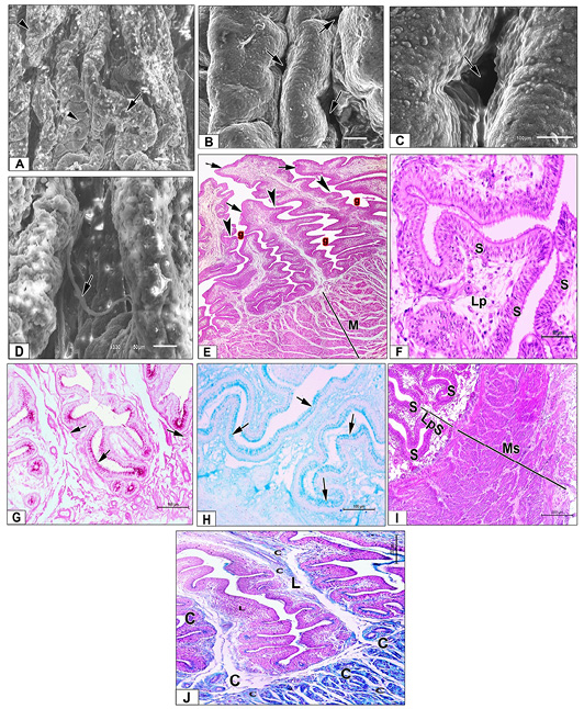
The vagina was the short and S-shaped tube and was the last portion of the oviduct (Figure 1). The SEM study showed that the mucosal folds of this region were transversally oriented and carried secondary folds. The folds are narrower than in most other parts of the oviduct. Moreover, deep pits were localized between the longitudinal vaginal folds constituting the sperm host glands with remnants of sperms (Figure 8A-D).
Histologically, the vaginal mucosal folds were long primary folds with multiple secondary ones that were oriented in a regular or parallel manner (Figure 8E). They showed deep invaginations forming mucosal crypts. The surface epithelium was lined with pseudo-stratified columnar ciliated epithelium with goblet cells which were crowded at the mucosal crypts (Figure 8F) and strongly reacted with PAS (Figure 8G) and AB (Figure 8H) stains. The propria-submucosa consisted of highly vascular loose connective tissue intermingled with numerous fibroblasts, immunocompetent cells and devoid of tubular glands (Figure 8I). The tunica muscularis was a very thick well-developed layer compared to other parts of the oviduct consisting of an inner circular layer that was thicker than the outer longitudinal, forming the vaginal sphincter (Figure 8E, I) and surrounded by collagenic fibers (Figure 8J). The serosa was made of loose connective tissue covered by a single layer of mesothelium.
The current anatomical and histological investigation revealed five portions forming the turkey hen’s oviduct including infundibulum, magnum, isthmus, uterus (shell gland), and vagina. These results were similar to those obtained by most of the available literatures in domestic birds.
In agreement with Robert et al. (2011) in Emu birds (dromaius novaehollandiae) and Paul et al. (2016) in the hens, two portions; cranial (infundibular funnel) and caudal (tubulus infundibularis), form the turkey hen’s infundibulum. On the other hand, investigations of Mohammadpour and Keshtmandi (2008) in pigeons and Moraes et al. (2010) in ducksreported three regions: The fimbriated (infundibular fimbriae), funnel, and tubular. The fimbriated portion was not observed in the current study. The extensive spirally arranged finger-like folds with some anastomosis occupying the whole lumen of the infundibular funnel agreed with those reported in ostriches and indigenous geese (Anser anser) who noticed a spiral course manner of the infundibular mucosal folds. In contrast, Vijayakumar et al. (2016) reported the absence of mucosal folds in the Emu funnel-shaped part of the infundibulum.
Concerning the lining epithelium of the infundibular funnel, it consisted of simple tall columnar ciliated and non-ciliated (secretory) cells. Wani et al. (2017) found that the infundibular epithelium of the Kashmir faverolla chicken is a simple columnar ciliated epithelium that changed into simple columnar non-ciliated secretory cells at the bottoms of the grooves between folds. However, ostrich ; and duck have pseudo-stratified columnar ciliated epithelium lining of the infundibulum. In turkeys, a simple cuboidal lining epithelium was reported . Few mucosal glands with variable diameters were formed by invagination of the surface epithelium. In contrast to these results; the lamina propria of the infundibular funnel was devoid of any mucosal glands in sexually mature ducks , sexually mature quails , indigenous geese (Anser anser) , and in hens . In agreement with Wani et al. (2017), the apices of the infundibular epithelium and mucosal glandular cells showed a strong positive reaction for neutral mucopolysaccharides in Kashmir faverolla chicken. The presence of neutral mucopolysaccharides to facilitate the transferring of ovum , and incorporated in formation of the perivitelline membrane and chalaza around the egg yolk .
As reported by the current study, the absence of true tubular glands in the lamina propria-submucosa was previously reported by Patki et al. (2013); El-Sayed (2016) and Liebich (2019) who did not observe any glands in the lamina propria of the funnel region. In contrast, El- Gendy et al. (2016) noticed tubular glands in the lamina propria-submucosa of both the funnel and the neck portion of the Balady duck infundibulum. In accordance with Mohammadpour et al. (2012), the examined tunica muscularis was very thin and composed of inner circular and outer longitudinal SMFs. Unlike these results, El- Gendy et al. (2016) in Balady duck observed inner circular and outer oblique layers of SMFs, and El-Sayed (2016) in adult female turkey noticed widely separated bundles of smooth muscle with different orientations forming the infundibular tunica muscularis. Moreover, Liebich (2019) reported that smooth muscle in the funnel wall is necessary for picking up the liberated oocytes during ovulation by its contractile properties.
The current results concerning the simple columnar ciliated and non-ciliated (secretory) cells lining the surface epithelium of the tubulus infundibularis coincided with Mohammadpour and Keshtmandi (2008) in turkeys who reported ciliated simple columnar in the middle and lower infundibular ends. However, Vijayakumar et al. (2016) stated that the tubular part of the Emu infundibulum is covered with pseudo-stratified ciliated columnar epithelium with numerous goblet cells between the secretory cells. Similar to the mature ostrich , the intense PAS and AB positive reactions of the supra-nuclear portion of the infundibular neck surface epithelium indicate secretory activities of the infundibular neck in building up the first albumen layer around the ovum, which is then completed in the magnum. On the other hand, the negative PAS reaction of proprial glands indicate that these glands are devoid of any acid mucopolysaccharids , and suggesting their role in the secretion of the inorganic matrix of the laid egg .
The tubular glands were numerous, closely aggregated in the lamina propria-submucosa These results partially agreed with Parizzi et al. (2008); Bansal et al. (2010), who observed few tubular glands in the caudal part of the infundibulum of rhea and quail, respectively. Furthermore, Patki et al. (2013) detected small tubular glands in the neck region of the duck infundibulum, and Liebich (2019) reported numerous alveolar invaginations in the mucosa of the caudal infundibular portion.
In addition, the simple columnar ciliated and non-ciliated (secretory) cells lining the proprial glands are different from the previously reported simple cuboidal cells of the emu birds , or the pyramidal or tall columnar epithelial cells lining the tubular glands in the ostrich infundibular end .
Similar to findings of Deka et al. (2014) in duck, the tunica muscularis was slightly thicker than that of the funnel part formed from ill-defined inner circular and outer longitudinal SMFs and unlike El-Gendy et al. (2016) who reported inner circular and outer oblique SMFs forming the muscularis of the Balady duck infundibulum.
Similar to the results of Vijayakumar et al. (2016), the current study revealed the presence of numerous well-developed closely aggregated tubular glands in the magnum lamina propria yielded several long, broad, and thick primary folds occupying most of the magnum-thickness. Similar results were also reported in Kashmir faverolla chicken , ostrich , and Emu . In addition, the internal magnum mucosal folds were found to be the longest and thickest folds in the oviduct of Japanese quail and rhea .
Regarding the surface epithelium, it was composed of tall columnar ciliated and secretory cells. This result agreed with that observed by Bansal et al. (2010) in quail. However, Saber et al. (2009) in ostrich noticed a pseudo-stratified columnar epithelium with more ciliated cells than secretory cells. Similar to the magnum of hens , the turkey magnum glands of the current investigation showed three morphological phases of secretory activity; regenerating, secretory and exhausting “resting.” The properties of the magnum proprial glands varied according to the phase of the reproductive cycle , and responsible for albumen synthesis and secretion during the stage of egg production where coiled tubular glands in the lamina propria of the magnum secrete the main components of egg albumen; ovalbumin, ovomucoid, and ovotransferrin. As in ostrich , the muscular tunica of turkey magnum is formed of well-defined inner circular and outer longitudinal layers of SMFs. However, El-Gendy et al. (2016) reported inner longitudinal and outer oblique, and Fouad (1970) detected only a single circular muscular layer in Fayoumi fowl.
The detected shorter and less broad primary folds in the isthmus compared to the magnum were previously reported where the isthmus mucosal folds are not as eminent as those of the magnum, less voluminous and narrower. In contrast, Lakshmi et al. (2018) considered the isthmus primary folds of Emu (Dromaius novaehollandiae) to be the largest folds among all oviduct mucosal folds. In contrast, Robert et al. (2011) reported that the isthmus mucosal folds were similar to those of magnum in Emu. Moreover, Mehta and Guha (2012) in hens noticed branching of the leaf-like isthmus primary folds. The isthmus surface epithelium was tall columnar ciliated and secretory cells. In contrast, Parto et al. (2011) noticed a simple columnar with ciliated and goblet cells in the isthmus of laying turkeys and Vijayakumar et al. (2016) reported pseudo-stratified ciliated columnar epithelium in Emu. The surface epithelial cells strongly reacted with PAS and AB stains like hens (Ozen et al., 2009) and Pekin duck . In addition, Wani et al. (2017) reported that the epithelium lining the isthmus of faverolla chicken showed both acidic and neutral mucopolysaccharides. The lamina propria submucosa of the isthmus was housed by branched tubular gland “glandulae isthmi” ; . This result partially coincided with Sawsan (1994) who noticed tubular glands in laying goose and tubular coiled in laying pigeons. On the contrary, Das and Bisawal (1968) defined no glands in the isthmus of domestic duck (Anas boscas).
In the current study, the isthmus glands showed three different morphological phases of secretory activity; regenerating, secretory and exhausting (resting) phases. These results agreed with Lakshmi et al. (2018) who documented that the isthmus lamina propria had densely packed glands at different secretory activities. Similar to previous studies, the muscular tunica of the isthmus consists of well-defined inner circular and outer longitudinal layers of SMFs ; ; . Moreover, the blood vessels rich loose connective tissue layer, stratum vasculare, lying between the two layers of tunica muscularis was noticed as previously reported in Japanese quail .
The current investigation revealed complex thin, long primary uterine mucosal folds intermittent with some secondary ones. These results were in harmony with Wani et al. (2017) who reported that the uterine folds of Kashmir faverolla chicken are not as broad as those of isthmus and comparatively less glandular. In addition, Lakshmi et al. (2018) in Emu birds (Dromaius novaehollandiae) reported that the uterine mucosal folds are the longest among all regions of the oviduct and branched into primary and secondary folds. The uterine surface epithelial cells in turkeys were columnar ciliated and non-ciliated secretory cells. Similarly, Ozen et al. (2009) reported a single layer of ciliated and secretory cells in the duck uterine mucosa. However, previous studies in Punjab white quails , guinea fowl , and turkeys reported a single layer of pseudo-stratified columnar ciliated epithelium. Moreover, Das and Bisawal (1968) detected alternating columnar ciliated and goblet cells, and Wani et al. (2017) in Kashmir faverolla chicken noticed ciliated and non-ciliated cells of the pseudostratified epithelium, as well as intermittent ciliated columnar cells, contained secretory granules.
The lamina propria was packed with loosely arranged simple tubular uterine glands with narrow lumina lined by simple cuboidal epithelium inducing thickening of the uterine mucosa. These results were in harmony with Sharaf et al. (2013) who reported that the thickness of the uterine lamina propria of ostrich increases gradually to reach its maximal size during the laying period (stage of egg production); subsequently, the uterine glandular content massively increases. However, Kamel et al. (1987) noticed pigment cells in the lamina propria of Fayoumi hens’ uterus; these pigments were not observed in the current study due to the colorless nature of turkey hen’s eggs.
The uterine surface epithelial cells strongly reacted with PAS, while the uterine glands moderately reacted. This result coincided with Ibrahim et al. (2015) who proved that the uterine surface epithelium and tubular glands located in the lamina propria significantly contributed to the uterine secretions. The uterine tunica muscularis was composed of thick well-developed inner circular and outer longitudinal smooth muscle bundles separated by highly vascularized connective tissue. Similar results were reported in Japanese quail and Emu bird “Dromaius novaehollandiae” where the tunica muscularis was highly vascular and the thickest layer in the uterine wall. In turkeys, the uterine tunica muscularis is the thickest among all oviduct segments to provide the uterus with the necessary amounts of calcium during egg laying . Moreover, the very thick uterine muscular wall terminated as a pouch was reported to womb the egg during the stage of shell formation .
The capability of female birds to store sperm in their genital tract and the presence of a system that maintains the reserved sperm in a live state till the time of release is an advantageous for avian reproduction . This storing ability is achieved by the presence of specialized invaginations in the female oviduct named sperm host glands or sperm storage tubules “SST” . The most prominent features of the uterovaginal junction as reported in current work are the abrupt change from the expanded uterine mucosal folds into short vaginal folds and the presence of sperm host glands in the lamina propria which are not demonstrated in the remaining vaginal folds. Many researchers ; described the occurrence of sperm host glands in the utero-vaginal lamina propria. Moreover, Liebich (2019) reported the presence of the uterovaginal sperm host glands in the lamina propria of hens near the muscle sphincter vaginae. He added that they serve as a reserve site for the ejaculated sperm, keeping them viable for weeks and allowing a hen to lay fertilized eggs for two weeks after the last copulation. On the contrary, sperm host glands are not uncounted in the emu vagina . However, Khan et at. (1999) in chicken reported the presence of SSTs in the the lamina propria of three portions of the oviduct; infundibulum, utero-vaginal portion, and the vagina. Similar to the results of Das (2002), the SSTs were lined by a single layer of columnar cells with basal rounded nuclei and some luminal tubules contained spermatozoa. Moreover, Khan et at. (1999) reported in UVJ of White Leghorn chicken presence of some tubules contained spermatozoa and other empty ones. The presence of luminal tubules devoid of spermatozoa may be attributed to moving of spermatozoa to the follicle for fertilization or insufficient ejaculated semen to fill all tubules .
As reported by the current study, the thick tunica muscularis at the uterovaginal in addition to strong vaginal sphincter might retain the spermatozoa inside the sperm host glands and prevent their descending caudally to the vaginal lumen . They added that the sperm host gland tubules have the ability to stores the ejaculated spermatozoa till reaching the infundibulum (site of fertilization) for further fertilization, secrete albumen-like material forming an additional fibrous protein surrounds the laid ovum and prevents the excessive spermatozoal penetration of the ovum at the initial end of the oviduct during fertilization.
Contrary to the observed long primary and secondary vaginal mucosal folds in turkeys, Mirhish and Nsaif (2013a) and Robert et al. (2011) recorded that the vagina has the narrowest mucosal folds in the oviduct except for the infundibulum due to the absence of glands in the vagina. In consistence with Bezuidenhout et al. (1995), the vaginal lining epithelium was pseudo-stratified columnar ciliated epithelium with goblet cells which were crowded at the mucosal crypts, and unlike that of Sharaf et al. (2013) who observed ciliated, non-ciliated columnar and basal cells, and Das and Biswal (1968) who noticed neither goblet nor non-ciliated cells in the vaginal epithelium. Previous studies reported no neutral mucopolysaccharides in the quail vagina ; however, a strong PAS reaction of the turkey vaginal mucosal surface epithelium. This is probably because the quail study was conducted at earlier laying periods in quail compared to the current research. The highly vascular loose connective tissue and numerous immunocompetent cells that were observed in the vaginal lamina propria, suggests a high degree of immunological efficacy . Consistent with the current result, Ozen et al. (2009) and Mohammadpour et al. (2012) pointed out that the vaginal lamina propria had no tubular secretory glands.
Like the native chicken in Bangladesh, the turkey’s vaginal tunica muscularis was a well-developed layer compared to other parts of the oviduct. The inner circular layer was notably thicker than the outer longitudinal, which formed the vaginal sphincter . Moreover, Liebich (2019) reported thickening of the circular muscle at the junction portion between the uterus and vagina, forming a muscle sphincter vaginae. However, El-Gendy et al. (2016) showed that the vaginal muscular coat consists of thick inner longitudinal and thin outer circular muscle fibers; El-Sayed (2016) in turkey showed mainly inner circularly arranged muscle bundles and outer longitudinal with some oblique bundles. Histochemically, the current investigation results revealed that the epithelial lining of the whole oviductal wall of turkey hens secretes PAS-positive secretory materials. These results coincided with Mohammadpour et al. (2012) who decided that the mucous membrane along the entire oviduct produces slimy mucous secretions forming a soft resilient passage for the laid eggs during the egg production period. Furthermore, the proprial glands along the whole oviductal wall reacted negatively with AB stain in Pekin ducks , and duck, goose, and pigeons . The negative reactions indicate the absence of acid mucopolysaccharides within the oviductal proprial glands and suggesting that these glands are not involved in the production of oviductal mucins but share only in the secretion of the inorganic matrix of the laid eggs during formation . The obtained results would contribute to a better understanding of the significance of all parts of the oviduct in physiological aspects of turkey reproduction, and the multiple histopathological disorders affecting their reproductive tract.
CONCLUSIONS and Recommendations
1. Anatomical, histological, histochemical, and morphometric studies of the turkey hen’s oviduct under investigation showed five portions; infundibulum, magnum, isthmus, uterus (shell gland), and vagina with a prominent sperm host gland (sperm storage tubules) at the uterovaginal junction.
2. Each portion of the turkey’s oviduct varied concerning the length, shape, number, height of mucosal folds, and glandular contents as well as the thickness of muscular coat which allow the turkey hens to be adaptive for egg formation and transportation along their reproductive tract during the egg-laying period.
3. Absence of lamina muscularis mucosa, a continuation of the lamina propria with tunica submucosa forming lamina propria-submucosa filled with tubular glands, collagen fibers, and immunocompetent cells, was apparent along the entire turkey oviduct except for the vagina.
4. Histochemically, the neutral mucopolysaccharides were predominant in all portions of the oviduct, including their surface epithelium and proprial glands, while acid mucopolysaccharides dominated the mucosal epithelium of the infundibulum, magnum, isthmus, and vagina.
Novelty Statement
Due to leakage of publications on the histomorphology, morphometry and scanning electron microscopy of the Turkey hen’s oviduct during the egg production period particularly the sperm host glands. This manuscript tried to shed light on the morphological, histological, histochemical structure of the oviduct in turkey hens and the potential role of the sperm host glands (SHGs) to produce fertile egg in poultry industries.
Author’s Contribution
All listed authors have made a substantial contribution to the research design, specimens’ collection and preparation, analysis and interpretation of data; and drafted the manuscript and revised it critically. All authors have approved the published version.
Data availability statement
All data generated or analysed during this study are included in this published article.
Conflict of interest
The authors have declared no conflict of interest.
Figures
Figure 1. Figure 1.
Genital apparatus of a laying turkey hen (gastrointestinal tract removed) showing the left ovary and oviduct. A: closed and B: opened oviduct. 1: Ovary with mature and developing follicles, 2: Infundibulum funnel part, 3: Infundibulum tubular part, 4: Magnum, 5: Isthmus, 6: Uterus (with egg), 7: Vagina, 8: Rectum reflected, 9: Dorsal mesentery of the oviduct.
Figure 2.
Photomicrographs of the infundibular funnel part of adult turkey hen’s oviduct showing. (A): Tunica mucosa thrown into extensive anastomosing finger-like folds (arrows) occupying the whole lumen, submucosa (S), and muscularis (M), H and E stain, scale bar: 200µm. (B): Lamina epithelialis mucosa (S) is composed of simple tall columnar ciliated and non-ciliated cells with oval vesicular nuclei and acidophilic cytoplasm, H and E stain, scale bar: 50µm. (C): The apices of the surface epithelial cells (arrows) and that of the mucosal glands (arrowheads) strongly reacted with PAS stain, PAS stain, scale bar: 100µm. (D): The surface epithelium (arrows) and mucosal glands (arrowheads) reacted intensely with AB stain, AB stain, scale bar: 200µm. (E): Lymph nodule (L), fine collagen fibers (arrows), tunica muscularis (M) and tunica serosa (Se), Crossmon’s trichrome stain, scale bar: 200µm.
Figure 3.
SEM image (A) and photomicrographs (B-F) of the tubulus infundibularis of adult turkey hen’s oviduct showing. (A): Spirally oriented longitudinal ridges (arrows), and definite mucosal crypts (arrowheads). (B): Folded tunica mucosa (arrows), lymph nodule (L) in the lamina propria, tunica submucosa (S), tunica muscularis (M), and tunica serosa (Se), H and E stain, scale bar: 200µm. (C): The surface epithelium (arrowheads) consists of simple columnar ciliated and non-ciliated cells, numerous mucosal glands (G), diffuse immune competent cells (D), and lymph nodule (L) in the lamina propria, H and E stain, scale bar: 100µm. (D): The apices of the surface epithelium (arrowheads), the mucosal glands (arrows) and their secretions (S) reacted intensely with PAS stain, PAS stain, scale bar: 100µm. (E): The apical portions of the surface epithelium (arrows) reacted strongly, while the mucosal glands reacted negatively with AB stain, AB stain, scale bar: 100µm, (F): Fine collagen fibers (arrows), and Lymph nodule (L) in the lamina propria. Crossmon’s trichrome stain, scale bar: 200µm.
Figure 4.
SEM images (A-B) and photomicrographs (C-J) of the magnum of adult turkey hen’s oviduct showing. (A): Voluminous mucosal folds (arrows) and narrow clefts (arrowheads). (B): By higher magnification, transverse secondary mucosal folds (arrows) with numerous pits (arrowheads). (C): Four tunics forming the magnum wall; mucosa (M), submucosa (S), muscularis (Ms), and serosa (Se). Note: narrow lumen (L), H and E stain, scale bar: 200µm. (D): Long broad leaf-like mucosal folds (arrows) with deep surface invaginations (arrowheads), H and E stain, scale bar: 500µm. (E): The surface epithelium (S), exhausted glands (Eg) and secretory glands (Sg), H and E stain, scale bar: 50µm. (F): The surface epithelium (S) and mucosal tubular glands (G) occupied the most thickness of the magnum wall, H and E stain, scale bar: 100µm. (G): The surface epithelial cells (arrows) and the secretory gland (Sg) reacted strongly positive, while the exhausted glands (Eg) reacted negatively with PAS stain, PAS stain, scale bar: 200µm. (H): Higher magnification of figure 5E showed the surface epithelial cells (arrows) and the secretory gland (Sg) reacted strongly positive, while the exhausted glands (Eg) reacted negatively with PAS stain, PAS stain, scale bar: 50µm. (I): The surface epithelial cells (arrowheads) reacted positively, while the magnum glands (Mg) reacted negatively with AB stain. AB stain, scale bar: 50µm. (J) Collagen fibers (arrows), smooth muscle fibers (arrowhead) forming the core of submucosa (S), the tunica muscularis (M) and tunica serosa (Se), Crossmon’s trichrome stain, scale bar: 200µm.
Figure 5.
SEM image (A) and photomicrographs (B-G) of the isthmus of adult turkey hen’s oviduct showing. (A): Isthmus mucosal folds (arrows), and parallel rows of discontinuous furrows (arrowheads). (B): The wall composed of mucosa (M), submucosa (S), muscularis (Ms), and serosa (Se). Note: wide lumen (L), H and E stain, scale bar: 500µm. (C): Short broad primary mucosal folds (F) giving the lumen its prominent star-shaped. Note: the submucosa (S) and muscularis (Ms), H and E stain, scale bar: 200µm. (D): The surface epithelial cells (S), the glands (Gs), and lamina propria (Lp), H and E stain, scale bar: 100µm. (E): The apices of the mucosal surface epithelium (arrowheads) reacted strongly, while the exhausted glands (Eg) reacted moderately with PAS, PAS stain, scale bar: 100µm. Insert image: Secretory glands (SE) reacted strongly, while exhausted glands reacted moderately with PAS, PAS stain: scale bar, 50 µm. (F): The surface epithelium (S) reacted positively, while the isthmus glands reacted negatively with Alcian blue, Alcian Blue stain, scale bar: 200µm. (G): The core of the lamina propria-submucosa composed of smooth muscle fibers (SM) and collagen fibers (arrows). Note: the tunica muscularis (MS), stratum vasculare (Sv), blood vessels (arrowheads) and tunica serosa (Se), Crossmon’s trichrome stain, scale bar: 200µm.
Figure 6.
SEM image (A) and photomicrographs (B-F) of the uterus of adult turkey’s oviduct showing. (A): Uterus mucosal folds (arrows), and interfold spaces (arrowheads). (B): Four tunics forming the uterine wall; mucosa (M), submucosa (S), muscularis (Ms), and serosa (Se). Note: The uterine mucosal folds are separated by deep grooves (arrows), H and E stain, scale bar: 500µm. (C): The uterine surface epithelium (S) is composed of columnar ciliated and non-ciliated secretory cells, and the uterine glands (G) are lined by simple cuboidal epithelium, H and E stain, scale bar: 50µm. (D): Strong PAS-positive reactions in the apical portions of the uterine epithelium (arrowheads) and connective tissue (arrows) surrounds the uterine glands (G) that reacted moderately, PAS stain, scale bar: 50µm. (E): Fine collagenic fibers (arrows) surround the uterine glands (G) in the core of uterine folds, Crossmon’s trichrome stain, scale bar: 100µm. (F): Thick tunica muscularis (Ms) and collagen fibers in between (arrows), Crossmon’s trichrome stain, scale bar: 200µm.
Figure 7.
Photomicrographs of the uterovaginal junction of adult turkey hen’s oviduct showing. (A): Short thick mucosal folds (arrows) with broad apices surrounded by deep grooves (arrowhead). Note: the thick tunica muscularis (Ms), H and E stain, scale bar: 500µm. (B): The mucosal epithelium (S) is composed of columnar ciliated and non-ciliated secretory cells, and the sperm host glands (G) in the underlined lamina propria (Lp), H and E stain, scale bar: 50µm. (C): The apices of the mucosal epithelium (arrows) reacted strongly, while that of the glands (G) reacted moderately with PAS stain, PAS stain, scale bar: 100µm. (D): The mucosal epithelial cells reacted strongly with AB stain, AB stain, scale bar: 50µm. (E and F): The sperm host glands (G) surrounds with fine collagen fibers (arrows) in the lamina propria, Crossmon’s trichrome stain, scale bar: 200µm and 100 µm, respectively.
Figure 8.
SEM images (A-D) Photomicrographs (E-J) of the vagina of adult turkey hen’s oviduct showing. (A): The vagina mucosal folds (arrows), and secondary folds (arrowheads). (B-C): Pits of sperm host glands. (D): Remnants of the sperms (arrow). (E): Long primary folds (arrows) and multiple secondary ones (arrowheads) separated by deep grooves (g). Note: very thick tunica muscularis (Ms), H and E stain, scale bar: 500µm. (F): The vaginal epithelium (S) is composed of pseudo-stratified columnar ciliated epithelium and the core of lamina propria (LP) is devoid of any glands, H and E stain, scale bar: 50µm. (G): The apical portions of the vaginal epithelium reacted intensely with PAS stain, PAS stain, scale bar: 100µm. (H): The apices of the vaginal epithelium reacted strongly with AB stain, AB stain, scale bar: 100µm. (I): The vaginal epithelium (S), the lamina propria-submucosa (LPS), and a very thick tunica muscularis (MS), H and E stain, scale bar: 200µm. (J): Lymph nodule (L) in the lamina propria and collagen fibers (C) around the muscle bundles of the tunica muscularis, Crossmon’s trichrome stain, scale bar: 200µm.
References
Al-Mahmud AM, Das SK (2013). Sperm-Host Gland (SHG) in native chicken (Gallus domesticus): A review. Int. J. Bio Res., 1(5): 10-17.
Artan ME, Daghoglu S (1984). Tavuk, keklik ve bildircinda yumurta yolunum mikroskopik yapis uzerinde karsilastirmali bircalisma. J. Fac. Vet. Med., 10: 17-28.
Bakst MR, Cecil H (1997). Artificial insemination of poultry: Procedures for the collection, evaluation, storage, and insemination of semen. Poult. Sci. Assoc., Inc. Savoy, IL.
Bakst MR (2011). Role of the oviduct in maintaining sustained fertility in hens. J. Anim. Sci., 89: 1323–1329. https://doi.org/10.2527/jas.2010-3663[CrossRef]
Bakst MR, Donoghue AM, Yoho DE, Moyle JR, Whipple SM, Camp MJ, Liu GQ, Bramwell RK (2010). Comparisons of sperm storage tubule distribution and number in 4 strains of mature broiler breeders and in turkey hens before and after the onset of photostimulation. Poult. Sci., 89: 986–992. https://doi.org/10.3382/ps.2009-00481[CrossRef]
Balash J, Al-Baghdady EF (2013). Histological study of the isthmus segment of the oviduct in female turkey at egg laying stage Al-Qadisiya J. Vet. Med. Sci., 12: 2. https://doi.org/10.29079/vol12iss2art250[CrossRef]
Bansal N, Uppal V, Pathak D, Brah GS (2010). Histomorphometrical and histochemical studies on the oviduct of Punjab white quails. Indian J. Poult. Sci., 45(1): 88-92.
Barrie GMJ (2007). Reproductive biology and phylogeny. Sci. Publ. Enfield, NH, USA. 6A: 150-151.
Baumel JJ, King AS, Breazile JE, Evans HE, Berge JCV (1993). Handbook of Nomina anatomica avium. 2nd edition, prepared by the Int. Cormmittee on Avian Anatomical Nornenclature, a comrnittee of the World Association of Veterinary Anatornists. Cambridge, Massachusetts, Published by the Club.
Bezuidenhout AJ, Soley JT, Groenewald HB, Burger WP (1995). Sperm storage tubules in the vagina of the ostrich (Struthio camelus). Ondersteeport J. Vet. Res., 62(3): 193–199.
Blendea A, Cazimir I, Cornilă N, Irimescu I, Damian A (2012). Anatomohistological study regarding the ovary and oviduct in different age groups in the chicken (Gallus domesticus). Sci. Works C Ser. Vet. Med., 8(3): 18-27.
Bozzola J, Russel L (1998). Electron microscopy, principles and techniques for biologists. 2nd Edn., Boston, Toronto, London, Singapore: Jones, Artlett Publishers; 2(Sub Edn): 148–201.
Breen PC, De Bruyn PPH (1969). The fine structure of the secretory cells of the uterus (shell gland) of the chicken. J. Morphol., 128(1): 35–65. https://doi.org/10.1002/jmor.1051280103[CrossRef]
Brillard JP (1993). Sperm storage and transport following natural mating and artificial insemination. Poult. Sci., 72(5): 923–928. https://doi.org/10.3382/ps.0720923[CrossRef]
Das LN, Bisawal G (1968). Micro-anatomy of the reproductive tract of domestic duck (Anas boscas). Indian Vet. J., 45(12): 1003-1009.
Das SK (2002). Artificial insemination in native chicken with special reference to the role of immunocompetent cells in the sperm host glands to the spermatozoa. MS Thesis, Department of Anatomy and Histology, Faculty of Veterinary Science, Bangladesh Agricultural University, Mymensingh-2202, Bangladesh.
Deka A, Baishya G, Sarma K, Bhuyan M (2014). Comparative anatomical study on infundibulum of Pati and Chara-Chemballi ducks (Anas platyrhynchos domesticus) during laying periods. Vet. World, 7(4): 271-274. https://doi.org/10.14202/vetworld.2014.271-274[CrossRef]
El-Gendy EM, El-Bably AMI, Shaker NA, Shaimaa HH (2016). Morphological and histological studies on the female oviduct of Balady duck (Anas boschas domesticus). Int. J. Adv. Res. Biol. Sci., 3(7): 171-180.
El-Habbak HAM (1990). Histological and some histochemical studies on the oviduct of Pekin ducks. M.V.Sc. thesis, Facult. Vet. Med., Cairo University.
El-sayed AHMA (2016). Morphological Study on the female genital system of adult female turkey with special reference to its vascular supply. M.V.Sc. Thesis, Facult. Vet. Med., Cairo University.
Ferdous KA, Parvez MNH, Rahman MT (2011). Effects of the age on growth and development of vagina in ISA brown chickens. Bangladesh Vet., 28: 75-79. https://doi.org/10.3329/bvet.v28i2.10676[CrossRef]
Fertuck HC, Newstead JD (1970). Fine structural observations on magnum mucosa in quail and hen oviduct. Z. Zellforsch. Mikrosk. Anat., 103(4): 447-459. https://doi.org/10.1007/BF00337520[CrossRef]
Fouad SM (1970). Histological studies of the female genital system of the fayoumi fowl, broiler, and adult. Ph.D. thesis, Fac. Vet. Med., Cairo University.
Ghule PM, Gaikwad SA, DH and E PL, Lambate SB, Patil AD, Tiwari SS, Ayana R (2010). Histomorphological study of the oviduct in Japanese quail (Coturnix coturnix japonica). Indian J. Vet. Anat., 22(1): 40-42.
Ibrahim IA, Ahmed AK, Mokhtar DM, Desoky MME (2015). Gross and microanatomical studies on the uterus of Japanese quail (Coturnix japonica) during the post-hatching period with special emphasis on sperm host gland. J. Vet. Sci. Technol., 6(6): 272. https://doi.org/10.4172/2157-7579.1000272[CrossRef]
Kadhem AZ (2014). Histomorphological study of infundibulum and magnum of indigenous geese (Anser anser). Mirror Res. Vet. Sci. Anim (MRSVA), 3(1): 53-58.
Kamel G, Hassan AHS, Ali AMA, Nafady A (1987). Light and scanning electron microscopical observation on the shell gland of fayoumi fowl. Assiut. Vet. J., 18: 58–62. https://doi.org/10.21608/avmj.1987.189473[CrossRef]
Kanchana R (2006). Gross anatomical, histological and biometrical studies on oviduct of guinea fowl (Numidameleagris). Thesis submitted G.B. Pant University of Agriculture and technology Pantanagar (Uttaranchal),
Khan MZI, Hashimoto Y, Iwami Y, Iwanaga T (1997). Postnatal development of B lymphocytes and immunoglobulin-containing plasma cells in the ckicken oviduct: Studies on cellular distribution and influence of sex hormones. Vet. Immunol. Immunopathol., 56(3-4): 329-338. https://doi.org/10.1016/S0165-2427(96)05767-4[CrossRef]
Khan MZl, Asaduzzaman M, Rahman A, Islam KhN (1999). Distribution and quantification of large granular lymphocytes (LGLs) in the sperm-host glands of chicken oviduct. Bang. J. Anim. Sci., 28: 47-51.
Lakshmi MS, Naik KR, Kumar DP (2018). Microanatomical studies on the uterus (shell gland) of Emu bird (Dromaius novaehollandiae). Pharm. Innov., 7(4): 372-374.
Liebich HG (2019). Veterinary Histology of domestic mammals and birds. Text and color atlas. 5th Edn, Published by 5M Publishing Ltd, ISBN: 9781789180091.
Lucy KM, Harshan KR (2000). Structure and postnatal development of magnum in Japanese quail (Cotumtx coturnix japonica). J. Vet. Anim. Sci., 31: 40-43.
Lucy KM, Harshan KR (2011). Distribution of lymphoid tissue in the oviduct of Japanese quail (Coturnix coturnix japonica). J. Indian Vet. Assoc. Kerala, 9(1): 33-35.
Mehta S, Guha K (2012). Comparative histological study on the oviduct of developing and laying hens (Gallus domesticus). Indian J. Vet. Anat., 24(2): 92-94.
Mirhish SM, Nsaif RH (2013a). Histological study of the magnum and vagina in turkey hens (Meleagris gallopavo). Glob. J. Biosci. Biotechnol., 2(3): 382-385.
Mirhish SM, Nsaif RH (2013b). Histological study of the isthmus and uterus of turkey hens (Meleagris Gallopavo). Glob. J. Biosic. Biotechnol., 2: 337-340.
Mishra D, Sultana N, Masum MA, Rahman S (2014). Gross and histomorphological studies of the oviduct of native chicken of Bangladesh. Bangladesh J. Vet. Med., 12(1): 9‒15. https://doi.org/10.3329/bjvm.v12i1.20458[CrossRef]
Mohammadpour AA, Keshtmandi M (2008). Histomorphometrical study of infundibulum and magnum in Turkey and Pigeon. World J. Zool., 3: 47-50.
Mohammadpour AA, Zamanimoghadam A, Heidari M (2012). Comparative histomorphometrical study of genital tract in adult laying hen and duck. Vet. Res. Forum, 3(1): 27–30.
Moraes C, Baraldi-Artoni SM, Pacheco MR, Nishizawa M, Nakaghi LSO (2010). Morphology and histology of Ana Boschas female ducks oviduct. Arq. Bras. Med. Vet. Zootec., 62(2): 293-301. https://doi.org/10.1590/S0102-09352010000200008[CrossRef]
Özen A, Ergün E, Kürüm A (2009). Light and electron microscopic studies on the oviduct epithelium of the Pekin duck (Anas platyrhynchos). Ankara Üniv. Vet. Fak. Derg., 56(3): 177-181. https://doi.org/10.1501/Vetfak_0000002218[CrossRef]
Parizzi RC, Santos JM, Oliveira MF, Maia MO, Sousa JA, Miglino MA, Santos TC (2008). Macroscopic and microscopic anatomy of the oviduct in the sexually mature Rhea (Rhea americana). Anat. Histol. Embryol., 37(3): 169–176. https://doi.org/10.1111/j.1439-0264.2007.00822.x[CrossRef]
Parto P, Khaksar Z, Akramifard A, Moghisi B (2011). The microstructure of oviduct in laying turkey hen as observed by light and scanning electron microscopies. World J. Zoo., 6(2): 120-125.
Patki HS, Lucy KM, Harshan KR, Chungath JJ (2009). Morphological studies on the infundibulum of Kuttanad duck (Anas platyrhynchos domesticus) during postnatal life. J. Vet. Anim. Sci., 40: 22-25.
Patki HS, Lucy KM, Chungath JJ (2013). Histological observation on the infundibulum of Kuttanad duck (Anas platyrhynchos domesticus) during postnatal life. Int. J. Sci. Res. Publ., 3(1): 1-8.
Paul B, Sarkar S, Islam MN, Aktaruzzaman M, Chowdhury SK (2016). Comparative histomorphology of the oviduct of three lines of chicken namely: sonali, deshi and hyline chicken. J. Sylhet Agric. Univ., 3(2): 173-180.
Robert B, Jr Reed Lee, James CA, Blackford T (2011). Macroscopic anatomy of the reproductive tract of the reproductively quiescent Emu (Dromaius novaehollandiae). Anat. Histol. Embryol., 40(2): 134-141. https://doi.org/10.1111/j.1439-0264.2010.01051.x[CrossRef]
Saber AS, Emara SAM, Abosaeda OMM (2009). Light, scanning and transmission electron microscopical study on the oviduct of the ostrich (Struthio camelus). J. Vet. Anat., 2: 79-89. https://doi.org/10.21608/jva.2009.42315[CrossRef]
Sari ML, Noor RR, Hardjosworo PS, Nisa C (2014). Characteristic morphology female reproductive system Pegagan duck. Int. J. Chem. Eng. Appl., 5(4): 307-310. https://doi.org/10.7763/IJCEA.2014.V5.399[CrossRef]
Sasanami T, Matsuzaki M, Mizushima S, Hiyama G (2013). Sperm storage in the female reproductive tract in birds. Reprod. Dev., 59(4): 334-338. https://doi.org/10.1262/jrd.2013-038[CrossRef]
Sawsan ME (1994). Histological and histochemical studies on the ovary and oviduct of laying and non laying geese, ducks and pigeons. Ph.D. thesis, Fac. Vet. Med. Zagazig University.
Sayed AHA (2000). Post-hatching development of the pars distalis of the adenohypophysis of the quail (Coturnix coturnix) in relation to the oviduct. Ph.D. thesis, Fac. Vet. Med. Cairo University.
Sharaf A, Eid W, Abuel-Atta AA (2012). Morphological aspects of the ostrich infundibulum and magnum. Bulgarian J. Vet. Med., 15(3): 145-159.
Sharaf A, Eid W, Abuel-Atta AA (2013). Age-related morphology of the ostrich oviduct (isthmus, uterus and vagina). Bulg. J. Vet. Med., 16(3): 145−158.
Suvarna SK, Layton C, Bancroft JD (2019). Bancroft’s theory and practice of histological techniques. Elsevier, ISBN: 978070206864. https://doi.org/10.1016/B978-0-7020-6864-5.00005-0[CrossRef]
Vijayakumar K, Paramasivan S, Madhu N (2016). Histological and histochemical observations on oviduct of laying and non-laying emu birds (dromaius novaehollandiae). Int. J. Agricult. Sci. Res., 6(4): 89-96.
Wani H, Darzi MM, Kamil SA, Wani SAZ, Munshi H, Shakoor A, Raja TA, Shoukat S, Kashani B, Shah A (2017). Histological and histochemical studies on the reproductive tract of Kashmir faverolla chicken. J. Entomol. Zool. Stud., 5(6): 2256-2262.
Wishart GJ, Horrocks AJ (2000). Fertilization in birds. Fertilization in protozoa and metazoan animals, cellular and molecular aspects, Tarin JJ and Cano A (eds). Springer, Verlag Berlin Heidelberg GmbH, pp. 193–222. ISBN: 9783642635304. https://doi.org/10.1007/978-3-642-58301-8_5[CrossRef]