The present study discloses that cabergoline led to a significant decrease in serum PRL level, while haloperidol increased PRL level. These results coincide with those of (Gill-Sharma, 2008; Johnson, 2012) who reported the necessity of pituitary D2 and D4 receptors for the dopaminergic inhibitory mechanism.
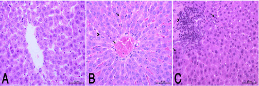
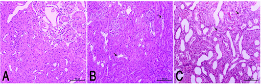
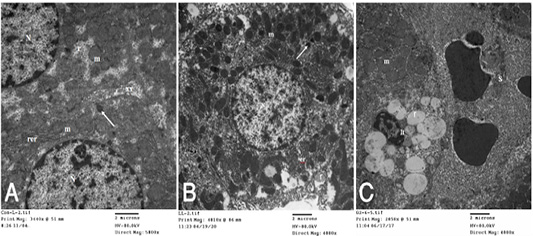
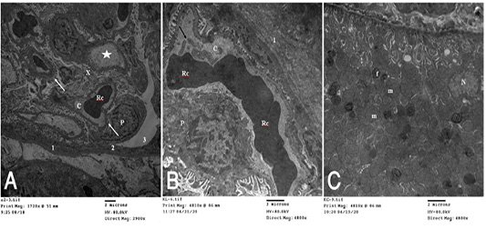
On the liver, hypoP led to increase AST and ALT without affecting any of the other serum parameters while hyperP led to increase MDA, liver enzymes and bilirubin values as compared with controls. AST and ALT are found in high concentrations in hepatocytes, so liver injury causing destruction of hepatocytes increases both enzymes in the blood but ALT is more specific (McGill, 2016; Kasper et al., 2018). Thus, in the current study elevation of both enzymes in hyperP and to a lesser extent in hypoP reveals incidence of hepatocellular injury. Moreover, the raised bilirubin values in hyperP clarifies the more drastic effect of hyperP to induce hepatobiliary disease (Moyer and Balistreri, 2011; VanWagner and Green, 2015). The aforementioned results are supported by the histopathological and TEM findings as the liver of hypoP group showed mild degenerative changes in hepatocytes while hyperP showed degenerative and necrotic changes of hepatocytes associated with leuckocytic infiltration, proliferation of Van Kupffer cells and congestion of central veins. By TEM, the liver of hypoP appeared of nearly normal structure while hyperP showed liver with vaculations and bundles of collagen fibers perisinusoidal as well as congestion of sinusoids. The hepatic stellate cells (HSC or ITO) were increased overloaded by fat globules and hepatocytes showed swelling of mitochondria. The ITO cells are the main source of activated fibroblasts (Brenner et al., 2012). In liver injury, there is an expansion of ITO cells which differentiate into a myofibroblast-like cell with marked expression of alpha-smooth muscle actin. Myofibroblast-like cells have a high fibrogenic capacity in the diseased liver and are also involved in matrix degradation. Moreover, the transition of ITO cells into myofibroblast-like cells is regulated by an intricate network of intercellular communication between ITO cells and Kupffer cells, damaged hepatocytes, endothelial and inflammatory cells, cytokines and reactive oxygen species (ROS). The ITO cells also plays an active role in a number of liver diseases, with a particular reactivity pattern in fibrotic liver disorders. Moreover, the swollen mitochondria due to hyperP, in the present study, is an indicator that the outer membrane of the mitochondria is going to rupture and the release of the pro-apoptotic proteins such as cytochrome C from mitochondria into the cytosol (Petronilli et al., 2001). Therefore, mitochondrial swelling is a sign of cellular death (Davis and Jeffery, 2002). From the current results, early signs of hyperP in liver are increased enzymes, bilirubin and MDA values besides degeneration of hepatocytes, presence of abundant ITO cells, collagen fibers and swollen mitochondria. The aforementioned results did not appear either in the control or the hypoP. Thus, it could be suggested that hyperP induces liver injury during early stages. In addition, elevated PRL levels may be a causative factor to enhance the activity of ITO cells resulting in liver cirrhosis and induce swollen mitochondria leading to apoptosis. This suggestion receives evidence from the previous findings of a series of studies (Gonzales et al., 2007; Cuifang and Miaoyan, 2009; Jha and Kannan, 2016; Khalil et al., 2017) who recorded elevated plasma PRL levels in males with liver cirrhosis, hypogonadism and portal hypertension. In present results, it appears that neither hypo nor hyperprolactinemia affect serum creatinine and urea. Histopathological examination of hypoP disclosed mild degeneration of renal lining epithelium while hyperP showed moderate degeneration. Under TEM, kidney glomeruli of hypoP illustrated thickening of the fibrous layer of the Bawman’s capsule and swelling of podocytes while hyperP showed presence of bundles of collagen fibers periglomerular, tubular casts, debris and swelling of the mitochondria. Bhatia et al. (2020) reported that swelling of renal mitochondria is associated with decreased mitochondrial membrane potential and is considered a response of increased ROS in renal tissue. Also, mitochondrial dysfunction plays a critical role in the progression of acute and chronic kidney injury. The presence of bundles of collagen fibers periglomerular as well as tubular casts and debris is an indication of glomerulonephrosis (El-Safi and Mohamed, 2017). Therefore, it was investigated, in the current study, that high PRL level is a stressor and elevates serum MDA content. Thus, it could be concluded that high PRL level, although it does not change serum creatinine or urea, but it results in injury-induced oxidative stress and inflammation. Also, the presence of hyperP could be considered as an early sign that the kidney is exposed to stress and going to acute or chronic renal dysfunction.
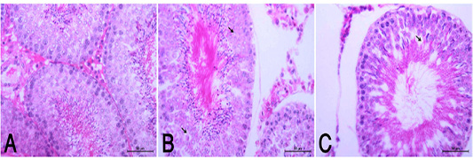
Results of the current study show that hyperP led to marked drop in serum T; a finding that coincides with that of Grattan et al. (2007) and Henderson et al. (2008) who reported that excess PRL induces infertility in males due to the inhibiting effect on GnRH release and consequently pituitary gonadotropins. Therefore, it could be suggested that excess PRL suppresses GnRH, gonadotropins and Leydig cells to secrete T. HypoP did not influence the spermiogram while hyperP led to increase sperm abnormalities. The histopathological examination disclosed that hyperP appeared with moderate degree of atrophy and necrosis of spermatogonial cell layer as well as distortion of seminiferous tubules and disorganized germinal epithelia. These alterations did not affect GSI. Laszczynska et al. (2002) stated that hyperP associated with apical Sertoli cell cytoplasmic degeneration, increase in percentage of convoluted tubules with apoptotic spermatogonia and spermatocytes as well as abnormalities in reproductive behavior in rodents. MDA result disclosed that high PRL levels could be considered as a stressor. Therefore, the appearance of significant sperm abnormalities could be attributed to the stressor effect of high PRL and drop of T.