Special Issue: Emerging and Re-Emerging Animal Health Challenges in Low and Middle-Income Countries · Volume 13, Issue 1, 16 October 2025 · Open Access
Haemato-Biochemical Markers for Brucellosis Infection of Dairy Water Buffalo
· · ·
Affiliations & Notes ⌄
1Department of Veterinary Public Health, College of Veterinary Medicine, University of Basrah, Iraq. *Correspondence | Nameer A. Khudhair, Department of Veterinary Public Health, College of Veterinary Medicine, University of Basrah, Iraq
The results of the complete blood pictures of non-infected buffaloes and infected animals with brucellosis showed a significant increase (p ≤0.05) in monocyte in brucellosis infected buffaloes compared to non- infected animals.
While RBC, HGB, HCT and platelets indices appeared non-significantly difference (p ≤0.05) between infected and non-infected buffaloes with brucellosis (Table 2).
The data analysis of biochemicals parameters for metabolic markers revealed significant decrease (p ≤0.05) of T. protein, globulin and decline in mineral concentration (calcium and phosphors) in brucella infected buffalo compared to non-infected animals.
Nevertheless, the results showed non-significant changes (p ≤0.05) in albumin, glucose and cholesterol levels when compared between brucella non-infected and infected animals (Table 3).
Selected from the article's own text.
Abstract
The marshes are rich lands for raising thousands of Euphrates water buffalo, which is a major source of income for most of the region’s residents, benefiting from milk and its products. However, they suffer from a lack of veterinary health care, despite the recording of repeated cases of abortion. Therefore, the study aims to use blood and biochemical indicators, in addition to clinical signs, to diagnose brucellosis and prevent its spread among buffalo breeders and users of animal products. A total of 184 dairy water buffalos were tested for brucella infection by ID Screen® Brucellosis Serum Indirect Multi-species ELISA kit (ID. Vet Company/France). Then buffalos were divided equally into brucella non-infected and brucella infected groups (n=49 for each). Complete blood count was analyzed by autoanalyzer hematological device (Exigo H400, Sweden). Biochemical parameters were measured by using veterinary dry chemical auto-analyzer, health checker panel (EXIGO C200/Sweden). Indirect ELISA analysis of brucellosis discovered 36 infected buffalos (39.13%) and 13 doubtful brucellosis (14.13%). The total number of dairy buffalos that recorded infected by brucella are 49 (53.26%) from the total number of animals tested (n=184). The hematological reports revealed significant elevation of monocyte in buffalos infected brucella. While, total protein, globulin, ca, and p were significantly increased in dairy buffalos infected by brucella compared with non-infected animals. Nevertheless, hepatic and renal function markers appeared non-significant difference between infected and non-infected animals by brucella, except for ALT activity that showed elevation in their values of infected buffalos when compared with brucella non-infected buffalos. Taken together, a survey of 184 water dairy buffalos recorded brucellosis infection, and assessed haemato-biochemical parameters may helpful as markers for brucella diagnosis in addition to case history.
Keywords | Buffalos, Brucellosis, Hematological and biochemical, ELISA brucellosis kit
Introduction
Water buffalo have long served as a vital source of income for the inhabitants of the wetlands . Insufficient vaccination programs and a lack of a serological surveillance system to track the geographical spread of brucellosis are hindering efforts to eradicate the disease in water buffaloes in the Marshlands.
The genus Brucella, consisting of six species, is responsible for causing brucellosis, a severe illness that affects all species of farm animals, with ruminants being the most affected . The illness has an effect on all kinds of farm animals, although it has the biggest impact on ruminants. The smooth or classical species include Brucella abortus, B. melittis, B. suis, and B. neotomae, while the rough species consist of B. ovis and B. canis. Every species has a preference for a particular host. Brucella abortus is the main causative agent of brucellosis in cattle and buffalo. Brucella melitensis infects ovine and caprine species and causes serious illnesses in humans ; .
Abortion is the primary manifestation seen in all species of farm animals, leading to the loss of pregnancy and a decline in milk supply. This disorder predominantly affects the reproductive and skeletal systems, leading to decreased fertility in both males and females As a result, the occurrence of abortions decreases, but there may be other medical conditions that arise, such as the retention of placenta, stillbirth, or the delivery of weak calves ; . Additionally, there may be the development of metritis or chronic endometritis, which may lead to reduce fertility, infertility, or sterility on expansion to regenerative and mammary organs. Brucella disease influences most of the crucial organs counting heart, liver, kidneys and muscles driving to impedance of their ordinary work through modification of their biochemical constituents depending on the arrange of disease and harm .
The diagnosis of brucellosis may include both direct and indirect methods. Serological assays, such as ELISA, which specifically target the identification of anti-Brucella spp. antibodies, are a very advantageous choice because of their affordability, mobility, and rapidity .
Although the use of ELISA is considered the most effective method for detecting brucellosis, but it is not at hand of veterinarians in the field of wetlands, in addition to the work effort and high cost of test involved. In other hand, depending on clinical signs for diagnosis of brucellosis is very important, but there is other causative agent may interact in diagnosis with brucella such as Escherichia coli ; , Salmonella or Malaria that may distribute in this low hygienic and rural area .
Therefore, the current study is attempted to identify brucella infection through uses of blood indices and biochemical parameters together with clinical signs and case history as indicator to detecting brucellosis infection in dairy buffaloes of Al-Chibayish wetlands.
Materials and Methods
Based on randomly selected individual animal samples taken in randomly selected herds, the study will enable to determine if there is significant infection, the study will enable to determine if there is significant infection percentage that affected buffalo and enable diagnosis throughout hematological and biochemical indicators. Therefore, 184 blood sample were collect randomly from buffalos in Al-Chibayish area. Buffaloes who were above 2 years old on each chosen farm, as recommended by their owners, were selected for sample collection. Each sample were labeled by number and owner name to detect the brucella infection later.
Sample collection
A total of 5 milliliters of blood was obtained from each animal by collecting it from the jugular/coccygeal vein using disposable syringe. The blood bought in EDTA containing tube (2 ml), and a gel and clot activator tube (3 m). The sample tubes were inclined on a table in a shed for about 1 hour at the ambient temperature, after which the serum was separated from the other clots by centrifugation on 3000rpm/minute for 10 minutes. Then it was carefully poured into another tube specifically designed for serum and marked with the same code. The sera were then delivered in a refrigerated container to the Bio-Vet Laboratory for veterinary and molecular diagnostics for further analysis.
Determination of Brucella infection by ELISA technique
ID screen® brucellosis serum Indirect multi-species ELISA kit
Indirect ELISA for the detection of antibodies against Brucella abortus, melitensis, or suis in bovine, ovine, caprine, and porcine serum and plasma (individual samples or pools of up to 10) from ID. Vet Company/France. This product is certified according to OIE specifications and Annex C of European Directive CEE 64/432 to correctly detect the OIEELISAspISS standard serum for multi-species testing for ruminants and swine. Test individual serum or plasma samples, or pools of up to 10.
Description and principle
Wells are coated with purified Brucella abortus LPS. We added the diluted specimens and the control at a 1/20 ratio to the microwells for testing. Anti-Brucella antibodies, if present, form an antibody-antigen complex. We added a multi-species horseradish peroxidase (HRP) conjugate to the microwells. It binds to the anti-Brucella antibodies, forming an antigen-antibody conjugate-HRP complex. After washing to remove excess conjugate, we added the substrate solution (TMB).
The color of the results depends on the quantity of specific antibodies present in the spacemen to be tested at 450nm.
Hematological profile test
After determination of Brucella infection by ELISA technique, blood sample were chosen to analyzed the brucella infected animal and same number of samples for non-infected animals to compared between them. This complete blood count estimation which includes (RBC, HBG, HCT, MCV, MCH, MCHC, WBC, LYM, MONO, GRANU., PLT) measured by autoanalyzer hematological device (Exigo H400, Sweden).
Biochemical parameters evaluation
Biochemical parameters were measured using a modern veterinary chemical autoanalyzer EXIGO C200/Sweden, which uses the dry disc principle of analysis by adding blood, plasma or serum. This system uses the Lambert-Beer law, based on the principle of spectroscopic absorption or turbidity measurement of transmitters, and uses adapted test methods, based on end-point, rate and two-point reactions, in addition to eight simultaneous wavelength tests. The parameters included total protein, albumin, globulin, glucose, total cholesterol and calcium and phosphor as metabolic indicators. In addition, liver enzymes activities (ALT, AST, total bilirubin and ALP) and renal function markers (blood urea nitrogen, creatinine) were measured.
Results
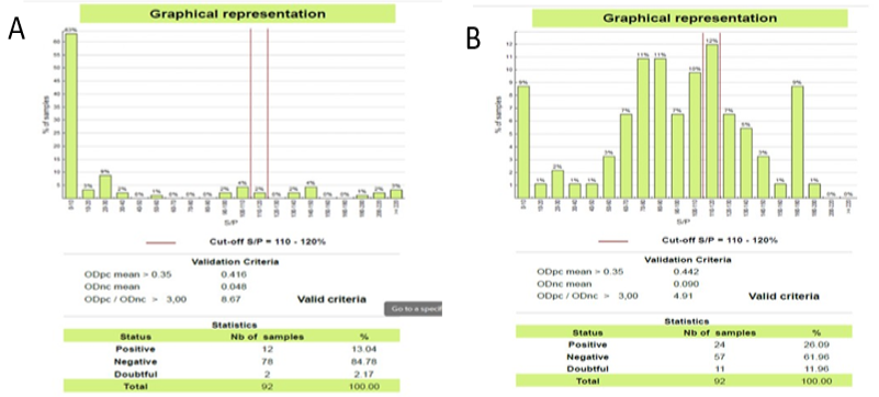
The results analysis of brucellosis in serum of water buffalos by ELISA device are outlined in Figure 1A, B. The graphical picture of ELISA device results showed a data of brucella infected and non-infected animals. This data summarized in Table 1.
The data of ELISA reading showed 12 (13.4%) brucella positive sample and 2 (2.17%) doubtful out of 92 samples in the first plate while the second reading of ELISA showed 24 (26.09%) brucella positive sample with 11 (11.96%) out of 92 samples in the second plate as illustrated in Table 1.
Table 1: Serum ELISA reading of water buffaloes brucellosis test of Al-Chibaysh area.
Plate
Sample count
Positive brucellosis
Positive percent
Doubtful brucellosis
Doubtful percent
Negative
brucellosis
1
92
12
13.04
2
2.17
88
2
92
24
26.09
11
11.96
57
Total
184
36
39.13%
13
14.13%
145
The results of the complete blood pictures of non-infected buffaloes and infected animals with brucellosis showed a significant increase (p ≤0.05) in monocyte in brucellosis infected buffaloes compared to non- infected animals. Whereas total WBC count lymphocyte and granulocyte appeared non- significant difference (p ≤0.05) between infected and non-infected buffaloes with brucellosis. While RBC, HGB, HCT and platelets indices appeared non-significantly difference (p ≤0.05) between infected and non-infected buffaloes with brucellosis (Table 2).
The comparison of biochemical metabolic markers of brucellosis infected and non-infected buffaloes serum represented in Table 3. The data analysis of biochemicals parameters for metabolic markers revealed significant decrease (p ≤0.05) of T. protein, globulin and decline in mineral concentration (calcium and phosphors) in brucella infected buffalo compared to non-infected animals. Nevertheless, the results showed non-significant changes (p ≤0.05) in albumin, glucose and cholesterol levels when compared between brucella non-infected and infected animals (Table 3).
Table 2: Comparison of complete blood count for brucellosis infected and non-infected buffalo in Al-Chibaysh area.
Non-infected buffaloes (n= 49)
Brucella buffaloes (n=49)
P. value
WBC 109/L
7.70 ± 1.58
7.11 ±1.52
0.378
LYM 109/L
2.24 ± 0.57
2.21 ± 0.56
0.914
MON 109/L
0.54 ± 0.16
0.99 ± 0.42 *
0.006
NEUTRO 109/L
4.91 ± 1.14
4.26 ± 1.15
0.190
HGB g/dL
7.92 ± 1.95
8.74 ± 1.95
0.323
MCH pg
19.66 ± 0.60
19.42 ± 0.64
0.370
MCHC g/dL
36.75 ± 0.93
37.21 ± 0.87
0.228
RBC 1012/L
4.06 ± 1.12
4.54 ±1.13
0.320
MCV fl
53.58 ± 2.77
52.26 ± 2.79
0.266
HCT %
21.50 ± 4.78
23.45 ± 4.75
0.334
PLT 109/L
265.33 ± 44.86
279.38 ± 44.21
0.456
Table 3: Comparison of biochemical metabolic markers of brucella infected and non-infected buffaloes.
Non-infected buffaloes (n= 49)
Brucella buffaloes (n=49)
p. value
T. Protein (g/L)
68.44 ± 2.88*
54.50 ± 5.67
0.001
Albumin (g/L)
32.30 ±2.17
31.78 ± 0.93
0.482
Globulin (g/L)
35.03 ±4.97*
22.71 ± 5.66
0.020
Glucose (Mmol/L)
3.32 ±0.54
3.34 ± 0.25
0.932
T. Cholesterol (Mmol/L)
1.28 ±0.38
1.07 ± 0.43
0.267
Ca (Mmol/L)
3.05 ± 0.49*
2.75 ± 0.19
0.040
P (Mmol/L)
2.77 ± 0.39 *
2.18 ± 0.45
0.007
Table 4: Comparison of biochemical liver and kidney function markers of brucella infected and non-infected buffaloes.
Non-infected buffaloes (n= 49)
Brucella buffaloes (n=49)
P. Value
ALT(U/L)
41.33 ± 6.26
60.55 ± 4.41*
0.002
AST(U/L)
150.33 ±7.36
145.09 ±11.25
0.246
T-BIL
5.63 ± 1.12
5.46 ± 0.75
0.686
ALP(U/L)
75.22 ±13.89
81.27 ± 11.44
0.299
BUN Mmol/L)
6.01 ± 1.37
5.43 ± 0.99
0.078
CREA Mmol/L)
130.56±9.002
129.55 ±17.91
0.880
However, the results of biochemical analysis of liver enzymes activities and renal function markers in Table 4 revealed significant elevation (p ≤0.05) in ALT in infected animals compared to non-infected buffaloes, while ALP showed non-significant change although there was increase in their values compared with non-infected buffaloes. Whereas, AST, T-BIL appeared non-significant difference (p ≤0.05) between the two groups of the study. Therewith, blood urea nitrogen (BUN) and creatinine (CREA) in infected and non-infected animals were not affected by the infection and appeared non-significant difference (p ≤0.05) (Table 4).
Discussion
Dealing with dairy production, it is crucial to know of the productive herd situation as regards Brucella infection, due to the zoonotic characteristic of this disease, liable to contaminate and severely affect human beings. Brucellosis is a major Public Health concern, worldwide.
Owing to the importance of buffalo raising in the marshes, Al-Chibayish district hosts a large number of ruminants, producing significant quantities of dairy commodities for human consumption. In addition to the low level of awareness among breeders about the risks of contracting this disease, lack of adequate vaccination campaigns and a serological monitoring system that can map the brucellosis geographical specific targets is one of the obstacles to eliminating brucellosis in water buffaloes in the Marshlands. There is evidence referred to brucellosis is an endemic disease in Dhiqar province, a total of 147 blood sample of cattle population recorded 32 (21.8%) cases of positive brucellosis by using the rose Bengal test . Furthermore, detecting of brucellosis through the clinical signs regarded non-specific, and presence of drawback of laboratory diagnostic test (like Rose Bengal test) in addition to expensive and unavailability of the other diagnostic methods (like ELISA and PCR techniques).
The indirect ELISA kit to detection of brucellosis in 184 water buffaloes investigated the infected animal’s percent about 39.13% (36 infected animals), while if estimation the percent of doubtful brucellosis may approached to 53.36%. These results agreed with the results of Dheyab and Abdulhameed (2023), when evaluate brucellosis infection in cattle and human of Dhiqar province. But the ELISA technique is difficult to doing in this rear area and may loss the financial ability of the breeder. Therefore, the present study aimed to find hematological or biochemical markers that useful to detect brucellosis infection in water buffaloes. Clinical signs of brucellosis are difficult to predict because it occurs as a symptomatic certain phase of disease, and the most clearly signs to expectation of brucellosis is abortion and infertility of animal .
The hematological indices are a vital tool for the clinician that help approach diagnosis of disease. Our results of these study found that most hematological indices appeared non-significant difference when compared infected and non-infected brucella in water buffalos. Monocyte values of infected animals showed significant elevation although the total leukocyte count, lymphocyte and granulocyte (mostly neutrophils) appeared decline in their values when compared with non-infected buffalos. Nevertheless, the erythrocyte, hemoglobin and platelets indices appeared non-significant differences between the infected and non-infected animals. High monocyte counts mostly inked to long term infection, blood disorder or autoimmune disease and it is normally rises in case of brucella infection . These came in agreement with when they compare the hematological indices between healthy and brucellosis groups. They found there was significant increase in monocyte counts and significantly decrease in eosinophils, Platelet distribution width, and Mean platelet volume. Total and differential leukocyte had a vital immune defense role in the body. Brucellosis is a bacterial disease that change certain type of leukocyte like monocyte, eosinophil and neutrophil . Also, there is evidence that brucella may share some component of typhoid and paratyphoid fever that established neutropenia, and these became clear to reduction in neutrophils when there is brucella infection in animals . In the other study done by Maruf et al. (2019) conducted to determine hematological and biochemical response of cattle suffered from brucella infection. This study revealed decrease in values of erythrocyte, total leukocyte, hemoglobin and neutrophils in Brucella-infected cows compared to non-infected healthy cows. These results relatively agreed with our results and also Compatible with Kushwaha et al. (2014). Whereas, Hb, PCV, RBC, WBC, lymphocytes and basophil values involved in the range of reference values in Brucella-infected cattle for the study done by Sikder et al. (2012). They attributed the causes of reduction of hematological markers to reduction of RBC as a result of erythropoietin hormone reduction.
In the same context, biochemical parameters recorded significant reduction in their values when compared to non-infected buffalos by brucella. Our results recorded decline in T. protein, globulin, calcium and phosphor with elevation in ALT activity.
results agreed with El-Boshy et al. (2009) that found increase AST and ALT activities, while non-significant difference recorded for the other biochemical parameters. In contrast, our results not compatible with Maruf et al. when they recorded high values for glucose, creatinine, total protein and AST in Brucella-infected cows than non-infected cows.
The elevation in ALT and reducing of albumin level were the most obvious indicator for the study of Mubaraki et al. (2022). These referred that the results did not establish for brucella infection although there is liver enzymes disorder .
Calcium and phosphor are the most important mineral in animal body, recorded in this study decline in their concentration in brucella infected animals when compared with non-infected buffalos. While Vlasenko and his team found there was decrease in serum calcium level in brucellosis infected cattle, but phosphor recorded values same as control animals never the less, no change were recorded in serum calcium and phosphor levels in brucellosis infected buffalos when compared with healthy animals, and they attributed the insignificant values to the change of pH in small intestine that constrain the absorption of calcium and phosphor .
CONCLUSIONS
The present study investigated that the survey of 184 dairy water buffalos in Al-Chibaysh wetland infected with brucella. The hemato-biochemical indicators are the aid tools for diagnosis of brucellosis in dairy buffalos in combination with clinical signs (if obvious) and case history. Elevation of monocyte and reduced total and differential WBC with the reduction of total protein, globulin, calcium and phosphor. From other hand, increase the activity of liver enzymes may referred to brucella infection if combatable with clinical signs.
Acknowledgement
This research couldn’t be executed unless the support of directorate of veterinary medicine in Dhiqar province via the assistance of Dr. Wisam Khadem Saleh to collect the samples. Thanks are extended to Bio-Vet laboratory for diagnosis of brucella and hematological and biochemical analysis.
Novelty Statement
The main challenge for the present study is diagnoses brucellosis in water buffaloes of marshland (this is main novelty), and uses of hemato-chemical parameters in addition to case history and clinical signs as indicator for suspected brucellosis.
Author’s Contribution
Battol A. Majeed: collect the sample of blood from buffaloes and contributed with writing of paper. Zainab A. Saud: responsible on hematological and biochemical analysis. Dalal K. Rahi: contributed by work on Eliza assay of brucellosis. Nameer A. Khudhair: occupaied the writing work of paper and monitoring the procedures of work.
Generative AI and AI-assisted technology statement
All research details were written with human intervention and no involvement of artificial intelligence.
Ethical approval
All blood sample were collected by the acceptance of the owners and the procedure were done by approval of animal ethics committee in college of Veterinary Medicine, University of Basrah.
Conflict of interest
The authors have declared no conflict of interest.
Figures
Figure 1:.
A and B: Graphical representation of Brucellosis in the serum of water buffalos.
References
Amin A, Hosny A, Shalabi N (2009). Some biochemical and haematological studies in brucellosis infected buffaloes, cows and sheep in el-sharkia governorate. Kafrelsheikh Vet. Med. J., 7(1): 352-368. https://kvmj.journals.ekb.eg/article_107196_f8cabdc24649172ec7f0e1b89cc4ee08.pdf https://doi.org/10.21608/kvmj.2009.107196[CrossRef]
Begum J (2023). Monocytes: What high and low levels mean. Retrieved November 06, 2023 from https://www.webmd.com/a-to-z-guides/what-to-know-about-high-monocyte-count
Deka RP, Lindahl J, Grace D (2018). Epidemiological research on brucellosis in India: Knowledge generated and gaps. International Livestock Research Institute. https://www.slideshare.net/ILRI/brucellosis-india
Dheyab AQ, Abdulhameed MF (2023). Seroprevalence of bovine brucellosis and its incidence in human in Thi-Qar province. Iraqi J. Vet. Sci., 37(4): 877-884. https://pdfs.semanticscholar.org/018c/7e7f51f077ef864ab2ce19f9f18dda9e3bc6.pdf https://doi.org/10.33899/ijvs.2023.136554.2592[CrossRef]
El-Bahgy H, Ali A (2017). Epidemiology and biochemical effects of brucellosis in Kafer-El Sheikh and Qualyobia cow’s farms. J. Agric. Vet. Sci., 10: 53-59. https://doi.org/10.9790/2380-1003015359[CrossRef]
El-Boshy M, Abbas H, El-Khodery S, Osman S (2009). Cytokine response and clinicopathological findings in Brucella infected camels (Camelus dromedarius). Vet. Med., 54(1): 25-32. https://doi.org/10.17221/3044-VETMED[CrossRef]
García CJC, Villalobos MW, Arias VSC, Fino SIM (2018). Acute liver failure complication of brucellosis infection: A case report and review of the literature. J. Med. Case Rep., 12: 1-5. https://doi.org/10.1186/s13256-018-1576-4[CrossRef]
Godfroid J, Nielsen K, Saegerman C (2010). Diagnosis of brucellosis in livestock and wildlife. Croatian Med. J., 51(4): 296-305. https://doi.org/10.3325/cmj.2010.51.296[CrossRef]
Hull NC, Schumaker BA (2018). Comparisons of brucellosis between human and veterinary medicine. Infect. Ecol. Epidemiol., 8(1): 1500846. https://doi.org/10.1080/20008686.2018.1500846[CrossRef]
Jassim HY, Saleh WMM, Habib HN, Ratton ZA, Al-Mousawi MD (2024). Genetic characterization and clinicopathological features of Escherichia coli in neonatal septicemic calves. J. Anim. Behav. Biometeorol., 12(2): 2024016-2024016. https://doi.org/10.31893/jabb.2024016[CrossRef]
Jiang W, Chen J, Li Q, Jiang L, Huang Y, Lan Y, Li Y (2019). Epidemiological characteristics, clinical manifestations and laboratory findings in 850 patients with brucellosis in Heilongjiang Province, China. BMC Infect. Dis., 19: 1-6. https://bmcinfectdis.biomedcentral.com/articles/10.1186/s12879-019-4081-5 https://doi.org/10.1186/s12879-019-4081-5[CrossRef]
Jin M, Fan Z, Gao R, Li X, Gao Z, Wang Z (2023). Research progress on complications of B rucellosis. Front. Cell. Infect. Microbiol., 13: 1136674. https://doi.org/10.3389/fcimb.2023.1136674[CrossRef]
Kushwaha N, Rajora V, Mohan A, Singh J, Shukla S (2014). Assessment of haemato-biochemical parameters and therapeutics on Brucella infected cattle. J. Microbiol. Experiment., 1(2): 00012. https://doi.org/10.15406/jmen.2014.01.00012[CrossRef]
Maruf A, Yasmin F, Yeasmin F, Alam M, Rahman M, Hasan M, Alam M, Alam M, Rahman A, Rahman M (2019). Assessment of haemato-biochemical and therapeutic responses of chronic brucellosis in crossbred dairy cows in Bangladesh. J. Vet. Med. One Health Res., 1(2): 211-229. https://doi.org/10.36111/10.36111/jvmohr.2019.1(2).0013[CrossRef]
Mohammad AL, Esraa AWMA, Salih WM (2012). Investigation of antibodies of Brucella melitensis in sheep. Univ. Thi-Qar J., 8(1). https://scholar.google.com/scholar?hl=enandas_sdt=0%2C5andq=Investigation+of+antibodies
Mubaraki MA, Sharahili AI, Elshanat S, El-Khadragy MF, Thagfan FA, Al-Megrin WA, Abdel-Gaber R, Bauomy AA, Alkhudhayri A, Dkhil MA (2022). Biochemical and hematological markers as brucellosis indicators in the najran region of Saudi Arabia. J. King Saud Univ. Sci., 34(6): 102138. https://doi.org/10.1016/j.jksus.2022.102138[CrossRef]
Naji HA, Saleh W, Saud ZA, Mhalhal TR, Alhasson FA, Reddy G, Abebe W (2023). Prevalence of resistance and virulence genes in Escherichia coli isolates from diarrheic dogs. Iraqi J. Vet. Sci., 37(2): 355-361. https://doi.org/10.33899/ijvs.2022.133514.2243[CrossRef]
Naji HA, Saud ZAH, Saleh WMM, Alsaad IAW (2021). Sero prevalence of Schmallenberg virus antibodies in buffalo from north Basra Governorate-Iraq. Vet. Pract., 22(2): 14-17. https://vetpract.in/themes/uploads/contents/158f0b6e44b6f06e2a5a91806a16f6b6.pdf
Naji H, Saleh W, Hanoon M, Imad I, Salim Y (2019). Serotyping, virulence gene expression and phenotypic characterization of E. coli O157: H7 in colibacillosis affecting buffalo calves in Basra governorate. Iraqi J. Vet. Sci., 33(2): 447-451. https://doi.org/10.33899/ijvs.2019.163198[CrossRef]
Qureshi KA, Parvez A, Fahmy NA, Abdel-Hady BH, Kumar S, Ganguly A, Atiya A, Elhassan GO, Alfadly SO, Parkkila S (2023). Brucellosis: Epidemiology, pathogenesis, diagnosis and treatment–a comprehensive review. Ann. Med., 55(2): 2295398. https://doi.org/10.1080/07853890.2023.2295398[CrossRef]
Segwagwe BE, Samkange A, Mushonga B, Kandiwa E, Ndazigaruye G (2018). Prevalence and risk factors for brucellosis seropositivity in cattle in Nyagatare District, Eastern Province, Rwanda. J. S. Afr. Vet. Assoc., 89(1): 1-8. https://doi.org/10.4102/jsava.v89i0.1625[CrossRef]
Sikder S, Rahman S, Alim M, Das S (2012). Haematological variations in Brucella abortus antibody positive cross-bred cattle at chittagong, bangladesh. YYU Vet. Fakultesi Dergisi, 23: 125-128. https://dergipark.org.tr/tr/download/article-file/146388
Vlasenko V, Baiseitov S, Pleshakova V, Alekseeva I (2020). Changes in blood chemistry values of the cattle with associative course of leukemia and brucellosis. BIO Web. Conf., 27: 00137. https://doi.org/10.1051/bioconf/20202700137[CrossRef]
Winter SE, Winter MG, Poon V, Keestra AM, Sterzenbach T, Faber F, Costa LF, Cassou F, Costa EA, Alves GE (2014). Salmonella enterica serovar Typhi conceals the invasion-associated type three secretion system from the innate immune system by gene regulation. PLoS Pathog., 10(7): e1004207. https://doi.org/10.1371/journal.ppat.1004207[CrossRef]