
Zinc oxide nanoparticles are widely and safely used due to their characteristic nature. The current study is focused at microbial synthesis of Zinc oxide nanoparticles (ZnO NPs) and their applications in biomedical and environmental areas. Intracellular ZnO NPs were developed utilizing Bacillus subtilis (CP002905.1) which is not only cost-effective but eco-friendly method too. The UV-Vis spectrum of the synthesized ZnO NPs displayed a distinct peak at 275 nm while FTIR analysis exhibited a major peak at 497.18 cm-1 revealing the ZnO bond. Size and zeta potential of ZnO NPs was 451 nm and -47.5 mV, respectively. Nevertheless, SEM revealed nanoflowers assembly of ZnO NPs. The results further disclosed antimicrobial potential of NPs against Staphylococcus aureus (BTCB02), Salmonella typhimurium (BTCB23)and Escherichia coli (BTCB03)at varying intensities and a maximum zone of inhibition (ZOI) of 15 mm was established with S. aureus at 125 µL. Moreover, the ZnO NPs also showed photocatalytic activity of 80.1% against dye (methylene blue). Hence these NPs can be used in various biomedical and environmental applications in days to come.
Research Article · Volume 70, Issue 2, 21 March 2025 · Open Access
Green-Synthesis-Zinc-Oxide-Nanoparticles-Bacillus
· · · ·
Affiliations & Notes ⌄
- 1Department of Biotechnology, Lahore College for Women University, Lahore. Pakistan. , *Correspondence | Mehwish Iqtedar, Department of Biotechnology, Lahore College for Women University, Lahore. Pakistan
- 2Email: mehwish.iqtedar@lcwu.edu.pk
Article Info ⌄
- Journal:
- Biologia (Lahore)
- Published:
- 21 March 2025
From the article
- Conclusively the bio-synthesized ZnO NPs by Bacillus subtilis (CP002905.1) was a low-cost, green and efficient approach.
- The SEM analysis of NPs indicated nanoflower-shaped arrangement.
- The ZnO NPs had antimicrobial activity against different multidrug resistant pathogens.
- Additionally intracellular ZnO NPs also showed photocatalytic activity against commercial dye.
Selected from the article's own text.
Abstract
Introduction
The term nano was described as being one billionth part of a meter (10-9 m) and it establishes a scale for molecules and structures that lie between 1 and 100 nanometers (nm) . Lately, researchers have been emphasizing on the green route of synthesis for developing metal and their oxides nanoparticles . Bio-mediated nanoparticles show potential in a number of fields, such as microbiology, biotechnology, drug delivery, nanomedicine, nano-catalysis and biosensors . Hence research on green method of producing metallic nanoparticles is increasing by utilizing different natural sources like plants, microorganisms (bacteria and fungi) together with varying metabolites. The green approach is in fact safer and eco-friendly without involving or generating high temperatures and pressures or releasing hazardous compounds. Many microbes most preferably bacteria, has been used to produce safe and hassle-free NPs and they are known for their significant antibacterial characteristics that can be applied in bio-medical sectors . They enable controlled production of biocompatible nanoparticles with uniform size and shape, enhancing their applicability as antimicrobial and photocatalytic agents Mehta et al. (2009).
Zinc is crucial for maintaining important physiological functions in a human body which chiefly includes DNA repair and DNA replication. ZnO-NPs have grabbed lot of attention due to their less toxicity and enhanced biocompatibility. Moreover according to reports they are great antibacterial agents and they possess significant anticancer and antioxidant properties . ZnO is a metal oxide that exhibits pyroelectric and piezoelectric properties that enables enhanced wound healing, anti-inflammatory benefits, and high catalytic activity. As a result, they are extensively utilized in the biomedical field . It was noted in another investigation that at remarkably low concentrations, biosynthesized zinc oxide nanoparticles exhibit significant antibacterial potential against both gram-negative and gram-positive bacteria. It was also described that these antibacterial properties are stronger than those of ZnO NPs made by chemical synthesis techniques .
Multiple techniques such as Field Emission Scanning Electron Microscopy (FESEM), Transmission Electron Microscopy (TEM), X-ray diffraction (XRD), UV-Vis absorption spectroscopy and Fourier Transform Infrared Spectroscopy (FTIR) can be employed to characterize and analyze the nanoparticles .
This study focuses on green synthesis of ZnO NPs via Bacillus subtilis (CP002905.1), its characterization, antimicrobial properties and photocatalytic activity against dye. As the antibacterial resistance is building up these days, new alternatives need to be designed to tackle with the problem.
Materials and Methods
Microorganism
Bacillus subtilis (CP002905.1) strain was acquired from the research project lab, Department of Biotechnology and was revived in nutrient media and stored as 80% glycerol stocks for future use .
Synthesis of nanoparticles
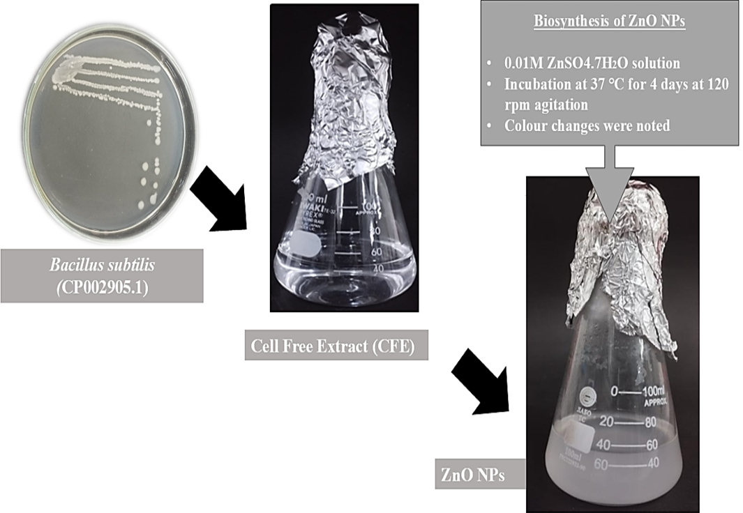
The NPs were prepared according to the method described after modifications . After the preparation of cell free extract (CFE) 0.01M ZnSO4.7H2O solution was added for the development of the NPs and incubated at 37 oC for 4 days at 120 rpm agitation. After 4 days of incubation NPs formation was monitored and colour changes were noted.
Characterization of ZnO NPs
Characterization of nanoparticles was conducted using UV-Visible spectrophotometer (OD200-600) (U28 Hitachi, USA) for verification of NPs formation whereas X-Ray Diffraction (XRD) ; (D8 Discover Bruker, USA) was utilized for studying the nature of NPs while zeta sizer was used to assess size and zeta potential of the NPs. Additionally, Fourier Transform infrared spectrophotometer (FTIR) (Shimadzu IR Tracer, Japan) was used for the evaluation of the functional groups and Scanning Electron Microscopy (SEM) (Zeiss EVO LS10, Germany) demonstrated shape of NPs at varying magnifications ; .
Antimicrobial potential
The antimicrobial properties of biosynthesized zinc oxide nanoparticles were measured against multi drug resistant bacteria (MDR) i.e., Escherichia coli (BTCB02), Staphylococcus aureus (BTCB03) and Salmonella typhimurium (BTCB23) respectively according to the method described . Sterilized discs were loaded with NPs solution with different volumes ranging from 15 µL, 25 µL, 50 µL, 75µl, 100 µL and 125 µL. The inoculated plates were incubated for 24 h at 37oC and zones of inhibition (ZOI) were measured. As a control, ZnSO4.7H2O was also tested for its antibacterial susceptibility.
Photocatalytic degradation
Photocatalytic degradation potential of biosynthesized Zinc oxide nanoparticles was estimated by preparing the 2.5 ppm methylene blue (MB) dye solution as described . Different volumes of NPs were employed (1 mL, 2 mL, 3 mL, 4 mL and 5 mL). Solution containing methylene blue and nanoparticles were exposed to UV light of 50W intensity for 30 min. After every 10 min intervals sample was taken out of every flask. Then UV-Vis spectrum was measured for each sample. Then the rise or fall in characteristic absorbance peak of methylene blue was noted and the percentage of dye degradation was calculated as:
E (%) = (Co - Ct/Co) × 100
Where; initial concentration is represented by Co, final concentration of methylene blue in solution is denoted by Ct and E is the efficiency.
Statistical analysis
Statistical analysis was performed using SPSS on antimicrobial assay. ANOVA was applied to check significant (p≤0.05) difference between different volumes of nanoparticles against pathogen as well as between strains on same volume. Post Hoc Duncan’s multiple range test was also applied.
Results and Discussion
Synthesis of nanoparticles
The biosynthesis of zinc oxide nanoparticles involves a mechanism where the enzyme NADPH-dependent reductase plays a crucial role and facilitates reduction by transferring electrons from NADPH to NADP+. This results in their reduction leading to the formation of elemental zinc oxide nanoparticles . In the current research the NPs were formed after the incubation of CFE of Bacillus subtilis (CP002905.1) and 0.01M ZnSO4.7H2O for the period of 4 days in dark condition in shaking incubator (120 rpm at 37 oC). A significant change in colour was noted which indicated the formation of nanoparticles. Transparent colour of ZnSO4.7H2O solution transitioned into milky colour confirming the development of NPs (Figure 1).
Characterization of ZnO nanoparticles
In the current study, Bacillus subtilis was used for the formulation of zinc oxide nanoparticles while some studies synthesized them by using B. licheniformis, Rhodococcus pyridinivorans and Aeromonas hydrophila ; ; . The change in color of the mixture from transparent to white (Figure 1) indicated development of biosynthesized zinc oxide nanoparticles nevertheless the color change denotes reduction of zinc ions. Additionally, Surface Plasmon Resonance (SPR) phenomenon was also involved in colour transitions which was made possible by the production of bio-metabolites within ZnO NPs .
Further characterization of biosynthesized ZnO nanoparticles was performed by UV-Vis spectrophotometer, FTIR, Zeta sizer, (size and zeta potential), XRD and SEM. A characteristic absorption peak was observed at 275 nm which is characteristic peak of ZnO nanoparticles (Figure 2a). Previous study on intracellular ZnO nanoparticles reported almost similar characteristic peak at 270- 365 nm range which corroborate with our findings . Likewise, extracellular synthesis of ZnO NPs from Bacillus cereus exhibited a peak at 352 nm . Similarly, in another study, ZnO NPs produced using Aeromonas hydrophila gave a characteristic peak at 374 nm . Zeta sizer analysis showed that ZnO NPs were of 451 nm in size (Figure 2b) while the zeta potential of biosynthesized ZnO NPs was -47.5 mV (Figure 2c) with Polydispersity index (PDI) of 1.0. Yet in another study, size of extracellular ZnO nanoparticles was reported to be approximately 500 nm whereas a size of 386.4 nm was also reported Hidayat Chai et al. (2018). We can see in the current study ZnO NPs seem to be quiet stable and the long-term steadiness of these biosynthesized nanoparticles is due to the presence of several biomolecules that prevent aggregation on its surface ; . This directs that a potential greater than -30 mV is indicative of improved colloidal stability for the nanofluid .
Additionally, FTIR analysis of biosynthesized ZnO NPs was also carried out and spectra revealed different peaks at varying wave number. Peak at wavelength 497.18 cm-1 is the characteristic peak showing the ZnO bond whereas the peak at wavelength 692.07 cm-1 corresponds to H-Br stretching and represents the alkyl halide class. The bands at 1032.73 cm-1, 1272.89 cm-1, 1471.61 cm-1 and 1646.54 cm-1 corresponds to C‒O, C‒N, C‒C and ‒C=C‒ stretching. While peak at 3375.19 cm-1 represents the –NH stretching and 3609.53cm-1 represents –OH stretching (Figure 2d). A related study using Enterobacter cloacae exhibited similar peaks at 1651 and 3304 cm-1 corresponding to O-H group and another at 2353 cm-1 corresponding to C = O group. Additionally, a peak at 1312 cm-1 was displayed that exposed C–H group. A distinct peak developed at 463 cm-1 showing Zn–O bond which is accordance with our findings . Similarly, other studies also reported peak between ranges of 421-559 cm-1 for zinc oxide bond . Similar investigations showed peaks at 466.77 cm−1, 482 cm−1, 513 cm−1, 515 cm−1, 584 cm−1 and 612 cm−1 ; ; ; . The slight variation in a peak might be due to the strain used to produce the nanoparticles and protocol adopted for synthesis.
The morphology of the biosynthesized nanoparticles in the current study was examined at magnifications of 500X (Figure 3a), 1000X (Figure 3b) and 2000X (Figure 3c) by SEM analysis. The outcome showed that the zinc oxide nanoparticles revealed nanoflowers assembly. Other studies also reported a variety of shapes with ZnO NPs such as nanoflower, hexagonal and spherical cluster ; ; . XRD analysis (Figure 3d) was also conducted in the current study that showed the synthesized nanoparticles were amorphous in nature. This outcome is supported by another finding that in nature, the nanostructures of ZnO are amorphous ; ; .
Antimicrobial activity of biosynthesized ZnO NPs against multidrug resistance bacteria
Various bacterial strains were tested against ZnO NPs at volumes ranging from 15 to 125 µL. However, a volume of 15 µL, 25 µL, 50 µL, 75 µL, 100 µL and 125 µL gave ZOI of 10 mm, 11 mm, 12 mm, 13 mm, 14 mm and 15 mm, against S. aureus (BTCB02) respectively (Figure 4). Significant (p ≤ 0.05) difference was obtained between the zones obtained at different volumes test against S. aureus whereas ZnSO4.H2O alone gave ZOI of diameter 9 mm against S. aureus (Figure 4). The results showed that against S. typhimurium (BTCB23), ZnO NPs at a volume of 15 µL, 25 µL, 50 µL, 75 µL, 100 µL and 125 µL gave zone of inhibition of diameter 10 mm, 10 mm, 11 mm, 12 mm, 13 mm and 14 mm, respectively. Significant (p ≤ 0.05) difference was obtained between the zones obtained at different volumes test against S. typhimurium. ZnSO4. H2O alone gave zone of inhibition of diameter 8 mm against S. typhimurium which shows the ZnO NPs were better antimicrobial agents than the metallic salt. The results demonstrated that for Escherichia coli (BTCB03), ZOI of 10 mm, 11 mm, 11 mm, 12 mm, 12 mm and 13 mm were established at ZnO NPs volume of 15 µL, 25 µL, 50 µL, 75 µL, 100 µL and 125 µL, respectively. Significant (p ≤ 0.05) difference was obtained between the zones obtained at different volumes test against E. coli while a ZOI of 10 mm was produced by ZnSO4.H2O (standard). Results also revealed that the antimicrobial potential increased with the increase in volume of NPs. S. aureus, S. typhimurium and E. coli established highest ZOI at 125 µL volume of NPs and a maximum ZOI of 15 mm was observed against S. aureus.
A related study also tested ZnO NPs synthesized with Calendula officinalis extract against S. aureus, Klebsiella pneumoniae and E. coli. Highest ZOI was displayed at 35.2 mm with S. aureus whereas minimum resistance was visible at 23.6 mm and 13.5 mm against Klebsiella pneumoniae and E. coli respectively. This shows that S. aureus possesses significant antibacterial properties against ZnO NPs . The current study showed that S. aureus was more sensitive towards ZnO than E. coli which could be due to the difference in their cell wall composition and structure, that explains its enhanced sensitivity. Moreover, S. aureus possess thicker peptidoglycan layer that may interact with ZnO more efficiently, increasing its antimicrobial activity . It was also explained that Gram negative bacteria are more resistant to NPs because of their extra outer membrane .
The current results showed that the increasing concentration of nanoparticles causes the increase in inhibition of growth of pathogenic bacteria resulting in bigger zones. This result is supported by the report which states that the zone of inhibition expands as zinc oxide nanoparticle concentration rises . Another theory suggests that ZnO NPs mainly restrict transport channels physically and cause abrasion induced damage to harmful bacteria cell membranes as part of its antibacterial action. This causes cellular processes to be disturbed and nutrition intake to be hindered, ultimately resulting in bacterial cell death. Reactive oxygen species (ROS), which further contribute to the cell damage and the overall antimicrobial impact, can also be produced by ZnO NPs . An electromagnetic attraction is created between the surface of the negatively charge microorganism and the positive surface of metal oxides. As a result of this interaction, ZnO and other metal oxide NPs stick better to the bacterial cell membrane. Once bonded, these nanoparticles have the capacity to cause oxidative stress, which produces ROS. The oxidation and eventual death of the microorganism are caused by the ROS, which also seriously damage lipids, proteins, and DNA within the cell. The dual mechanism of physical adhesion and subsequent chemical destruction is a crucial component of metal oxide nanoparticles antibacterial effectiveness .
Photocatalytic of biosynthesized ZnO nanoparticles
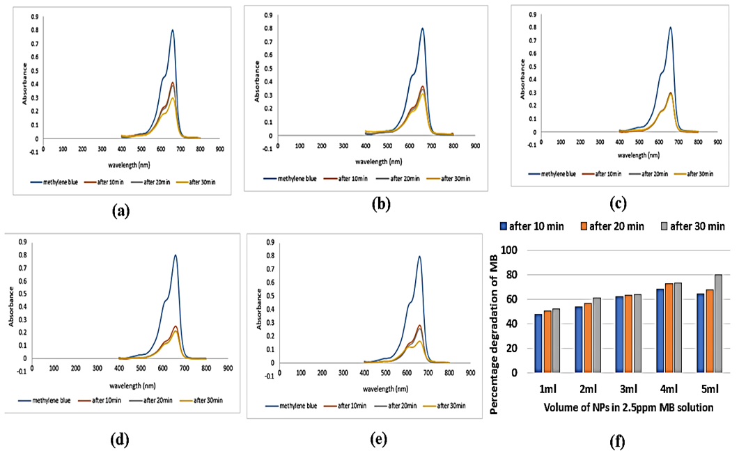
Photocatalytic performance of ZnO NPs against synthetic dye i.e., methylene blue was determined by exposing a sample containing dye solution along with biosynthesized nanoparticles to UV light of strength of 25W in laminar air flow chamber and measuring their absorbance by using spectrophotometer. By calculating the percentage degradation of methylene blue (Figure 5a-e), maximum degradation was observed in sample containing 2.5 ppm MB and 5 mL of biosynthesized ZnO NPs of 80.1%. The percentage degradation of MB in solution containing 2.5 ppm MB and 1 mL of ZnO NPs were 47.9%, 50.6% and 52.2% at intervals of 10 min, 20 min and 30 min, respectively. The percentage degradation of MB in solution containing 2.5 ppm MB and 2 mL of ZnO NPs were 53.6%, 56.3% and 61.2% at intervals of 10 min, 20 min and 30 min, respectively. The percentage degradation of MB in solution containing 2.5 ppm MB and 3 mL of ZnO NPs were 62.4%, 63.2% and 64% at intervals of 10 min, 20 min and 30 min, respectively. The percentage degradation of MB in solution containing 2.5 ppm solution of MB and 4 mL of ZnO NPs were 68.1%, 72.9% and 73.1% at intervals of 10 min, 20 min and 30 min., respectively. The percentage degradation of MB in solution containing 2.5 ppm MB (Figure 5f) and 5 mL of ZnO NPs were 64.2%, 67.6% and 80.1% at intervals of 10 min, 20 min and 30 min, respectively. Results showed that maximum degradation of methylene blue dye solution occurred at 30min time interval under UV irradiation of sample containing 5 mL of ZnO NPs that is 80.1% whereas minimum degradation was noted after 10 min under UV irradiation with sample containing 1 mL of ZnO NPs.
Photocatalytic efficacy of ZnO nanoparticles was also investigated in current study and under 30 min UV irradiation sample containing 5 mL nanoparticles showed maximum degradation of methylene blue dye up to 80.1%. This result is supported by another study in which ZnO nanoflowers were produced using Bacillus licheniformis under room temperature, followed by their photocatalytic potential evaluation. Then deterioration of methylene blue dye under UV radiation was examined . Furthermore, photocatalytic efficacy of zinc nanostructures was also investigated in study which reported that Jujube fruit extract was used as a reducing agent as well as stabilizer during the green technique of synthesis of zinc oxide nanoparticles. After that, the ability of these nanoparticles to break down two organic dyes, methylene blue and eriochrome black-T, in the presence of sunlight was examined.
Significant degradation efficiencies i.e., with 72% of methylene blue and 86% of eriochrome black-T degrading in five hours of exposure . According to the reported mechanism, the electrons of nanoparticles are triggered in the conduction band after leaving valence band by UV irradiation. Pairs of electron-hole are produced by this process, with the conduction band having free electrons and with holes in the valance band. After interacting with oxygen (O₂) or water (H₂O) molecules, these electron-hole pairs can create reactive oxygen species, including hydrogen peroxide, superoxide radicals and hydroxyl radicals. Because of their extreme reactivity, these ROS are essential to the photocatalytic destruction of dye molecules .
Conclusions and Recommendations
Conclusively the bio-synthesized ZnO NPs by Bacillus subtilis (CP002905.1) was a low-cost, green and efficient approach. The UV-Vis spectrum displayed a distinctive peak at 275 nm where as a size of 451 nm with a zeta potential of -47.5 mV was revealed by Zeta sizer. The SEM analysis of NPs indicated nanoflower-shaped arrangement. The ZnO NPs had antimicrobial activity against different multidrug resistant pathogens. Additionally intracellular ZnO NPs also showed photocatalytic activity against commercial dye. Hence the study shows safer and cost-effective way of NPs synthesis and highlights their application in environmental remediation processes. The outcome ensures that ZnONPs exhibited potent antimicrobial property and proved to be an efficient photocatalyst. In future, further steps must be considered to improve and control the size and syntheis of NPs to further improve their applications.
Acknowledgements
The authors are thankful to the Central Laboratory Facility at Lahore College for Women University, Pakistan.
Novelty Statement
Intracellular ZnO NPs were produced via Bacillus subtilis that is a cost-effective and an eco-friendly technique with antibacterial and photocatalytic characteristics.
Author’s Contribution
Mehwish Iqtedar, Mahnoor Siddique and Asma Shahzad: Data curation, conceptualization, methodology, formal analysis, supervision, writing original draft, editing.
Afshan Kaleem and Roheena Abdullah: Methodology, review and editing.
Conflict of interest
The authors have declared no conflict of interest.
Figures


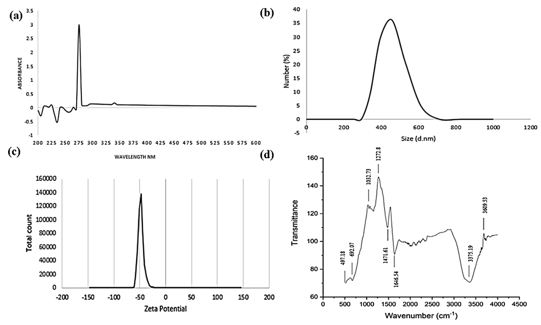
Characterization of intracellular synthesis of ZnO NPs from Bacillus subtilis (a) UV-Vis spectrum evaluation (b) Size assessment by zeta sizer (c) Zeta potential analysis (d) FTIR spectral analysis.
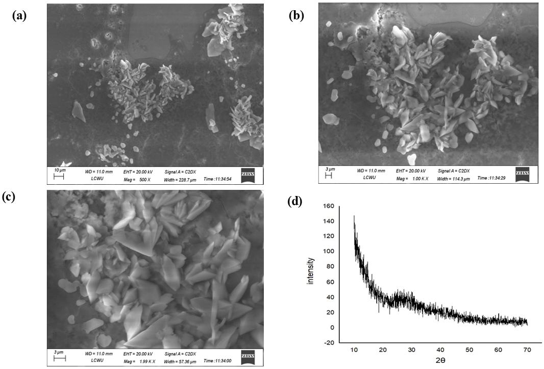
SEM analysis of ZnO NPs (nanoflowers) at varying magnifications (a) 500X (b) 1000X (c) 2000X (d) XRD analysis displaying nature of NPs.
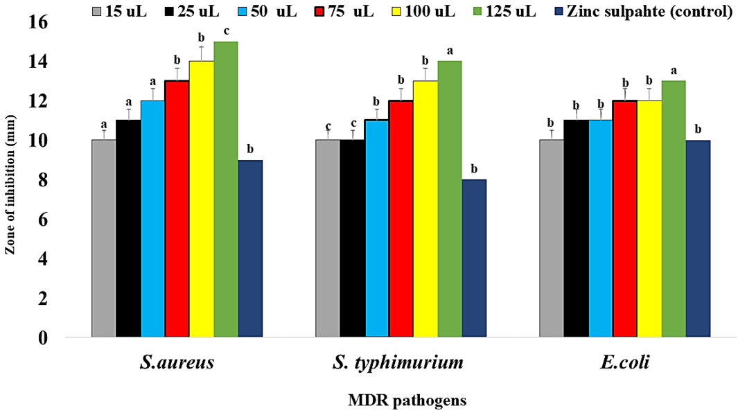
Antimicrobial potential of ZnO NPs against three pathogenic strains S. aureus, S. typhimurium and E. coli with varying intensities including ZnSO4.H20 (control).
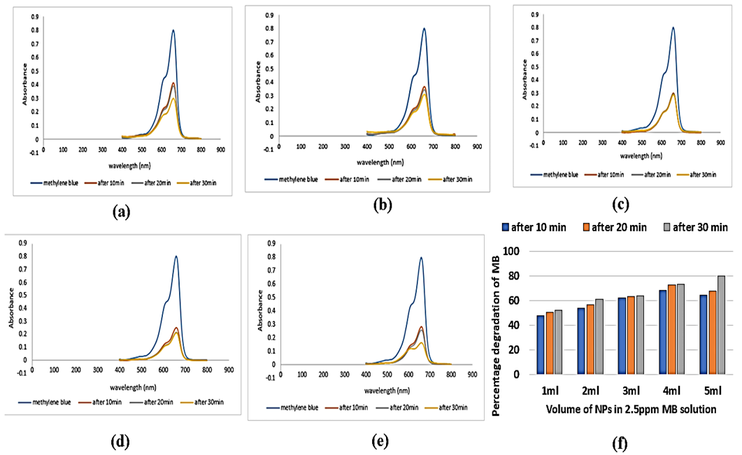
UV-Vis spectra of methylene blue solution (a)1 mL (b) 2 mL (c) 3 mL (d) 4 mL (e) 5 mL (f) Photocatalytic degradation of MB dye under various volumes of ZnO NPs and UV exposure.
UV-Vis spectra of methylene blue solution (a)1 mL (b) 2 mL (c) 3 mL (d) 4 mL (e) 5 mL (f) Photocatalytic degradation of MB dye under various volumes of ZnO NPs and UV exposure.
References
- Abdulgafour, H.I., Hassan, Z., Al-Hardan, N. and Yam, F.K., 2010. Growth of zinc oxide nanoflowers by thermal evaporation method. Phys. B: Condensed Matter., 405(11): 2570-2572. https://doi.org/10.1016/j.physb.2010.03.033[CrossRef]
- Agarwal, H., Venkat, K.S. and Rajeshkumar, S., 2017. A review on green synthesis of zinc oxide nanoparticles. An eco-friendly approach. Resour. Efficient Technol., 3: 406–413. https://doi.org/10.1016/j.reffit.2017.03.002[CrossRef]
- Aravind, M., Amalanathan, M. and Mary, M.S.M., 2021. Synthesis of ZnO nanoparticles by chemical and green synthesis methods and their multifaceted properties. SN Appl. Sci., 3: 1-10. https://doi.org/10.1007/s42452-021-04281-5[CrossRef]
- Awwad, A.M., Amer, M.W., Salem, N.M. and Abdeen, A.O., 2020. Green synthesis of zinc oxide nanoparticles (ZnO NPs) using Ailanthus altissima fruit extracts and antibacterial activity. Chem. Int., 6: 151-159.
- Dobrucka, R. and Długaszewska, J., 2016. Biosynthesis and antibacterial activity of ZnO nanoparticles using Trifolium pratense flower extract. Saudi J. Biol. Sci., 23(4): 517–523. https://doi.org/10.1016/j.sjbs.2015.05.016[CrossRef]
- El-Masry, R.M., Talat, D., Hassoubah, S.A., Zabermawi, N.M., Eleiwa, N.Z., Sherif, R.M., Abourehab, M.A.S., Abdel-Sattar, R.M., Gamal, M., Ibrahim, M.S. and Elbestawy, A., 2022. Evaluation of the antimicrobial activity of ZnO nanoparticles against enterotoxigenic Staphylococcus aureus. Life, 12(10): 1–13. https://doi.org/10.3390/life12101662[CrossRef]
- Golmohammadi, M., Honarmand, M. and Ghanbari, S., 2020. A green approach to synthesis of ZnO nanoparticles using jujube fruit extract and their application in photocatalytic degradation of organic dyes. Spectrochim. Acta A: Mol. Biomol. Spectrosc., 229(2): 11-19. https://doi.org/10.1016/j.saa.2019.117961[CrossRef]
- Hidayat-Chai, M.H., Amir, N., Yahya, N. and Saaid, I.M., 2018. Characterization and colloidal stability of surface modified zinc oxide nanoparticle. J. Phys. Conf. Ser., 23(1): 14-22. https://doi.org/10.1088/1742-6596/1123/1/012007[CrossRef]
- Hudzicki, J., 2012. Kirby-Bauer disk diffusion susceptibility test protocol author information. Am. Soc. Microbiol., 12(9): 1–13.
- Iqtedar, M., Riaz, H., Kaleem, A., Abdullah, R., Aihetasham, A., Naz, S. and Sharif, S., 2020. Biosynthesis, optimization and characterization of ZnO nanoparticles using Bacillus cereus MN181367 and their antimicrobial activity against multidrug resistant bacteria. Rev. Mexicana Ingeniería Química, 19(1): 253-266. https://doi.org/10.24275/rmiq/Bio1605[CrossRef]
- Jayaseelan, C., Rahuman, A.A., Kirthi, A.V., Marimuthu, S., Santhoshkumar, T., Bagavan, A., Gaurav, K., Karthik, L. and Rao, K.V.B., 2012. Novel microbial route to synthesize ZnO nanoparticles using Aeromonas hydrophila and their activity against pathogenic bacteria and fungi. Spectrochim. Acta A: Mol. Biomol. Spectrosc., 90(3): 78–84. https://doi.org/10.1016/j.saa.2012.01.006[CrossRef]
- Kalaba, M.H., El-Sherbiny, G.M., Ewais, E.A., Darwesh, O.M. and Moghannem, S.A., 2024. Green synthesis of zinc oxide nanoparticles (ZnO-NPs) by Streptomyces baarnensis and its active metabolite (Ka): A promising combination against multidrug-resistant ESKAPE pathogens and cytotoxicity. BMC Microbiol., 24: 254. https://doi.org/10.1186/s12866-024-03392-4[CrossRef]
- Kavitha, S., Dhamodaran, M., Prasad, R. and Ganesan, M., 2017. Synthesis and characterisation of zinc oxide nanoparticles using terpenoid fractions of Andrographis paniculata leaves. Int. Nano Lett., 7(9): 141-147. https://doi.org/10.1007/s40089-017-0207-1[CrossRef]
- Kundu, D., Hazra, C., Chatterjee, A., Chaudhari, A. and Mishra, S., 2014. Extracellular biosynthesis of zinc oxide nanoparticles using Rhodococcus pyridinivorans NT2: Multifunctional textile finishing, biosafety evaluation and in vitro drug delivery in colon carcinoma. J. Photochem. Photobiol. B: Biol., 140(11): 194–204. https://doi.org/10.1016/j.jphotobiol.2014.08.001[CrossRef]
- Lin, K.H., Sun, S.J., Ju, S.P., Tsai, J.Y., Chen, H.T. and Hsieh, J.Y., 2013. Observation of the amorphous zinc oxide recrystalline process by molecular dynamics simulation. J. Appl. Phys., 113(7): 1-10. https://doi.org/10.1063/1.4789956[CrossRef]
- Madkour, L.H., 2023. Eco-friendly green biosynthesized metallic nanoparticles and biotechnological applications in pharmaceuticals sciences. J. Mater. Sci. Eng. B, 13(1): 22-31. https://doi.org/10.17265/2161-6221/2023.1-3.001[CrossRef]
- Mansoori, G.A. and Soelaiman, T.A.F., 2005. Nanotechnology, an introduction for the standards community. J. ASTM Int., 2(6): 17–38. https://doi.org/10.1520/JAI13110[CrossRef]
- Maruthupandy, M., Anand, M., Maduraiveeran, G., Suresh, S., Beevi, A.S.H. and Priya, R.J., 2016. Investigation on the electrical conductivity of ZnO nanoparticles decorated bacterial nanowires. Adv. Natl. Sci. Nanosci. Nanotechnol., 7: 1-9. https://doi.org/10.1088/2043-6262/7/4/045011[CrossRef]
- Mehta, S.K., Kumar, S., Chaudhary, S., Bhasin, K.K. and Gradzielski, M., 2009. Evolution of ZnO nanoparticles via facile CTAB aqueous micellar solution route: A study on controlling parameters. Nanosc. Res. Lett., 4(1): 17–28. https://doi.org/10.1007/s11671-008-9196-3[CrossRef]
- Mohan, A.C. and Renjanadevi, B., 2016. Preparation of zinc oxide nanoparticles and its characterization using scanning electron microscopy (SEM) and X-Ray diffraction (XRD). Proc. Technol., 24(4): 761–766. https://doi.org/10.1016/j.protcy.2016.05.078[CrossRef]
- Muthuvel, A., Jothibas, M. and Manoharan, C., 2020. Effect of chemically synthesis compared to biosynthesized ZnO NPs using Solanum nigrum leaf extract and their photocatalytic, antibacterial and in-vitro antioxidant activity. J. Environ. Chem. Eng., 8(2): 10-27. https://doi.org/10.1016/j.jece.2020.103705[CrossRef]
- Ngom, I., Ngom, B.D., Sackey, J. and Khamlich, S., 2019. Biosynthesis of zinc oxide nanoparticles using extracts of Moringa oleifera: Structural and optical properties. Mater. Today Proc., 36: 526–533. https://doi.org/10.1016/j.matpr.2020.05.323[CrossRef]
- Ovais, M., Khalil, A.T., Ayaz, M., Ahmad, I., Nethi, S.K. and Mukherjee, S., 2018. Biosynthesis of metal nanoparticles via microbial enzymes: A mechanistic approach. Int. J. Mol. Sci., 19(12): 1–20. https://doi.org/10.3390/ijms19124100[CrossRef]
- Raliya, R. and Tarafdar, J.C., 2013. ZnO nanoparticle biosynthesis and its effect on phosphorous-mobilizing enzyme secretion and gum contents in Clusterbean (Cyamopsis tetragonoloba L.). Agric. Res., 2(1): 48–57. https://doi.org/10.1007/s40003-012-0049-z[CrossRef]
- Reddy, K.M., Feris, K., Bell, J., Wingett, D.G., Hanley, C. and Punnoose, A., 2007. Selective toxicity of zinc oxide nanoparticles to prokaryotic and eukaryotic systems. Appl. Phys. Lett., 90(21): 10–13. https://doi.org/10.1063/1.2742324[CrossRef]
- Safar, R., Doumandji, Z., Saidou, T., Ferrari, L., Nahle, S., Rihn, B.H. and Joubert, O., 2019. Cytotoxicity and global transcriptional responses induced by Zinc Oxide nanoparticles NM 110 in PMA-differentiated THP-1 cells. Toxicol. Lett., 308(19): 65–73. https://doi.org/10.1016/j.toxlet.2018.11.003[CrossRef]
- Selvarajan, E. and Mohanasrinivasan, V., 2013. Biosynthesis and characterization of ZnO nanoparticles using Lactobacillus plantarum VITES07. Mater. Lett., 112(1): 180–182. https://doi.org/10.1016/j.matlet.2013.09.020[CrossRef]
- Shabani, M.H., Jafari, A., Manteghian, M. and Mousavi, S.M., 2024. Green synthesis of zinc oxide nanoparticles using Enterobacter cloacae microorganism and their application in enhanced oil recovery. Sci. Rep., 14: 29409. https://doi.org/10.1038/s41598-024-80819-y[CrossRef]
- Thongam, D.D., Gupta, J. and Sahu, N.K., 2019. Effect of induced defects on the properties of ZnO nanocrystals: Surfactant role and spectroscopic analysis. SN Appl. Sci., 1(9): 1-12. https://doi.org/10.1007/s42452-019-1058-3[CrossRef]
- Tiwari, A.K., Jha, S., Tripathi, S.K., Shukla, R., Awasthi, R.R., Bhardwaj, A.K., Singh, A.K. and Dikshit, A. 2024. Spectroscopic investigations of green synthesized zinc oxide nanoparticles (ZnO NPs): Antioxidant and antibacterial activity. Discover Appl. Sci., 6: 399. https://doi.org/10.1007/s42452-024-06049-z[CrossRef]
- Tripathi, R.M., Bhadwal, A.S., Gupta, R.K., Singh, P., Shrivastav, A. and Shrivastav, B.R., 2014. ZnO nanoflowers: Novel biogenic synthesis and enhanced photocatalytic activity. J. Photochem. Photobiol. B: Biol., 141: 288–295. https://doi.org/10.1016/j.jphotobiol.2014.10.001[CrossRef]
- Venkatachalam, P., Jayaraj, M., Manikandan, R., Geetha, N., Rene, E.R., Sharma, N.C. and Sahi, S.V., 2017. Zinc oxide nanoparticles (ZnO NPs) alleviate heavy metal-induced toxicity in Leucaena leucocephala seedlings: A physiochemical analysis. Plant Physiol. Biochem., 110(2): 59–69. https://doi.org/10.1016/j.plaphy.2016.08.022[CrossRef]
- Zhang, H. and Chen, G., 2009. Potent antibacterial activities of Ag/TiO2 nanocomposite powders synthesized by a one-pot sol-gel method. Environ. Sci. Technol., 43(8): 2905–2910. https://doi.org/10.1021/es803450f[CrossRef]
- Zhang, L., Jiang, Y., Ding, Y., Daskalakis, N., Jeuken, L., Povey, M., O’Neill, A.J. and York, D.W., 2010. Mechanistic investigation into antibacterial behaviour of suspensions of ZnO nanoparticles against E. coli. J. Nanopart. Res., 12(5): 1625–1636. https://doi.org/10.1007/s11051-009-9711-1[CrossRef]
- Zhou, G., Wang, Q., Wang, Y., Wen, X., Peng, H., Peng, R., Shi, Q., Xie, X. and Li, L., 2023. Outer membrane porins contribute to antimicrobial resistance in gram-negative bacteria. Microorganisms, 11(7): 1-19. https://doi.org/10.3390/microorganisms11071690[CrossRef]
Services
Article usage statistics
Loading…