Gamal Younis1, Amal Awad1*, Mona Maghawry2, Fatma Selim1
1Department of Bacteriology, Mycology and Immunology, Faculty of Veterinary Medicine, Mansoura University, Mansoura, 35516; 2Animal Health Research Institute, Dokki, Egypt.
*Correspondence | Amal Awad, Mansoura University, Mansoura, Egypt; Email: dr_amalvet@yahoo.com
Abstract
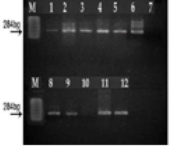
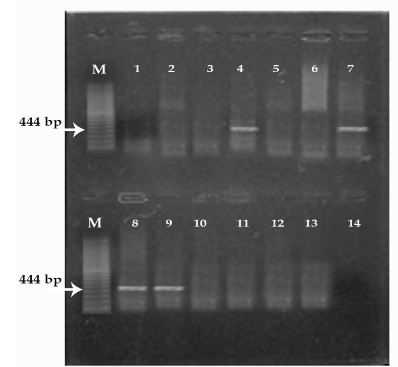
Pseudomonas aeruginosa produces large array of extracellular toxins that play a crucial role in virulence potential. In this study, the virulence profile of 35 clinical isolates of P. aeruginosa distinctly recovered from pneumonic cattle, mastitic milk samples, and wound infections was determined using phenotypic and genotypic methods. The overall incidence rates from pneumonic cattle, mastitic milk samples, and wound infections were 6%, 25% and 13.3%, respectively with a total incidence of 14%. Recovered isolates were then assayed for their enzymatic activity and the results revealed that out of these 35 isolates, 28 isolates produced haemolytic activity on blood agar plates, 27 isolates showed lecithinase enzyme activity on egg yolk agar media and 12 isolates showed proteolytic enzyme activity on brain heart infusion agar medium supplied with gelatin. Three virulent genes (las B, tox A and exoS) were screened by polymerase chain reaction (PCR) and the results showed that las B, tox A and exoS genes were amplified from 14.7%, 8.57%, and 17.14% of tested isolates, respectively. In conclusion, the present studies confirmed previous observations that the virulence of P. aeruginosa is multifactorial; however, the results indicate that certain exoproducts may play a more important role in certain types of infection.
Keywords | P. aeruginosa, Phenotypic, Genotypic, Virulent genes, PCR
Editor | Kuldeep Dhama, Indian Veterinary Research Institute, Uttar Pradesh, India.
Received | June 11, 2015; Revised | July 29, 2015; Accepted | July 30, 2015; Published | August 07, 2015
Citation | Younis G, Awad A, Maghawry M, Selim F (2015). Extracellular enzymes and toxins of pseudomonas aeruginosa strains isolated from clinically diseased Egyptian cows. Adv. Anim. Vet. Sci. 3(10): 522-526.
ISSN (Online) | 2307-8316; ISSN (Print) | 2309-3331
Copyright © 2015 Younis et al. This is an open access article distributed under the Creative Commons Attribution License, which permits unrestricted use, distribution, and reproduction in any medium, provided the original work is properly cited.