
The present study is aimed at determining the teratogenic effects of different doses of Cr VI, administered to pregnant mice. LD50 of Cr VI, was calculated as 88mg/kg of B.W in pregnant mice. Among 05 groups of pregnant females (10 females in each group), three were treated with 50%, 25% and 12.5% of the LD50 of Cr VI, respectively, with one control and one vehicle control group. Fetuses were recovered on 18th day of gestation and fixed for morphometric and morphological studies. Experimental groups showed variable degree of fetal abnormalities, along with reduction in body weights parallel to increase in dose concentration, as compared to control and vehicle control. Morphometric studies showed significant differences in nervous, integumentary and skeletal systems against control. Exencephaly, Omphalocoel, hygroma and limb abnormalities were recorded. It is shown that Cr VI causes teratogenic effects in mice embryos.
Research Article · Volume 49, Issue 4, 18 July 2017 · Open Access
Exposure of Pregnant Mice to Hexavalent Chromium Causes Fetal Defects
· · · · · ·
Affiliations & Notes ⌄
- 1Department of Zoology, University of the Punjab, Quaid-i-Azam Campus, Lahore, Pakistan 2Department of Environment Sciences, COMSATS Institute of Information Technology, Vehari Campus, Vehari, Pakistan 3Department of Veterinary Clinical Sciences, The Royal Veterinary College, Hawkshead Lane, North Mymms, Hatfield, Hertfordshire, AL9 7TA, UK 4University of Veterinary and Animal Sciences, Pattoki Campus, Pattoki, Pakistan , * Corresponding author: naveedahmad@ciitvehari.edu.pk
Article Info ⌄
- Journal:
- Pakistan Journal of Zoology
- Published:
- 18 July 2017

From the article
- Cr VI exposure can cause significant damage to pubertal development through alteration of antioxidants, anaemia, and altered hormone levels in utero through pubertal period.
- Cr VI causes teratogenic effects in gross external morphology, by passing through placenta.
- The results of our studies showed a high significance of exencephaly, anophthalmia, microcephaly, hydrocephaly, facial defects, and limbs defects omphalocoel intra growth retardation and resorptions.
- After administration of CrVI to the pregnant mice 6-12 days of gestation results were examined as there is incomplete or totally absent ossification in vertebral and facial as well as pelvic regions.
Selected from the article's own text.
Abstract
INTRODUCTION
Atmospheric deposition of heavy metals in soil is a major source of their exposure to living organisms . Among these metals chromium (Cr) is being sixth most abundant in earth crust. According to WHO normal value of Cr VI in drinking water is 50 µg/L but in New Caledonia, California, Italy, Zimbabwe, Mexico and in Pakistan it is above the prescribed limit (73 µg Cr VI/L) , Cr level is shown to be high in different parts of Pakistan, specially, in big urban areas like Lahore and Gujranwala ; . Its level was much higher than set levels of FAO and EPA. Cr becomes part of ground water by leaching and used for drinking and other household jobs .
Heavy metals including Cr are being involved in developmental toxicity besides general toxicity . It is present in blood (4.58±0.62 ng/ml of serum) and can pass through placenta . Its placental transfer was studied in rats using atomic absorption spectroscopy. Intragastric administration of 51Cr, incorporated into brewer’s yeast, to pregnant rats led to significant labeling of the newborn .
Reproductive physiology, of both males and females, is affected prior to effecting embryonic development i.e. damage to convoluted seminiferous tubules epithelium, reduction of spermatozoa formation and increase in prevalence of teratospermia, atrtic follicles congestion in stromal tissue, decrease in follicle and oocyte number, increased level of Cr(VI) in blood, increased duration of estrus cycle, disintegrated cell membranes of two layered follicular cells and altered villiform mitochondria in thecal cells ; ; .
Certain exposure levels of Cr(VI) cause teratogenic effects in avian and mammalian models at various stages of embryonic development e.g. distorted embryos, reduction in the number of implantations and number of fetuses and reduced ossification ; .
The present study describes the teratogenetic effects of Cr VI administered to pregnant mice.
MATERIALS AND METHODS
Six weeks old Swiss Webster albino male and female mice (Weight: 30±2) free from any viral, bacterial or parasitic agent were purchased from Veterinary Research Institute, Lahore, Pakistan and were allowed to mate freely (1 male:2 female) in steel cages. Cages were sanitized prior to placing mice in them and were placed in well ventilated animal house with maintained temperature (20 ±02°C). Cages contained food pellets (National Feeds Ltd, Feed # 13, Crude Protein: 16.5±1%, Energy: 2700 Kcal/Kg ±100) and water ad libitum. Safe and healthy condition was provided by continuous cleaning and maintaining temperature at 20±02°C.
Estimation of gestation period
Mated females were identified on the basis of deposition of semen at vaginal opening as sperm plug. It was considered as Day “0” of gestation. Females with sperm plug were separated from males and housed in different cages with minimum disturbance.
Determination of LD50 of K2Cr2O7
LD50 was determined using Probit analysis . Sixty (60) pregnant females were divided into six groups, each with ten dams, were force fed orally via rubber tube cut from butterfly needle on 6th day of gestation. Mortality of only those females were considered, which died on or before 8th day of gestation. Potassium dichromate (K2Cr2O7, BDH Prolabo Chemicals), was used as chemical source for Cr (VI). Six random doses used for all six groups of pregnant mice were prepared by dissolving 5, 25, 50, 100, 150 and 200 mg/kg of B.W. concentrations of K2Cr2O7 in water. LD50 was calculated as 88 mg/kg of B.W.
Procedure adopted
In three experimental groups, females were force fed with 50% (44 µg/g B.W.), 25% (22 µg/g B.W.) and 12.5% of LD50 (11 µg/g B.W.) of K2Cr2O7 dissolved in 0.1 ml of water on 6th day of gestation. Doses were given singly on GD6. In addition one control and one vehicle control groups were also maintained.
All dams were euthanized and given cesarean section on 18th day of gestation. Fetuses were recovered and fixed in Bouin’s fluid for 48 h for easy morphometric and morphological studies . Wet weight, crown rump length, head circumference, eye circumference, fore and hind limbs lengths and tail length was measured for morphometric analyses with digital balance and vernier caliper for each fetus recovered. For morphology, external structures were observed with the help of binoculars according to Makris et al. (2009).
Histological sections were proceeded and stained by the method of Khadija et al. (2011). Method of Kawamura et al. (1990) was used for fetal skeletal preparation.
Statistical analysis
One-way ANOVA was applied on means of all parameters in micrometery with 10 females in each group, further group wise comparison was done by applying Tukey’s HSD (Honestly Significant Difference) test using IBM SPSS 20.
RESULTS
Morphological studies
Morphologically, fetuses in all experimental groups showed variable degree of abnormalities, as compared to control and vehicle control (Table I). All experimental groups had fetuses having abnormalities like exencephaly, open eyes, sub cutaneous hemorrhages, macroglossia, hyperextension of limbs and runt fetuses. Fetuses from highest (44µg/g B.W.) and lowest (11µg/g B.W.) dose groups shared abnormalities like microcephaly, synotia, microtia, distended thoracic cavity, limb micromelia kinked and short tail. Hydrocephaly, resorptions and edema were found in highest (44µg/g B.W.) and middle (22µg/g B.W.) dose groups (Figs. 1, 2).
Abnormalities found exclusive to experimental groups are as follows: Craniostenosis, short snout, anophthalmia, microphthalmia, exophthalmia, palpabral coloboma, kyphosis, lordosis, spina bifida, prognathia, scoliosis, limb malrotation and deformation, clinodactyly, ectrodactyly, apodia, ankylodactyly, fused digits, flexed paw and hooked tail, at 44µg/g B.W.
Anotia, branchignathia, archinia, limb hyperflexion, laparoschisis, paw dysplasia, amelia and branched tail Omphalocoele at 22µg/g B.W. and 11µg/g B.W.
Table I.- Descriptive data of experiment, observed from 18 days old fetuses from different dose groups of hexavalent chromium. Litter size varied among groups, all fetuses were observed (normal and abnormal) including resorptions. Table also shows abnormalities present in each dose group.
| Dose µg/g B.W. | Control | VC | 11 | 22 | 44 |
| No. of females | 10 | 10 | 10 | 10 | 10 |
| Litter size | 102 | 88 | 83 | 50 | 74 |
| Normal | 102 | 88 | 72 | 30 | 38 |
| Abnormal | 0 | 0 | 6 | 4 | 11 |
| Resorptions | 0 | 0 | 2 | 6 | 12 |
| No. of abnormalities | |||||
| Anophthalmia | - | - | - | - | 1 |
| Limb hyperextension | - | 3 | 2 | ||
| Limb hyperflexion | - | - | - | - | 2 |
| Limb malrotation | - | - | 2 | ||
| Limb micromelia | - | - | 3 | - | 4 |
| Spina bifida | - | - | - | - | 4 |
Morphometric studies
All morphometric parameters, except eye circumference and forelimb length, in 44µg/g B.W. exposure group, including mean fetal weight and head circumference in 22µg/g B.W. dose group and hind limb length and tail length in both 22µg/g B.W. and 11µg/g B.W. dose groups were significantly (p <0.05) different from controls. Vehicle control group had no difference from normal control group (Table II).
Histological and skeletal studies
Anatomical sections of control and vehicle control mice fetuses showed normal left and right ventricles of brain, serous glands, intra-retinal space, lens, neural layer of retina, mandibular gland and cerebral aqua duct,
Table II.- Various morphometric parameters (mean±SEM) of 18 days old fetuses from different dose groups of hexavalent chromium. Litter size varied among groups, all fetuses were observed (normal and abnormal) excluding resorptions.
| Dose µg/g B.W. | Weight (mg) | CR length (mm) | Head circumference (mm) | Eye circumference (mm) | Fore limb length (mm) | Hind limb length (mm) | Tail length (mm) |
| C | 1342 ± 27 | 21.68 ± 0.28 | 25.93 ± 0.29 | 6.73 ± 0.16 | 7.15 ± 0.11 | 9.58 ± 0.24 | 11.19 ± 0.14 |
| VC | 1355 ± 19 | 21.41 ± 0.2 | 25.60 ± 0.18 | 6.66 ± 0.08 | 7.13 ± 0.16 | 9.28 ± 0.08 | 10.96 ± 0.13 |
| 11 | 1322 ± 58 | 21.32 ± 0.32 | 25.44 ± 0.43 | 6.47 ± 0.08 | 7.1 ± 0.03 | 8.94 ± 0.09* | 10.38±0.08* |
| 22 | 1117 ± 38* | 21.14 ± 0.22 | 24.75± 0.23* | 6.42 ± 0.01 | 6.96 ± 0.07 | 8.61 ± 0.11* | 10.3 ± 0.19* |
| 44 | 995 ± 26* | 20.07 ± 0.26* | 23.55*± 0.23 | 6.36 ± 0.1 | 6.96 ± 0.04 | 8.11 ± 0.09* | 10.36±0.12* |
* =p<0.05.
hyaloid cavity, nasal septum and nasal cavity, primordium of frontal bone, follicle of vibrissae (Fig. 3), while selected sections from dose group 44µg/g B.W exhibit misshapen spinal cord, left atrium, right atrium, exencephaly, misshapen ventricle and hind brain. Fetal sections from 22µg/g B.W revealed defects like spina bifida, right atrium, left atrium, mandibular gland and fused pinna. Defects like exencephaly, misshapen subcorticle region, misshapen spinal cord and omphalocoele were detected in sections of dose group 11µg/g B.W (Fig. 3).
Normal degree of ossification was observed in the skeleton of control and vehicle control mice fetuses (Fig. 4). However, the degree of ossification was decreased with increase in dose concentrations (Fig. 4).
DISCUSSION
This study is focused on developmental toxicity of Cr VI. Different dose groups were established with control and vehicle control group to check toxic effects of Cr VI on various anatomical parameters of fetuses.
Cr VI exposure can cause significant damage to pubertal development through alteration of antioxidants, anaemia, and altered hormone levels in utero through pubertal period. Induction of oxidative stress can be one of the mechanisms, causing Cr VI based cellular deteriorations. It was reported that Cr VI exposure adversely affects reproductive function by crossing through placenta barrier and even breast feeding. Higher levels of H2O2 and lipid peroxidation were seen with low levels of specific activities of antioxidant enzymes . Cr VI causes oxidative stress and production of reactive oxygen species, which cause DNA damage, lipid peroxidation and alteration in calcium metabolism ; .
It is now well established that oxyanions like Cr VI can pass through placenta as being in blood plasma may be with bounded form . Three major signalling pathways were found to be active during Cr VI toxicity: i) activation of detoxification genes; ii) induction of signal transduction effectors; and iii) epigenetic modification of chromatin marks. Cr VI causes teratogenic effects in gross external morphology, by passing through placenta. Further, in vitro studies would be helpful for checking functions of oxidation scavengers during the embryo toxicity production of Cr VI .
The results of our studies showed a high significance of exencephaly, anophthalmia, microcephaly, hydrocephaly, facial defects, and limbs defects omphalocoel intra growth retardation and resorptions. Marouani et al. (2010) performed experiments on rats and found similar results that are intrauterine growth retardation, facial defects, and resorptions. According to them the fetal defects may be caused by change in placental histology due to decidualcells atrophy and hypertrophy of blood lacunae. After administration of CrVI to the pregnant mice 6-12 days of gestation results were examined as there is incomplete or totally absent ossification in vertebral and facial as well as pelvic regions. Our findings are similar to those of Bailey et al. (2006), who observed cervical malformations after administration of Cr.
The difference in ossification of vertebrae may possibly be due the reduced enzymatic activity of alkaline phosphatase which alters calcium concentrations resulting in change in bone morphology and ossification .
The present studies reveal that the administration of K2Cr2O7 during organo-genetic period can damage the development of embryo. Further studies are of course required for understanding the mechanism of malformations.
ACKNOWLEDGEMENTS
This research was supported financially by Higher Education Commission (HEC) of Pakistan. We are highly obliged for their support.
Statement of conflict of interest
Authors have declared no conflict of interest.
Figures
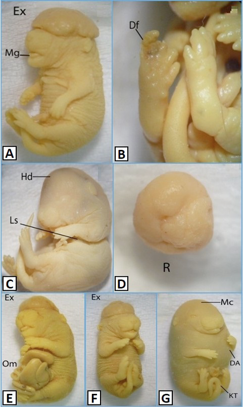
Morphological features of 18 days oldfetuses of mice administration with K2Cr2O7 at a dose of 22µg/g B.W. (A, B), and 11µg/g B.W. (C-G) Control, Vehicle control (VC); Df, deformed; Hd, hydrocephaly; Ls, laproschisis; Mg, macroglossia; R, resorption. DA, dilated abdomen; Ex, exencephaly; KT, kinked tail; Mc, Microcephaly; Om, Omphalocoel. Magnificaton: A=10X, B&D=25X, C, E&G=8X, F=9X
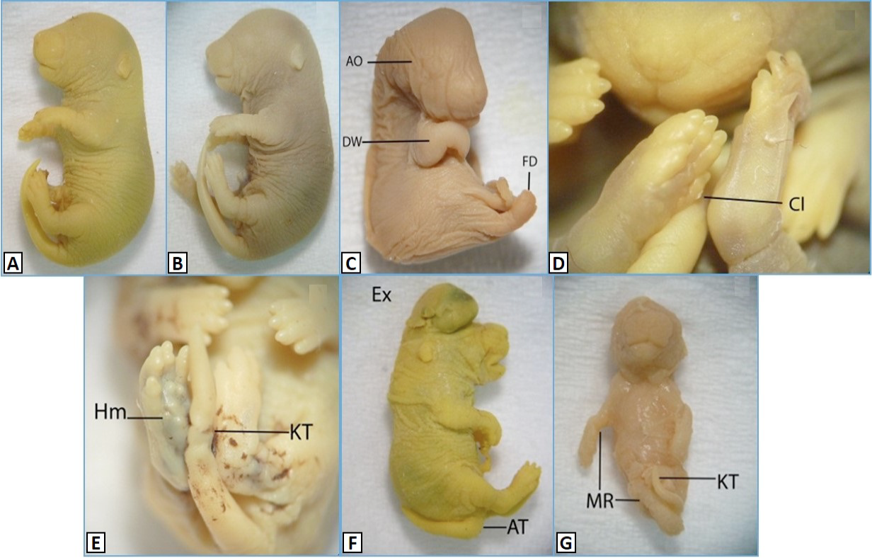
Morphological features of 18 days old fetuses of mice, administered with K2Cr2O7 at a dose of 44µg/g B.W. A, Control; B, VC, vehicle control; AO, anophthalmia; AT, abnormal tail; Cl, clinodactyly; DW, drooped wrist; Ex, exencephaly; FD, fused digits; Hm, hemorrhage; KT, kinked tail; MR, malrotation. Magnification: A=7X, B&C=8X, D=25X, E=20X, F=10X, F=9X.
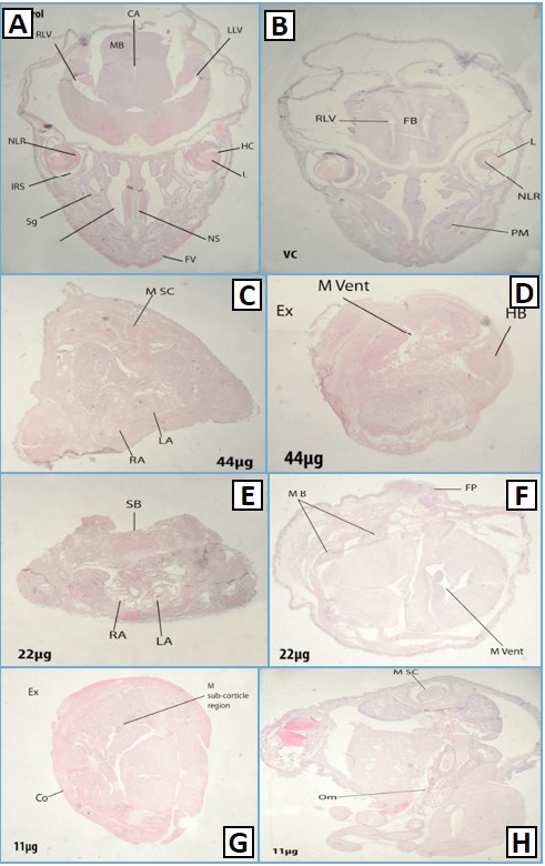
Histological transverse sections of Control (A), Vehicle control (B) of different dose groups (44,22,11µg/g B.W) (C-H), 18 days old fetus from brain, Ex: exencephaly, M Vent: misshapen ventricle, HB: hind brain, MB, mandibular gland; FP, fused pinna, M sub corticle region: misshapen sub corticle region; Co, cortex; RA, right atrium; LA, left atrium; RA, right atrium; M Sc, misshapen spinal cord; SB, spina bifida; Om, Omphalocoel; NC, nasal cavity; Sg, serous gland; IRS, intra retinal space; RLV, right lateral ventricle, MB, mandibular gland; CA, cerebral aquaduct; LLV, left lateral ventricle; HC, hyaloid cavity; NS, nasal septum; FV, follicles of vibrissae; L, lens; FB, primordium of 1frontal bone; NLR, neural layer of retina; PM, pre molar.
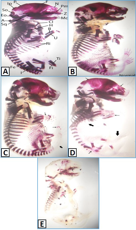
Skeletons of control (A) and vehicle control (B), and selected sections from different dose groups (11,22,44µg/g B.W) (C-E). Simple arrows show less degree ossification while bold arrows show no ossification in three different dose groups. F, Frontal; N, Nasal; Pm, Pre Maxila; Z, Zygomatic; Md, Mandible; C, Clavicle; H, Humerus; R, Radius; U, Ulna; Ri, Ribs; Ti, Tibia; Fi, Fibula; I, Ilium; Fe, Femur; Sq, Squamosal; A, Atlas; Eo, Exocipital; So, Supraocipiltal; Ip, Interperitonial; P, Parietal.
References
- Aftab, T., Shafiq, T., Khan, B. and Chaudhry, M.N., 2011. Physicochemical properties, contamination and suitability of canal water for irrigation, Lahore Branch, Pakistan. Pak. J. analyt. environ. Chem., 12: 88-94.
- Bailey, M.M., Boohaker, J.G. and Sawyer, R.D., 2006. Exposure of pregnant mice to chromium picolinate results in skeletal defects in their offspring. Birth Def. Res., 77: 244–249. https://doi.org/10.1002/bdrb.20081[CrossRef]
- Boncompagni, E., Muhammad, A., Jabeen, R., Orvini, E., Gandini, C. and Sanpera, C., 2003. Egrets as monitors of trace-metal contamination in wetlands of Pakistan. Arch. environ. Contam. Toxicol., 45: 399-406. https://doi.org/10.1007/s00244-003-0198-y[CrossRef]
- Bostan, N., Ashraf, M., Mumtaz, A.S. and Ahmad, I., 2007. Diagnosis of heavy metal contamination in agro-ecology of Gujranwala, Pakistan using cattle egret (Bubulcus ibis) as bioindicator. Ecotoxicology, 16: 247-251. https://doi.org/10.1007/s10646-006-0124-y[CrossRef]
- Domingo, J.L., 1994. Metal-induced developmental toxicity in mammals: A review. J. Toxicol. environ. Hlth. Part A Curr. Iss., 42: 123-141.
- Elbetieha, A. and Al-Hamood, M.H., 1997. Long-term exposure of male and female mice to trivalent and hexavalent chromium compounds: effect on fertility. Toxicology, 116: 39-47. https://doi.org/10.1016/S0300-483X(96)03516-0[CrossRef]
- Erdélyi, M., Kertész, V., Balogh, K. and Mézes, M., 2006. Effect of chromium on the development of the chicken embryo. Trace elements in the food chain. Proceedings of an International Symposium on Trace Elements in the Food Chain, Budapest, Hungary, pp. 416-421. http://w3.chemres.hu/tefc2006/document/PB.pdf
- Fan, Y., Ovesen, J.L. and Puga, A., 2012. Long-term exposure to hexavalent chromium inhibits expression of tumor suppressor genes in cultured cells and in mice. J. Trace Elem. Med. Biol., 26: 188-191. https://doi.org/10.1016/j.jtemb.2012.04.009[CrossRef]
- Kanojia, R.K., Junaid, M. and Murthy, R.C., 1996. Chromium induced teratogenicity in female rat. Toxicol. Lett., 89: 207-213. https://doi.org/10.1016/S0378-4274(96)03812-X[CrossRef]
- Kawamura, S., Kirohashi, A., Kato, T. and Yasuda, M., 1990. Cone-staining technique for fetal rat specimens without skinning and removing adipose tissue. Congen. Anom., 30: 93-95. https://doi.org/10.1111/j.1741-4520.1990.tb00498.x[CrossRef]
- Naeem, K., Ahmad, N. and Asmatullah, 2011. Embryotoxicity of orally administered misoprostol to pregnant mice. Pakistan J. Zool., 43: 511-521.
- Li, H., Chen, Q., Li, S., Xu, Y., Yao, W. and Chen, C., 1999. Studies on male reproductive toxicity caused by hexavalent chromium. Chinese J. Preven. Med., 33: 351-353.
- Makris, S.L., Solomon, H.M., Clark, R., Shiota, K., Barbellion, S. and Buschmann, J., 2009. Terminology of developmental abnormalities in common laboratory mammals (version 2). Congr. Anom., 49: 123-246. https://doi.org/10.1111/j.1741-4520.2009.00239.x[CrossRef]
- Marouani, N., Tebourbi, O., Mokni, M., Yacoubi, T.M., Sakly, M., Benkhalifa, M. and Rhouma, B.K., 2010. Embryotoxicity and fetotoxicity following intraperitoneal administrations of hexavalent chromium to pregnant rats. Zygote, 19: 229-235. https://doi.org/10.1017/S0967199410000274[CrossRef]
- Mertz, W., Roginski, E.E., Feldman, F.J. and Thurman, D.E., 1969. Dependence of Chromium transfer into the rat embryo on the chemical form. J. Nutr., 99: 363-367.
- Miyauchi, S, Srinivas, S.R., Fei, Y.J., Gopal, E., Umapathy, N.S., Wang, H., Conway, S.J., Ganapathy, V. and Prasad, P.D., 2006. Functional characteristics of NaS2, a placenta-specific Na+-coupled transporter for sulfate and oxyanions of the micronutrients selenium and Chromium. Placenta, 27: 550-559. https://doi.org/10.1016/j.placenta.2005.05.004[CrossRef]
- Murthy, R.C., Junaid, M. and Saxena, D.K., 1996. Ovarian dysfunction in mice following chromium (VI) exposure. Toxicol. Lett., 89: 147-154. https://doi.org/10.1016/S0378-4274(96)03803-9[CrossRef]
- Nicholson, F.A., Smith, S.R., Alloway, B.J., Carlton-Smith, C. and Chambers, B.J., 2003. An inventory of heavy metals inputs to agricultural soils in England and Wales. Sci. Total Environ., 311: 205-219. https://doi.org/10.1016/S0048-9697(03)00139-6[CrossRef]
- Oze, C., Bird, D.K. and Fendorf, S., 2006. Non-anthropogenic hexavalent chromium genesis in soil and groundwater. Geol. Soc. Am. Abs. Prog., 38: 438.
- Samuel, J.B., Stanley, J.A., Sekar, P., Princess, R.A., Sebastian, M.S. and Aruldhas, M.M., 2012. Persistent hexavalent Chromium exposure impaired the pubertal development and ovarian histoarchitecture in wistar rat offspring. Environ. Toxicol., https://doi.org/10.1002/tox.21810[CrossRef]
- Sankaramanivel, S., Jeyapriya, R. and Hemalatha, D., 2006. Effect of chromium on vertebrae, femur and calvaria of adult male rats. Human exp. Toxicol., 25: 311–318. https://doi.org/10.1191/0960327105ht627oa[CrossRef]
- Stohs, S.J. and Bagchi, D., 1995. Mechanisms in the toxicity of metal ions. Free Rad. Biol. Med., 18: 321-336. https://doi.org/10.1016/0891-5849(94)00159-H[CrossRef]
- Sundararaman, P.G., Sridhar, G.R., Sujatha, V. and Anita, V., 2012. Serum chromium levels in gestational diabetes mellitus. Ind. J. End. Met., 16: S70-S73. https://doi.org/10.4103/2230-8210.94266[CrossRef]
- Valko, M., Morris, H. and Cronin, M.T.D., 2005. Metals, toxicity and oxidative stress. Curr. Med. Chem., 12: 1161-1208. https://doi.org/10.2174/0929867053764635[CrossRef]
- Weinberg, G.L., Vadeboncouer, T., Ramaraju, G.A., Garcia-Amaro, M.F. and Cwik, M.J., 1998. Pretreatment or resuscitation with a lipid infusion shifts the dose-response to bupivacaine-induced asytole in rats. Anesthesiology, 88: 1071-1075. https://doi.org/10.1097/00000542-199804000-00028[CrossRef]
- Yoritaka, A., Hattori, N., Uchida, A., Tanaka, M., Stadtman, E.R. and Mizuno, Y., 1996. Immunohistochemical detection of 4-hydroxynonenal protein adducts in Parkinson disease. Proc. natl. Acad. Sci., 93:2696-2701. https://doi.org/10.1073/pnas.93.7.2696[CrossRef]
Services
Article usage statistics
Loading…