2Poultry Diseases Department, Veterinary Research Institute, National Research Centre, P.O. 12622, Giza, Egypt
3Department of Parasitology and Animal Diseases, Veterinary Research Institute, National Research Centre, P.O. 12622, Giza, Egypt. *Correspondence | M.M. Amer, Poultry Diseases Department, Faculty of Veterinary Medicine, Cairo University, P.O. 12211, Giza, Egypt
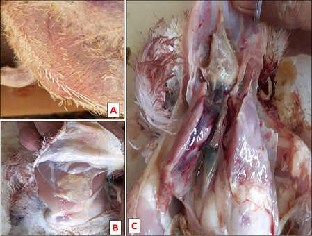
The obtained results revealed that, at the 1st DPI infected chickens showed low feed intake with ruffled feather followed by red swollen skin at site of infection with increase in thickness at the 3rd DPI (Figure 1A), and birds with severe lesions were reluctant to move.
In the current study treatment with doxycycline + colistin (0.5 gm/ liter in drinking water for 5 days), at the 2nd DPT birds of subgroups B, showed increased feed intake and activity than infected non-treated subgroups (A).
Our result pointed out that experimental infection of broiler chicken with S. aurous and/or E. coli resulted in induction of cellulitis with marked pathological and histological changes.
Doxycycline + colistin combination was used to treat infected birds where it helps in reduction of lesions, with somewhat improvement in measured parameters.
Selected from the article's own text.
Abstract
In the commercial broiler, cellulitis is considered one of the most economically prevalent problems due to the presence of the lesion leading to increased condemnations and carcass downgrading. Clinically cellulitis is a deep infection of broiler chicken skin caused by many bacterial species, mainly Escherichia coli (E. coli) and/ or Staphylococcus aureus (S. aureus), causing severe economic losses in poultry. This study was done on 14-day old broiler Ross 308 chickens subcutaneous (s.c) injected with E. coli and/ or S. aureus to induce cellulitis. Clinical signs, mortality, pathological lesion, and growth performance were determined. Hematological parameters, liver and kidney functions were also recorded. Colistin+ Doxycycline combination (Doxyforte®) was used to control the infection. Clinically, site of infection was appeared red, swollen accompanied with increased skin thickness, postmortem lesions in the 3rd day post infection with s.c. yellowish suppurative exudates, pericarditis and perihepatitis were prominent E. coli infected with hepatic subcapsular hemorrhage mostly in S. aureus groups. Hematological parameters were mostly affected in all infected non-treated groups compared to negative control without significant difference. Histopathological changes of infected non-treated groups showed inflammation of s.c tissue with massive heterophils and mononuclear cell infiltration, hydropic degeneration of the hepatocytes and congested splenic sinusoids. While treated groups showed limited skin inflammatory condition at the site of injection and return of skin to normal color and thickness. Doxycycline+ colistin combination helps in reduction of lesions in treat infected birds, with marked improvement in measured parameters. We recommended active actions to prevent causes and factors helping in including cellulitis, regular lowering bird density, enhancing restrict biosecurity, modulating the vaccination timing, improving management practices, as well as application of probiotics to improve and restore good gut health.
Avian cellulitis (AC) is a relatively recently recognized acute and diffuse suppurative inflammation, affecting all s.c tissues and sometimes extends to muscular tissue. It is frequently associated with abscess formation, discoloration and thickening of the broilers skin leading to an increase in condemnation rate at slaughterhouse . AC was detected on different parts of chicken body including the head, dorsum, thighs, breast, legs, and abdomen ; ; ; . Clinically, cellulitis may be observed in diseased chickens in case of occurrence the infection in the head area. Involvement of other parts of the body, on the other hand, is only discovered incidentally on post-mortem (PM) or slaughterhouse inspections ; . Skin traumas and scratches are considered the main predisposing factors for occurrence of cellulitis in broilers, facilitating invasion of pathogenic bacteria . Also, other factors like cannibalism, insect bites, poor litter conditions, immunodeficiency, foot problems in addition to systemic infections are thought to predispose AC ; .
Economic losses occur chiefly as a result of increased condemnation rate and/or downgrading of affected carcasses . Losses due to cellulitis in broilers were up to 30% in USA, 0.8% in Canada, at least 18 thousand tons in Brazil and, 0.9-1.7% of total carcass condemnation in Egypt ; ; ; .
The lesions showed varying-colored secretions from yellow to green, which were either serous, fibrous yellow, green, or suppurative. Many bacterial agents were involved to be the cause of AC as Aeromonas spp., Clostridia, Enterobacter spp., E. coli, Proteus mirabilis, P. aeruginosa, Staphylococci and Streptococci, where E. coli was the predominant ; . Recently, E. coli was recorded as the most prevalent in AC (45.2%), followed by Staphylococci (33.2%) . Immunosuppressed chickens were confirmed to possess more possibility for acquiring cellulitis. Experimental induction of AC was done via s.c injection of clostridia, E. coli, and S. aureus in 25-day-old broilers . In two separate experiments s.c injection of E. coli in 25 and 39-day-old broilers, 98% and 100% of birds developed characteristic cellulitis lesions as early as 28 and 18 hs post infection (PI) ; . Also, E. coli was recovered from > 75% of lesions ; ; . Both E. coli and Staphylococcus spp isolates from cellulitis are multidrug resistant .
Leukocyte profiles that deviate from normal are useful in conservation physiology because its changes are linked to the causes of stress and directly related to stress hormone levels as well as diseases and infections. Total white blood cell counts (TWBC) distributions and hematological parameters indicated leukocytosis, leukemoid reactions, and a high frequency of atypia . Polymicrobial bacteremia and fungemia are the reasons for the hematological observations, both of which could account for high TWBC and atypical cells .
The normal range of red blood cell (RBC) count is 3.5x10⁶ /mm 3, packed cell volume (PCV) is 22-35%, hemoglobin is 7-13 g/dL in broilers . In addition to causing relative neutrophilia and lymphopenia, infections commonly cause an increase in monocytes ; ; , and general increases in total WBC count , 1993; ; . Heterophils and lymphocytes are the most abundant white WBC in birds, they play an important role in innate and acquired immunity, respectively .
The utility of the avian Heterophils/lymphocytes (H/L) ratio was firstly realized by Gross and Siegel (1983) and used now to assess the welfare of chickens under rearing conditions ; ; ; ; , and infections or diseases causing increases in stress . The H/L ratio is an indicator of stress and welfare of hens caged in modern systems . The H/L ratio may reflect a readiness to cope with infection through injury (via heterophils) rather than with a communicable disease (via lymphocytes) . Blood H/L ratio reflects the status of immune system .
The usage of antibiotic or Bifidobacterium bifidum with avoiding immunosuppression can reduce cellulitis lesion and condemnation rate ; ; ; .
This study aimed to induce cellulitis experimentally by s.c infection of broiler chickens, recording clinical signs, pathological lesion, performance parameters and trial to use Colistin and Doxycycline in control of this infection.
MATERIALS AND METHODS
Bacterial strains
In the present study, E. coli (O78) and S. aureus cultures were purified and used. These strains were originally derived from cellulitis lesions in broiler chicken. E. coli strain O78 was molecular identified to be positive for 5 genes by PCR , 2020a). These isolates were sensitive to both Doxycycline and Colistin according to antibiotic susceptibility test using disc diffusion method according to Watts (2008) and CLSI (2016).
Experimental infection
E. coli O78 and S. aureus isolates were cultured and propagated as stated formerly by Matthijs et al. (2003). Both isolates were prepared for usage at a concentration of 4.5X10⁸ colony forming units (CFU)/ml. Each bird was infected by s.c injection of 0.5 ml of E. coli O78 and/or S. aureus over the left breast muscle .
Chickens
One hundred and fifty (150) 1-days old Ross 308 broiler chickens were purchased from a private commercial hatchery. The chickens were reared on straw deep litter.
Ration
The chickens were nourished on ready-made commercial pelleted rations free from feed additives (NRC, 1984) involving starter ration (CP not less than 23%) as well as growing ration (CP not less than 21%). Chickens were supplied ad libitum with drinking water and feed.
Vaccination
All chickens were immunized with ND+IBV vaccine at age of 5 days, IBD intermediate 228E vaccine at age of 10 days, in addition to La Sota vaccine at age of 16 day. All vaccines were given by ocular instillation route.
Drug
Doxyforte® is produced by Jordan Veterinary and Agriculture Medical Industry Company. P.O. Box: 2760 Amman 11953 JORDAN. Formula characteristics: Each gram contains: Doxycycline Hcl 240 mg and Colistin sulphate 500,000 IU. Formula: Water soluble powder. Dosage: 100 gm/ 200 liters drinking water for 5 days, Batch Number: 190326. Mfg. Date: 03/2019. Exp. Date: 03/2022.
Experimental design
At 14th days of life, chicks were randomly divided into 4 groups. Group 1 was kept as non-infected and non-treated (30 birds were kept as control negative group). Groups 2, 3 and 4 were (challenge groups, 40 chickens in each group) infected with S. aurous, S. aurous+ E. coli and E. coli; respectively. Chickens were s.c injected in the thigh fold with 1 ml of S. aureus (group 2) or E. coli (group 4), while chicks of group 3 were injected with 0.5 ml of both strains. Infected birds were observed daily for clinical signs, mortalities and examination of the inoculation site. At the 3rd day post infection (DPI) groups 2, 3 and 4 were subdivided into two subgroups (A and B), 20 chicken each. Subgroups A were kept as infected non-treated groups while subgroups B were infected treated groups with doxycycline + colistin in 1 gm/ liter drinking water for 5 days.
Broiler performance parameters
Feed consumption and conversion ratio were determined using the following Formula: Feed consumption (FC) g/bird = Feed intake in a replication/ No. of live birds in a replication. Feed conversion ratio (FCR) = Feed intake (g)/ Live weight (g). Parameters were recorded for each chicken in 4 groups at third and fourth week of age according to NCR (1984).
Daily examination of injection site with record of clinical signs, mortalities and post-mortem examination.
Measuring of skinfold thickness with skinfold meter of inoculation side (left) as compared with non-inoculated side (Right) at 3rd (DPI), 3rd (DPT) and the 7th DPT. Brest skin close to thigh web was drawn out far enough to apply the skinfold meter and measure thickness (Harpenden Skinfold Calipers).
Clinical signs: The infected and treated groups were observed daily for clinical signs just after infection till the end of the observation period.
Mortality rate: The number of positive dead birds/group was recorded daily till 35 days of age (end of the experiment).
Evaluation of deterioration in hematological parameters: Clotted blood for serum analysis and non-clotted blood for blood picture were collected from all groups at 3 DPI as well as 5 and 7 DPT. Blood samples from each group were collected for determination of hematological parameters such as RBC, WBC, PCV, Hemoglobin (Hb) concentration and platelets using Natt and Herrick blood diluent and hemocytometer. Blood smears were done for differential leucocyte count and H/L ratio .
Samples from each group for the assay of liver function parameters (GOT, GPT, ALP), kidneys function parameters (Urea, creatinine) using BioSystems S.A. Kits.
Histopathological Examination: Skin specimens were collected at 3 and 5 DPI; fixed in 10% neutral buffered formalin, paraffin embedded (FFPE) block of tissue . Paraffin tissue were sectioned at 4-6 μm thickness and stained with hematoxylin and eosin (H and E) .
Statistical analysis
The obtained results were statistically compared by ANOVA without detection of marked statistical significance.
RESULTS AND DISCUSSION
Avian cellulitis is a diseased condition affecting broiler chickens and characterized by inflammation of the s.c tissue, particularly in the thigh and abdomen, with the presence of suppurative exudates and fibrino-necrotic plaques ; ; , b). In the poultry industry, cellulitis represents one of the most essential reasons of partial or total condemnation of carcasses. The major causative agents for cellulitis are E. coli and S. aureus ; ; , b).
The obtained results revealed that, at the 1st DPI infected chickens showed low feed intake with ruffled feather followed by red swollen skin at site of infection with increase in thickness at the 3rd DPI (Figure 1A), and birds with severe lesions were reluctant to move. In postmortem lesions revealed s.c yellowish exudates, pericarditis and perihepatitis with subscapular hemorrhage in liver (Figure 1B, C). Mortality started at the 3rd DPI as 1 bird with ratio of 2.5% in 2nd and 3rd groups injected with S. aurous and S. aurous + E. coli respectively, and 2 birds in 4th group, E. coli infected group with ratio of 5%. After subgrouping at the 4th DPI, mortalities were 1 bird in S. aureus infected group (2a) with ratio (5%) and 3 birds in S. aurous + E. coli subgroup (3a) (15%). While, at 5th DPI, 2 birds in E. coli subgroup (4a). The highest mortality was 17.5% in subgroups (3a) followed by 15% in subgroup (4a) and the lowest mortality (7.5%) was in subgroup (2a). While control group and all treated subgroups showed no mortality. Our results were in accordance with that obtained by Norton et al. (1997) who administered an E. coli strain in broilers for experimental induction of cellulitis with suppurative plaques at 18 h pi. Macroscopic lesions induced by E. coli were distinguished by the existence of suppurative exudates, skin thickness and hemorrhages similar to those obtained in our study ; . Sanches et al. (2020) experimentally reproduce cellulitis in chickens by P. mirabilis and recorded cellulitis lesions (suppurative exudates and hemorrhages) in the chest region. Some birds showed additional congestion and infusions in the musculature with petechiae, bruises, and edema within the 24 h pi. Amer et al. (2020a) experimentally induced cellulitis in broiler chickens by s.c injection of E. coli O78 where yellowish to suppurative s.c exudate with thick yellowish red skin. The mortality in low and sporadic cases, without septicemia were recorded ; . The injected E. coli and /or S. aureus were successfully reisolated from the developed lesions ; ; ; ; .
In poultry farms, antibiotics are administered commonly for therapeutic applications and growth promotion ; ; . In the current study treatment with doxycycline + colistin (0.5 gm/ liter in drinking water for 5 days), at the 2nd DPT birds of subgroups B, showed increased feed intake and activity than infected non-treated subgroups (A).
Table 1: Average body weight gain, feed intake and feed conversion rate of infected non-treated and treated broiler chickens.
Group
Infection
Treatment
Age/weeks
Av. FI
ABWG
FCR
1
Control
Negative
1
145.69
139.42
1.04
2
386.75
283.20
1.37
3
840.60
511.80
1.64
4
1038.48
728.80
1.42
2a
S. aurous
-
3
697.25
489.70
1.42
4
956.9
510.40
1.87
2b
+
3
721.65
588.10
1.23
4
981.40
736.67
1.33
3a
S. aurous + E. coli
_
3
688.90
486.50
1.42
4
910.10
327.733
2.78
3b
+
3
722.40
632.30
1.14
4
726.75
577.90
1.26
4a
E. coli
_
3
686.35
394.90
1.74
4
975.80
416.80
2.34
4b
+
3
726.75
577.90
1.26
4
1014.60
590.47
1.72
The recorded ABW as well as FCR of control gr 1 (Table 1) at 1 and 2 weeks of age were 139.42 and 1.04 as well as 283.20 and 1.37, respectively. ABW and FCR of at the 3rd and 4thweek of age (2 and 3wpi) of group 1 (511.80, 1.64, 728.80 and 1.42) were the highest followed by those of S. aurous infected group 2a (489.70, 1.42, 510.40 and 1.87) followed by E. coli infected group 3a (394.90, 1.74, 416.80 and 2.34), while group 2a showed the lowest values (486.50, 1.42, 327.733 and 2.78). This result indicates that both E. coli or S. aurous solo infections resulted in lower ABW and FCR ; ; ; ; , and the dual infection were resulted in more severe losses. Infected treated an improvement in ABW and FCR was seen but still lower than control (Table 1). This result indicated that treatment can improve of ABW and FCR ; but not completely eliminate the pathological lesions ; ; ; .
Table 2: Thickness of skinfold in the site of infection (left) and the non-infected side (Right) and their difference.
Gr.
Infection
Treatment
Time
L
R
Difference
M ± SD
M ± SD
Mean
1
Control
Negative
3 DPI
13.2±1.1
14.6±0.55
1.4
3 DPT
20.6±0.89
18.2±2.05
2.4
7 DPT
21.4±2.07
20.6±2.61
0.8
2a
S. aurous
-
3 DPI
24.8±17.54
14±1
10.8
3DPT
39±17.36
18.8±2.68
20.2
7 DPT
42.2±23.66
20.4±0.89
21.8
2b
+
3 DPT
29.4±20.84
20.4±1.52
9.0
7 DPT
27.8±7.53
19±2.24
8.8
3a
S. aurous + E. coli
-
3 DPI
65.8±12.85
15.6±1.34
50.2
3 DPT
36.4±13.48
23.6±4.93
12.8
7 DPT
60.8±49.24
21±2.35
39.8
3b
+
3 DPT
48±41.92
14.6±1.67
33.4
7 DPT
34.6±19.71
20.2±0.45
14.4
4a
E. coli
-
3 DPI
55±15.05
15±0
40.0
3 DPT
61±40.45
23.4±6.84
37.6
7 DPT
57.2±52.03
25.6±6.66
31.6
4b
+
3 DPT
77.8±6.69
30±0
47.8
7 DPT
32.4±6.23
19.8±0.84
12.6
DPI: day post infection; DPT: day post treatment.
Skin fold thickness describes the s.c inflammation and deposits where the skin fold thickness is measured by specialized (Table 2). Results of skinfold thickness revealed that all infected groups show an increase in skin thickness at site of injection (left) as compared to control negative group. The highest thickness was in S. aurous+ E. coli non-treated group which was 60.8±49.24 at 7 DPI, then infected non-treated E. coli which was 57.2±52.03 at 7 DPI (Table 2). Then infected non treated S. aurous group which was 42.2±23.66 at 7 DPI, then followed by infected with S. aurous+ E. coli treated group which was 34.6±19.71 at 7 DPT, followed by infected treated E. coli which was 32.4±6.23 at 7 DPT, followed by infected treated S. aurous group which was 27.8±7.53 at 7 DPT, and the lowest was control negative group at 7 DPI as it was 21.4±2.07. The non-infected side (Right) shows no lesions; it was 20.6±2.61 while in infected non-treated E. coli group is much thicker (22.6±6.66) (Table 2). Increase of skin thickness in injected chicken’s groups indicate inflammation of s.c tissue and consequently increase skin thickness due to increase amount of s.c deposits, this was firstly recorded by Randall et al. (1984), later cellulitis was worldwide diagnosed in poultry ; ; . E. coli cause cellulitis with subsequently increase skin thickness ; . Amer et al. (2019) reported that infection with pathogenic E. coli causing cellulitis is due to excessive production of s.c exude and increase deposits in s.c region. The highest skin thickness was found in group injected with both E. coli and S. aureus, that can be attributed to co-infection which increase severity of the s.c inflammation. This result was parallel with that of Radwan et al. (2018) who found that E. coli and S. aureus are the most prevalent bacteria in poultry cellulitis.
The RBC count, Hb and (PCV% are helpful in diagnosing nutritional deficiencies, acute illnesses, and chronic medical conditions ; . The normal chicken blood parameters are arranged (RBC: 2.5-3.5 x10⁶ µl, PCV: 22-35 %, Hb: 7-13 g/dl and WBC: 12-30 x 103 µl) ; . At the 7th day, the values of RBC count, Hb and PCV% of control group were 2.91±0.37, 6.45±0.49 and 28±1.41 higher than 2.53±0.14, 6.55±0.35 and 30±4.24 in S. aurous infected group, 2.56±0.34,7.05±0.49 and 26.5±0.71 in S. aurous+ E. coli group, as well as in 2.14±0.37, 7.1±0.28 .and 29. 5±2.12 in E. coli group (Table 3) this result indicates that E. coli and/or S. aureus infections reduce RBC parameters.
Values of RBC, Hb and PCV% in doxycycline + colistin treated groups were 2.25±0.02, 6.2±0.14, and 28.5±2.12 in S. aureus, 2.28±0.11, 6.85±0.07, and 27.5±2.12 in S. aurous+ E. coli group, as well as 2.35±0.07, 6.3±0.71, and 27.5±0.71 in E. coli group. These values are nearly close to each other but still lower than control non infected non treated.
The TLC of control group (20.5±8.71) was nearly similar to E. coli (20.0 ±7.07) and both are higher than that of S. aureus (19.5±13.54), while that of S. aureus and E. coli showed the highest values (22.2±11.0) at the 7th day (Table 4). Values in treated groups were 17.5±3.54, 15.0±0.0, and 22.5±3.54 in S. aurous, S. aurous+ E. coli and E. coli infected group, respectively, but all were still lower than nontreated and control group. So, the result reflects the bad effect of treatment on measured TLC. Generally, blood platelets were nearly similar not in all treated or infected groups. Regarding differential leucocytes count the control negative group 1 showed Heterophils (H), Lymphocytes (L) and Monocytes (M) values of 16.5±0.50, 76.0±0.00, and 3.50±0.50; respectively. While the infected groups showed H, L, and M values of 16.0±1.00,77.5±0.50, and 3.00±1.00 in S. aurous, 18.0±0.00, 75.5±0.50, and 3.00±0.00 in S. aureus +E. coli group, while E. coli infected values were 17.5±0.50,76.0±1.00 and 3.50±0.50.
Table 3: Blood picture at 3 DPI (0 DPT), 3 and 7 DPT of infected non-treated and doxycycline + colistin treated groups of broiler chickens.
Gr.
Infection
Treatment
Time
/DPT
RBC X106
Hb mg/ml
PCV %
Platlets X10 3
TLC X 10 3
Mean± SD
Mean± SD
Mean± SD
Mean± SD
Mean± SD
1
Control
Negative
0
2.68±0.04
6.95±0.21
31±2.83
10.0±0.0
20.5±8.71
3
2.61±0.79
5.8±0.42
27.5±0.71
9.5±0.71
27.5±10.61
7
2.91±0.37
6.45±0.49
28±1.41
10.0±0.0
27.5±10.61
2a
S. aurous
_
0
2.26±0.08
6.65±0.07
28±0
10.0±0.0
21.0 ±14.0
3
2.31±0.34
6.85±0.92
26±2.83
10.0±0.0
17.5±13.54
7
2.53±0.14
6.55±0.35
30±4.24
10.0±0.0
19.5±13.54
2b
+
3
2.39±0.23
6.45±0.21
28±0
9.5±0.71
21±1.41
7
2.25±0.02
6.2±0.14
28.5±2.12
10.0±0.0
17.5±3.54
3a
S. aurous +
E. coli
-
0
2.35±0.07
5.15±0.35
25±1.41
10.5±0.71
20.0±8.0
3
2.60±0.28
7.15±0.78
31±2.83
10.0±0.0
22.5±3.54
7
2.56±0.34
7.05±0.49
26.5±0.71
10.0±0.0
22.2±11.0
3b
+
3
2.81±0.37
7±0.57
28.5±0.71
12.5±3.54
27.5±10.61
7
2.28±0.11
6.85±0.07
27.5±2.12
10.0±0.0
15.0±0.0
4a
E. coli
-
0
2.43±0.04
5.25±0.35
26.5±0.71
10.0±0.0
22.2±10.0
3
2.23±0.49
5.85±0.64
25±2.83
8.0±1.41
20.0±7.07
7
2.14±0.37
7.1±0.28
29.5±2.12
10.0±0.0
20.0 ±7.07
4b
+
3
2.85±0.32
6.3±1.56
28.5±0.71
12±4.24
27.5±10.61
7
2.35±0.07
6.3±0.71
27.5±0.71
10.0±0.0
22.5±3.54
RBC: red blood cell; Hb: Hemoglobin. PCV: Packed cell volume. TLC: Total leucocytes count.
Table 4: Leukocyte profiles of infected non-treated and doxycycline + colistin treated groups of broiler chickens.
Gr.
Infection
Treatment
DPT
Heterophils
Mean± SD
Lymphocytes
Mean± SD
H /L
ratio*
Monocytes
Mean± SD
Eosinophils
Mean± SD
Basophils
Mean± SD
1
Control
-
0
15.7±2.52
73.0±4.00
21.51
4.00±1.00
1.00±0.00
1.00±0.00
3
17.5±0.50
76.0±0.00
23.03
3.50±0.50
1.00±0.00
1.00±0.00
7
16.5±0.50
76.0±0.00
21.71
3.50±0.50
1.50±0.50
1.00±0.00
2a
Staph
-
0
15.7±1.53
79.0±1.00
19.87
3.00±0.00
0.67±0.58
1.00±0.00
3
17.0±0.00
77.5±0.5
21.94
3.00±0.00
1.00±0.00
0.50±0.50
7
16.0±1.00
77.5±0.50
20.65
3.00±1.00
1.50±0.50
1.00±0.00
2b
+
3
16.5±0.50
76.5±0.50
21.57
4.00±0.00
1.00±0.00
1.00±0.00
7
16.5±0.50
74.5±0.50
22.15
4.00±0.00
2.50±0.50
1.50±0.50
3a
Staph +
E. coli
-
0
15.7±0.58
78.7±1.53
19.95
3.67±0.58
1.00±0.00
0.67±0.58
3
17.5±0.50
76.0±0.00
23.03
3.00±0.00
2.00±0.00
1.00±0.00
7
18.0±0.00
75.5±0.50
23.84
3.00±0.00
1.50±0.50
1.00±0.00
3b
+
3
16.5±1.50
72.5±2.50
22.76
5.50±0.50
3.50±3.50
1.00±1.00
7
15.5±0.50
75.0±3.00
20.67
5.00±2.00
2.50±1.50
1.00±0.00
4a
E. coli
-
3
17.0±1.00
76.7±0.58
22.16
3.33±0.58
1.67±0.58
1.00±0.00
3
16.5±1.50
72.5±1.50
22.76
6.00±2.00
2.50±1.50
1.50±0.50
7
17.5±0.50
76.0±1.00
23.03
3.50±0.50
1.50±0.50
0.50±0.50
4b
+
3
15.0±0.00
76.0±3.00
19.74
5.00±2.00
2.00±1.00
1.00±0.00
7
16.5±0.50
76.0±1.00
21.72
3.50±0.50
2.00±0.00
1.00±0.00
* H/L Ratio: Heterophils/ lymphocytes ratio
Heterophils were higher in infection than control, while L was lower in infected. It was found that at infection site the S. aureus stimulates the immune cells to secrete chemokines, released WBC in the bone marrow into the blood, where the WBC and neutrophils are significantly increased, with a shift to the left in the nuclear index, toxic granules appeared in the cells, and decreased number of eosinophils . The detected inflammatory response at the inoculation sites showing a significant increase in absolute count and of pseudo-eosinophils, basophils%, and monocytes in infected group than in control group . Intracellular accumulation of antibacterial medicines in active form facilitates transformation of phagocytic activity of macrophages and neutrophils and affects viability of phagocytized bacteria ; ; ; .
Counts of H, L, and M in treated were 16.5±0.50, 74.5±0.50, and 4.00±0.00 in S. aureus, 15.5±0.50, 75.0±3.00, and 5.00±2.00 in S. aureus + E. coli, 15.5±0.50, 75.0±3.00, and 5.00±2.00 in E. coli (Table 5). It was observed that M count was H and L decreased, while M was increased in treated than non-treated and control groups. H/L ratios were 21.71 in control, infected with S. aureus (20.65), S. aureus + E. coli (23.84), and E. coli (23.03), while in infected treated was generally lower 22.15 in S. aureus, 20.67 in S. aureus + E. coli, and 21.72 E. coli (Table 4). Gross (1989) indicated that injected E. coli affects the H/L ratio. There is no detectable difference in the count of Eosinophils and Basophils in between groups.
Liver function test GPT, GOT, and ALP results in control group 1 were 8.5±0.71, 15± 4.24, and 887.5±17.68, respectively. GPT, GOT, and ALP for S. aureus infected group were 11±1.41, 22±5.66, and 632.5±519.72; in S. aurous + E. coli were 14.5±3.54, 22.5±4.95, and 1120±28.28; while in E. coli were12.75±5.3, 32.25±6.01, and 920±197.99 (Table 5), so results of infected groups are generally higher than control. GPT, GOT, and ALP results of doxycycline + colistin treated groups were 8.75±1.77, 16.25±2.47, and 975±49.5 in S. aurous; 8.75±1.77, 16.25±2.47, and 975±49.5 in S. aurous + E. coli; 9.5±2.12, 13.25±1.77, and 952.5±74.25 in E. coli. Results of treated groups showed lower values than non-treated and close to the control negative group. The raise in serum AST is signal for cellular damage to heart muscles and liver cells. Where the increase in serum ALT is mostly resulting from hepatic injuries ; ; . An increase in serum ALT, AST, LDH activities, globulin concentration and a decrease ALP activity in E. coli O78 at 107 CFU/0.5 ml intraperitoneally infected groups were recorded ; ; ; ; . The volume of AST and ALT rising alters according to the cause of liver cells injury . It was reported that there was no significant effect on level of ALT and creatinine in E. coli infected broilers after administration of colistin in feed . Rising in ALT can be occured through direct effect of bacterial toxins, drugs, chemicals on hepatocytes, particularlly those are near to the central vein ; . Colistin does not affect hematobiochemical serum on broilers .
Kidney function test represented by urea and creatinine values were 10.5±0.71 and 0.54±0.02 in control. While infected groups showed values of 9.5±0.71 and 0.51±0.08 in S. aurous; 9±1.41 and 0.6±0.06 in S. aurous + E. coli; as well as 9.75±2.47 and 0.48±0.04 in E. coli. The urea of infected groups was lower than control, while creatinine showed no marked difference. The urine and creatinine of
Table 5: Serum biochemical parameters at 3 DPI (0 DPT), 3 and 7 DPT of infected non-treated and doxycycline + colistin treated groups of broiler chickens.
treated S. aurous groups were 9.5±0.71 and 0.54±0.05; 9.75±0.35 and 0.54±0 for S. aurous + E. coli, as well as10.75±2.47 and 0.58±0.02 for E. coli. The values in treated groups were close to those of control. Marked reduction in serum albumin concentration and TP was seen in E. coli infected groups ; ; ; ; .
Histopathological examination of tissue section of control non-infected non-treated (control negative) group showed normal tissue structure in bursa, kidney, liver, muscles,
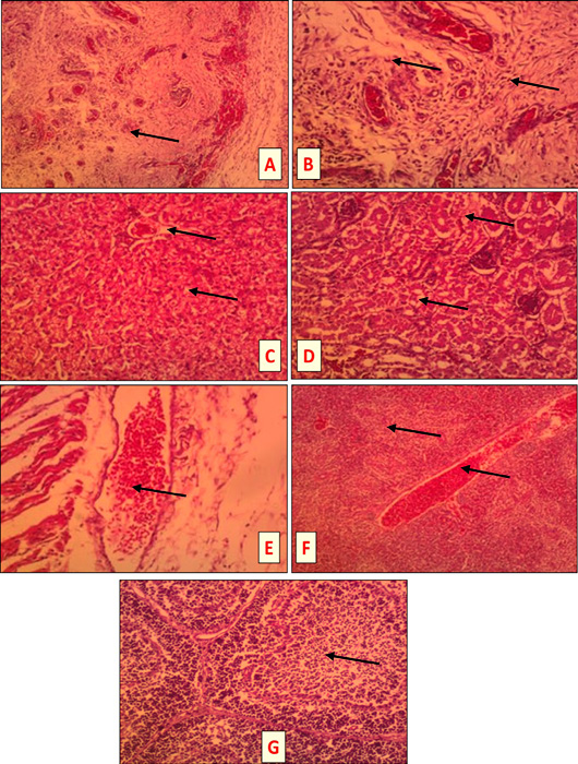
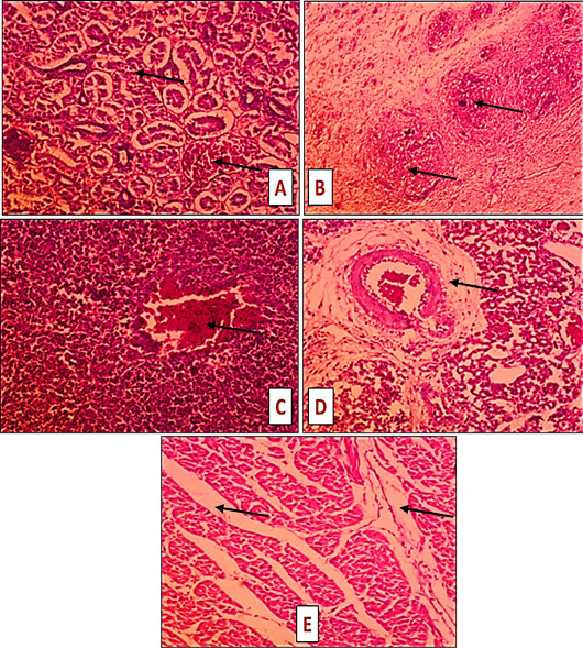
spleen and skin. Microscopical examination of tissues for infected non treated groups showed that, subcutis tissue of E. coli infected and mixed infected (S. aureus + E. coli) groups exhibited severe inflammatory reaction with accumulation of suppurative exudate with massive heterophils and mononuclear cells infiltration in s.c fatty tissue (Figures 2A, B and 3B) accompanied with muscle edema (Figure 3E) while showed milder s.c reaction in case of S. aureus infected group (Figure 2E). The liver of mixed infected group and S. aureus infected group showed hydropic degeneration (Figure 2C), while E. coli infected bird showed milder liver lesion. Kidney of mixed infected (S. aureus + E. coli) group exhibited severe hydropic degeneration (Figure 2D) while in case of E. coli or S. aureus infected group, kidney appeared hemorrhagic accompanied with coagulative necrosis of some tubules (Figure 3A). The bursa of all infected groups showed depletion of the lymphoid follicle (Figure 2G). The spleen of all groups appeared hemorrhagic (Figure 3C) but also accompanied with congested red bulb and blood vessels in addition to necrotic area in case of S. aureus infected group (Figure 2F). Also, the lung of S. aureus infected group exhibited perivascular edema of the pulmonary artery (Figure 3D). Similar histopathological lesions were detected ; ; ; ; .
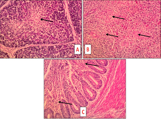
Microscopical examination of tissues for S. aureus and/or E. coli infected treated groups revealed slight depletion in both bursal tissue (Figure 4A) and spleen (Figure 4B) was seen in mixed infected treated group. Also, intestine of S. aureus infected treated group showed light inflammation (Figure 4C).
CONCLUSION AND RECOMMENDATIONS
Cellulitis in broilers composes an essential reason for carcass condemnation at slaughterhouses. Our result pointed out that experimental infection of broiler chicken with S. aurous and/or E. coli resulted in induction of cellulitis with marked pathological and histological changes. Skin lesions were measured. Effect on broiler performance was detected. Blood analysis and serum biochemical parameters were studied but were of low value in diagnosis. Doxycycline + colistin combination was used to treat infected birds where it helps in reduction of lesions, with somewhat improvement in measured parameters.
We recommended several actions can be taken to oppose or minimize cellulitis as reinforcing feather coverage, observing birds density, continuous enhancing of biosecurity, modulating the vaccinations timing, improving management practices, control immunosuppressive factors as well as application of probiotics in order to improve and restore good gut health.
ACKNOWLEDGEMENT
Authors acknowledged poultry disease departments in both faculty of Veterinary Medicine, Cairo University and Veterinary Research Institute, National Research Centre.
NOVELTY STATEMENT
The primary purpose of this research is to study the influences of experimentally induced cellulitis on broiler performance and recognize the gross and histopathological changes. In addition to investigate the effect of antibiotic treatment in control of this infection.
AUTHOR’S CONTRIBUTION
AMM, KME-B designed this study and supervised laboratory work. HSF, HMM, AAE-S collected samples and performed all laboratory work. BMA carried out histopathological examination. All authors shared manuscript writing, drafted, revised the manuscript, and approved the final manuscript.
Data availability
The authors affirm that the data bolstering the results of this research are accessible within the article in addition to its supplementary materials.
Ethical approval
All work design and procedures were approved by Medical Research Ethics Committee (MREC), National Research Centre, Egypt with Approval number 7427082021.
Abbreviations
ABWG, Average body weight gain; ALP, alkaline phosphatase; ALT, alanine transaminase; AST, aspartate transaminase; CFU, colony forming units; DPI, day post infection; DPT, Day post treatment; FC, Feed consumption, FCR, Feed conversion ratio; FI, feed intake; GOT, glutamic oxaloacetic transaminase; GPT, Glutamate pyruvate transaminase; HandE, hematoxylin and eosin; H/L, Heterophils/lymphocytes; Hb, Hemoglobin; LDH, lactate dehydrogenase; PCV, Packed cell volume; PM, postmortem; RBC, Red Blood Cell; TLC, Total Leukocyte Count; TWBC, Total white blood cell; WBC, White Blood Cell.
Conflict of interest
The authors have no conflict of interests regarding the publication of this paper. Also, the authors declare that the work was self-funded.
Figures
Figure 1:.
Lesion in broiler chicken s.c infected with S. aureus and /or E. coli showing. A: Thick rough dark colored skin. B: S.c lesion. C: S.c lesion, pericarditis and prehepatitis with subscapular hemorrhage in liver.
Figure 2:.
Tissue sections of S. aureus and /or E. coli infected birds at 3 DPI (HandEX, 200) showing the following lesions. A: Subcutis of E. coli infected bird: Severe suppurative inflammation characterized by heterophils and mononuclear cells infiltration in s.c tissues (head of arrow). B: Subcutis of S. aureus and E. coli infected bird: severe suppurative inflammation with marked accumulation of heterophils and mononuclear cells in s.c fatty tissue (head of arrow). C: Liver of S. aureus infected bird: hydropic degeneration of the hepatocytes (head of arrow). D: Kidney of S. aureus + E. coli infected bird: severe hydropic degeneration (head of arrow). E: Subcutis of S. aureus infected bird: subcutaneous hemorrhages (head of arrow). F: Spleen of S. aureus infected bird: congested red bulb and blood vessels with necrotic area (head of arrow). G: Bursa of E. coli and/or S. aureus infected bird: depletion of the lymphoid follicle (head of arrow).
Figure 3:.
Tissue sections of S. aureus and /or E. coli infected non treated birds (7 DPT) (HandE, X 200) showing. A: E. coli infected bird: kidney hemorrhages accompanied with coagulative necrosis of some tubules (head of arrow). B: E. coli infected bird: s.c severe suppurative inflammation with marked accumulation of heterophils and mononuclear cells in s.c fatty tissue (head of arrow). C: E. coli infected bird: spleen hemorrhages (head of arrow). D: S. aureus infected bird: perivascular edema of the pulmonary artery (head of arrow). E: S. aureus + E. coli infected bird: muscle edema (head of arrow).
Figure 4:.
Tissue sections of S. aureus and /or E. coli infected treated birds at 7 DPT (HandE, X 200) showing the following lesions. A: S. aureus + E. coli infected treated bird: bursa slight depletion of the lymphoid follicle (head of arrow). B: S. aureus + E. coli infected treated bird: spleen slight depletion of the lymphoid follicle (head of arrow). C: S. aureus infected treated bird: intestine light inflammation characterized by lymphocytic infiltration of the mucosa (head of arrow).
References
Abd Elatiff A, El-Sawah AA, Amer MM, Dahshan A-HM, Salam H, Shany SAS (2019). Pathogenicity of Escherichia coli O125 in commercial broiler chickens. J. Vet. Med. Res., 26(1): 1-8. https://doi.org/10.21608/jvmr.2019.43328[CrossRef]
Aksu DS, Aksu T, Qzsoy A (2010). The effects of lower supplementation levels of organically complexed minerals (Zinc, Copper and Manganese) versus inorganic forms on hematological and biochemical parameters in broilers. Kafkas Univ. Vet. Fak. Derg., 16(4): 553-559.
Altan O, Altan A, Cabuk M, Bayraktar H (2000). Effects of heat stress on some blood parameters in broilers. Turk. J. Vet. Anim. Sci., 24: 145–148.
Amer MM, Mekky HM, Fedawy HS, Amer AM, Elbayoumi KM (2020b). Review: Cellulitis in broiler chickens. Korean J. Food Health Convergence, 6(5): 1-10.
Amer MM, Mekky HM, Fedawy HS, Elbayoumi KM (2020a). Molecular and gene identification of E. coli isolated from cellulitis in broiler chickens. Vet. World, 13(12): 2703–2712. https://doi.org/10.14202/vetworld.2020.2703-2712[CrossRef]
Amer MM, Mekky HM, Fedawy HS, Elbayoumi KhM, Sedeek DM (2019). Antibiotic profile of bacterial species isolated from broiler chickens with cellulitis. World’s Vet. J., 9(4): 268-279. https://doi.org/10.36380/scil.2019.wvj34[CrossRef]
Andreasen CB (2020). Staphylococcosis. In diseases of poultry, 14th ed.; Swayne, D.E., Ed., John Wiley and Sons, Ltd.: Hoboken, NJ, USA. pp. 995–1003.
Apata DF (2009). Antibiotic resistance in poultry. Int. J. Poult. Sci., 8: 404-408. https://doi.org/10.3923/ijps.2009.404.408[CrossRef]
Asmaa AMM (2013). Studies on enterobacteriacae in chicken eggs. M.V.Sc. thesis (Poultry diseases dep), Faculty of Veterinary Medicine, University of Beni Suef.
Bancfort JD, Stevens A (1996). Theory and practice of histological technique. 4th Ed. New York: Churchill Livingstone.
Barbieri NL, de Oliveira AL, Tejkowski TM, Pavanelo DB, Rocha DA, Matter LB, Callegari-Jacques SM, de Brito BG, Horn F (2013). Genotypes and pathogenicity of cellulitis isolates reveal traits that modulate APEC virulence. PLoS One, 8(8): e72322. https://doi.org/10.1371/journal.pone.0072322[CrossRef]
Barnes HJ, Vaillancourt JP, Gross WB (2003). Colibacillosis. In Y.M. Saif, H.J. Barnes, J.R. Glisson, A.M. Fadley, L.R. McDougald and D.E. Swayne (Eds.), Diseases of Poultry 11th Edition, Ames Iowa State University Press. pp. 631-652.
Barros LSS, Silva RM, Silva IM, Baliza MD, Virgílio FF (2013). Escherichia coli from cellulitis lesions in broilers. J. Food Measur. Charact., 7(1): 40-45. https://doi.org/10.1007/s11694-013-9138-3[CrossRef]
Bianco C, Balanescu B, Cieslicka U, Balanescu P, Stefanov K, Lopez P, Hristova N (2016). The shades of avian cellulitis in meat-type chicken. J. Vet. Sci., 2(2): 49-52.
Bounous D, Stedman N (2000). Normal avian hematology: Chicken and turkey. In: Feldman BF, Zinkl JG, Jain NC, editors. Schalm’s veterinary hematology. New York: Wiley; pp. 1147-1154.
Campbell TW (1996). Clinical pathology. Reptile Medicine and Surgery (ed. D.R. Mader), W.B. Saunders Company, Philadelphia, PA. pp. 248–257.
Chen J, Tellez G, Escobar J (2016). Identification of biomarkers for footpad dermatitis development and wound healing. Front. Cell. Infect. Microbiol., 6: 26. https://doi.org/10.3389/fcimb.2016.00026[CrossRef]
Cirz RT, O’Neill BM, Hammond JA, Head SR, Romesberg FE (2006). Defining the pseudomonas aeruginosa SOS response and its role in the global response to the antibiotic ciprofloxacin. J. Bacteriol., 188(20): 7101-7110. https://doi.org/10.1128/JB.00807-06[CrossRef]
CLSI (2016). Performance standards for antimicrobial susceptibility testing. 26th ed. CLSI supplement M100S. Wayne, PA: Clinical and Laboratory Standards Institute.
Cookson K, Macklin K, Giambrone J, Haroldo TG (2007). The influence of E. coli inoculum titer and virally induced immune suppression on the incidence of cellulitis in a broiler skin challenge model. Proceedings of the fifty-sixth western poultry disease conference March 26-29, Las Vegas, Nevada.
Cotter PF (2015). An examination of the utility of heterophil-lymphocyte ratios in assessing stress of caged hens. Poult. Sci., 94: 512-517. https://doi.org/10.3382/ps/peu009[CrossRef]
Davis AK, Cook KC, Altizer S (2004). Leukocyte profiles of House Finches with and without mycoplasmal conjunctivitis, a recently emerged bacterial disease. EcoHealth Alliance, 1: 362-373. https://doi.org/10.1007/s10393-004-0134-2[CrossRef]
Davis GS, Anderson KE, Carroll AS (2000). The effects of long-term caging and molt of single comb white leghorn hens on heterophil to lymphocyte ratios, corticosterone and thyroid hormones. Poult. Sci., 79: 514–518. https://doi.org/10.1093/ps/79.4.514[CrossRef]
Derakhshanfar A, Ghanbarpour R (2002). A study on avian cellulitis in broiler chickens. Veterinarski. Arhiv., 72(5): 277-284.
Dhabhar FS, Miller AH, McEwen BS, Spencer RL (1996). Stress induced changes in blood leukocyte distribution role of adrenal steroid hormones. J. Immunol., 157: 1638-1644. https://doi.org/10.4049/jimmunol.157.4.1638[CrossRef]
Eleiwa NZH, Sayed EME, Nazim AA (2011). Prophylactic and therapeutic evaluation of the phytobiotic (Orego-stim)® in chicken experimentally infected with E. coli. J. Am. Sci., 7(8): 91-102.
Elfadil AA, Vaillancourt JP, Meek AH, Gyles CL (1996). A prospective study of cellulitis in broiler chickens in southern Ontario. Avian Dis., 40(3): 677-689. https://doi.org/10.2307/1592281[CrossRef]
El-Sawah AA, Dahshan AL-HM, El-Nahass E-S, Abd El-Mawgoud AI (2018). Pathogenicity of Escherichia coli O157 in commercial broiler chickens. Beni-Suef Univ. J. Basic Appl. Sci., 7(4): 620-625. https://doi.org/10.1016/j.bjbas.2018.07.005[CrossRef]
Elston JJ, Beck M, Alodan MA, Vega-Murillo V (2000). Laying hen behavior 2. Cage type preference and heterophil to lymphocyte ratios. Poult. Sci., 79: 477-482. https://doi.org/10.1093/ps/79.4.477[CrossRef]
Fallavena LCB, Moraes HLS, Salle CTP, Silva AB, Vargas RS, Nascimento VP, Canal CW (2000). Diagnosis of skin lesions in condemned or downgraded broiler carcasses, a microscopic and macroscopic study. Avian Pathol., 29(6): 557-562. https://doi.org/10.1080/03079450020016797[CrossRef]
Fidan ED, Nazlıgül A, Türkyılmaz MK, Aypak SÜ, Kilimci FS, Karaarslan S, Kaya M (2017). Effect of photoperiod length and light intensity on some welfare criteria, carcass, and meat quality characteristics in broilers. Rev. Brasil. Zoot., 46: 202-210. https://doi.org/10.1590/s1806-92902017000300004[CrossRef]
Fitri AN, Fitriana I, Rosetyadewi AW, Pratama AM, Septiana AI, Setiawan DCB, Wijayanti AD (2021). The effect of colistin administration as medicated feed on alanine aminotransferase and creatinine level in broiler infected with Escherichia coli. BIO Web Conf., 33, 03002. The 1st International Conference of Advanced Veterinary Science and Technologies for Sustainable Development (ICAVESS 2021). https://doi.org/10.1051/bioconf/20213303002[CrossRef]
Gomis S, Babiuk L, Godson DL, Allan B, Thrush T, Townsend H, Willson P, Waters E, Hecker R, Potter A (2003). Protection of chickens against Escherichia coli infections by DNA containing CpG motifs. Infect. Immun., 71(2): 857-863. https://doi.org/10.1128/IAI.71.2.857-863.2003[CrossRef]
Gomis SM, Watts T, Riddell C, Porter AA, Allan BJ (1997b). Experimental reproduction of Escherichia coli cellulitis and septicemia in broiler chickens. Avian Dis., 41(1): 234-240. https://doi.org/10.2307/1592464[CrossRef]
Gomis SM, Gomis AIU, Horadagoda NU, Wijewardene TG, Allan BJ, Potter AA (2000). Studies on cellulitis and other disease syndromes caused by Escherichia coli in broilers in Sri Lanka. Trop. Anim. Health Prod., 32(6): 341-351. https://doi.org/10.1023/A:1005293400605[CrossRef]
Gomis SM, Goodhope R, Kumor L, Candy N, Riddell C, Potter AA, Allan BJ (1997a). Isolation of Escherichia coli from cellulitis and other lesions of the same bird in broilers at slaughter. Can. Vet. J., 38(3): 159-162.
Gross WB (1989). Factors affecting chicken thrombocyte morphology and the relationship with heterophil: Lymphocyte ratios. Br. Poult. Sci., 30: 919-925. https://doi.org/10.1080/00071668908417218[CrossRef]
Gross WB, Siegel HS (1983). Evaluation of the heterophil/lymphocyte ratio as a measure of stress in chickens. Avian Dis., 27: 972-979. https://doi.org/10.2307/1590198[CrossRef]
Johnson LC, Bilgili SF, Norton RA, McMurtrey BL (1997). Source of Escherichia coli and age upon the development of cellulitis in broilers. Proceedings of the 38th Southern Conference on Avian Diseases (abstract S172). Atlanta, GA, USA
Kamel MF (2011). Bacteriological and immunological studies on bacterial causes of enteritis in broilers M.V.Sc. thesis (Bacteriology, Mycology and Immunology), Faulty of Veterinary Medicine, Beni Suef University.
Kariuki S, Gilks C, Kimari J, Obanda A, Muyodi J, Waiyaki P, Hart C (1999). Genotype analysis of E. coli strains from children and chicken living in close contact. Appl. Environ. Microbiol., 65(2): 472-476. https://doi.org/10.1128/AEM.65.2.472-476.1999[CrossRef]
Kumari M, Gupta RP, Sharma R (2014). Biochemical and immunological response of Ocimum sanctum in chickens experimentally infected with Escherichia coli. Indian J. Vet. Pathol., 38(2): 98-102. https://doi.org/10.5958/0973-970X.2014.01147.X[CrossRef]
Lala V, Zubair M, Minter DA (2023). Liver function tests. In: Stat Pearls [Internet]. Treasure Island (FL): Stat Pearls Publishing; 2023 Jan.
Latimer KS, Tang KN, Goodwin MA, Steffens WL, Brown J (1988). Leukocyte changes associated with acute inflammation in chickens. Avian Dis., 32: 760-772. https://doi.org/10.2307/1590996[CrossRef]
Lentfer TL, Pendl H, Gebhardt-Henrich SG, Frohlich EK, Von Borell E (2015). H/L ratio as a measurement of stress in laying hens. Methodology and reliability. Br. Poult. Sci., 56: 157–163. https://doi.org/10.1080/00071668.2015.1008993[CrossRef]
Leoni S, Tovoli F, Napoli L, Serio I, Ferri S, Bolondi L (2018). Current guidelines for the management of non-alcoholic fatty liver disease: A systematic review with comparative analysis. World J. Gastroenterol., 24(30): 3361-3373. https://doi.org/10.3748/wjg.v24.i30.3361[CrossRef]
Lindström K, Hawley D, Davis AK, Wikelski M (2005). Stress responses and disease in three wintering house finch (Carpodacus mexicanus) populations along a latitudinal gradient. Gen. Comp. Endocrinol., 143: 231-239. https://doi.org/10.1016/j.ygcen.2005.04.005[CrossRef]
Liu X, Boothe DM, Jin Y, Thungrat K (2012). In vitro potency and efficacy favour later generation fluoroquinolones for treatment of canine and feline Escherichia coli uropathogens in the United States. World J. Microbiol. Biotechnol., 9(2): 347-354. https://doi.org/10.1007/s11274-012-1188-x[CrossRef]
Matthijs MGR, van Eck JHH, Landman WJM, Stegeman JA (2003). Ability of Massachusetts-type infectious bronchitis virus to increase colibacillosis susceptibility in commercial broilers: A comparison between vaccine and virulent field virus. Avian Pathol., 32(5): 473-481. https://doi.org/10.1080/0307945031000154062[CrossRef]
Mellata M (2013). Human and avian extraintestinal pathogenic Escherichia coli: Infections, zoonotic risks, and antibiotic resistance trends. Foodborne Pathog. Dis., 10(11): 916-932. https://doi.org/10.1089/fpd.2013.1533[CrossRef]
Messier S, Quessy S, Robinson Y, Devriese LA, Hammez J, Fairbrother JM (1993). Focal dermatitis and cellulitis in broiler chickens: Bacteriological and pathological findings. Avian Dis., 37(3): 839-844. https://doi.org/10.2307/1592039[CrossRef]
Minias P (2019). Evolution of heterophil/lymphocyte ratios in response to ecological and life-history traits: A comparative analysis across the avian tree of life. J. Anim. Ecol., 88: 554-565. https://doi.org/10.1111/1365-2656.12941[CrossRef]
Moiseeva AA, Prisnyi AA, Gulyukin AM, Stepanova TV (2020). Leukocytes differential under effect of ciprofloxacin in experimental colibacillosis in chickens. IOP Conf. Ser. Earth Environ. Sci. 548: 042044. https://doi.org/10.1088/1755-1315/548/4/042044[CrossRef]
Nain S, Smits JE (2011). Validation of a disease model in Japanese quail (Coturnix coturnix japonica) with the use of Escherichia coli serogroup O2 isolated from a turkey. Can. J. Vet. Res. (Rev. Can. Res. Vet.), 75(3): 171-175.
Nicol CJ, Brown SN, Glen E, Pope SJ, Short FJ, Warriss PD, Zimmerman PH, Wilkins LJ (2006). Effects of stocking density, flock size and management on the welfare of laying hens in single-tier aviaries. Br. Poult. Sci., 47: 135-146. https://doi.org/10.1080/00071660600610609[CrossRef]
Norton RA, Bilgili SF, McMurtrey BC (1997). A reproducible model for the induction of avian cellulitis in broiler chickens. Avian Dis., 41: 422-428. https://doi.org/10.2307/1592198[CrossRef]
NRC (1984). National Research Council, National Requirement for Poultry. 9th ed. Washington DC: National Academy Press.
Odunitan-Wayas FA, Adeola F, Chimonyo M (2018). Haematological and serum biochemical responses of ovambo chickens fed provitamin a biofortified maize. Bras. Ciência Avícola, 20(3): 425-434. https://doi.org/10.1590/1806-9061-2016-0444[CrossRef]
Olkowski AA, Wojnarowicz C, Chirino-Trejo M, Wurtz BM, Kumor L (2005). The role of first line of defence mechanisms in the pathogenesis of cellulitis in broiler chickens: Skin structural, physiological and cellular response factors. J. Vet. Med. A Physiol. Pathol. Clin. Med., 52(10): 517-524. https://doi.org/10.1111/j.1439-0442.2005.00768.x[CrossRef]
Onbasilar EE, Aksoy FT (2005). Stress parameters and immune response of layers under different cage floor and density conditions. Livest. Prod. Sci., 95: 255-263. https://doi.org/10.1016/j.livprodsci.2005.01.006[CrossRef]
Paniago M (2009). Reduction of condemnations at processing plant through vaccination in the hatchery. CEVA Anim. Health Asia Pac., pp. 27.
Peralta OA, Carrasco C, Vieytes C, Tamayo MJ, Muñoz I, Sepulveda S, Tadich T, Duchens M, Melendez P, Mella A, Torres CG (2020). Safety and efficacy of a mesenchymal stem cell intramammary therapy in dairy cows with experimentally induced Staphylococcus aureus clinical mastitis. Sci. Rep., 10(1): 2843. https://doi.org/10.1038/s41598-020-59724-7[CrossRef]
Petrov V, Lyutskanov M, Kanakov D (2011). Effects of spontaneous and experimental colibacteriosis on some haematological and blood biochemical parameters in weaned rabbits. Bulgar. J. Vet. Med., 14(4): 238-246. https://doi.org/10.1136/vr.114.10.246[CrossRef]
Radwan IA, Abed AH, Abd Allah MM, Abd El-Latif MAA (2018). Bacterial pathogens associated with cellulitis in chickens. J. Vet. Med. Res., 25(1): 68-79.
Raheja S, Jakhar KK (2005). Studies on the effects of neem (Azadirachta indica) leaf extract on pathology of experimental fowl typhoid in broiler chickens. Indian J. Vet. Pathol., 29(2): 151.
Randall CJ, Meakins PA, Harris MP, Watt DJ (1984). A new skin disease in broilers? Vet. Rec., 114: 246.
Sadeghipour A, Babaheidarian P (2019). Making formalin-fixed, paraffin embedded blocks. Methods Mol. Biol., 1897: 253-268. https://doi.org/10.1007/978-1-4939-8935-5_22[CrossRef]
Saini V (2004). Studies on the effect of neem (Azadirachta indica) leaf extract on the pathology and pathogenesis of E. coli infection in broiler chickens. M.V.Sc thesis, CCSHAU, Hisar.
Saleemi MK, Hussan MFU, Khan MZ, Khan A, Abbas RZ, Ahmad A (2014). Hemato-biochemical effects of colistin administered intramuscularly in growing broiler birds. Pak. Vet. J., 34(1): 78-81.
Samour J (2011). Diagnostic value of hematology. In. Clinical Avian Medicine, Harrison G.J. and Lightfoot T.L. Ed., Spix Publishing, Inc., chapter 22 pp. 587-610.
Sanches MS, Baptista AAS, de Souza M, Menck-Costa MF, Justino L, Nishio EK, Oba A, Bracarense APFRL, Rocha SPD (2020). Proteus mirabilis causing cellulitis in broiler chickens. Braz. J. Microbiol., 51(3): 1353-1362. https://doi.org/10.1007/s42770-020-00240-1[CrossRef]
Santana PA, Murata LS, de Freitas CG, Delphino MK, Pimente CM (2008). Causes of condemnation of carcasses from poultry in slaughterhouses located in State of Goiás, Braz. Ciência Rural, Santa Maria, 38(9): 2587- 2592. https://doi.org/10.1590/S0103-84782008000900028[CrossRef]
Sarma PR (1990). Red cell indices in clinical methods: The history, physical, and laboratory examinations. Walker HK, Hall WD, Hurst JW, editors. Boston: Butterworths 3rd Ed.
Schulze RJ, Schott MB, Casey AC, Tuma LP, McNiven AM (2019). The cell biology of the hepatocyte: A membrane trafficking machine. J. Cell Biol., 218(7): 2096–2112. https://doi.org/10.1083/jcb.201903090[CrossRef]
Sharma A, Nagalli S (2023). Chronic liver disease. In: StatPearls [Internet]. Treasure Island (FL): StatPearls Publishing; 2023 Jan.
Sharma V, Jakhar KK, Nehra V, Kumar S (2015). Biochemical studies in experimentally Escherichia coli infected broiler chicken supplemented with neem (Azadira chat indica) leaf extract. Vet. World, 8(11): 1340-1345. https://doi.org/10.14202/vetworld.2015.1340-1345[CrossRef]
Suleiman A, Zaria LT, Grema HA, Ahmadu P (2013). Antimicrobial resistant coagulase positive Staphylococcus aureus from chickens in Maiduguri, Nigeria. Sokoto J. Vet. Sci., 11(1): 51-55. https://doi.org/10.4314/sokjvs.v11i1.8[CrossRef]
Szafraniec GM, Szeleszczuk P, Dolka B (2022). Review on skeletal disorders caused by Staphylococcus spp. in poultry. Vet. Quart., 42(1): 21-40. https://doi.org/10.1080/01652176.2022.2033880[CrossRef]
Thrall MA (2004). Hematology of amphibians, veterinary hematology and clinical chemistry: Text and clinical case presentations. Lippincott Williams and Wilkins, Philadelphia, PA
Ulfig A, Leichert LI (2021). The effects of neutrophil-generated hypochlorous acid and other hypohalous acids on host and pathogens. Cell. Mol. Life Sci., 78: 385-414. https://doi.org/10.1007/s00018-020-03591-y[CrossRef]
Wang C, Macklin KS, Krehling JT, Norton RA (2005). Influence of infectious bursal disease and chicken anemia vaccines on the development of cellulitis and myositis lesions in cage-reared broilers. J. Appl. Anim. Res., 27: 65-69. https://doi.org/10.1080/09712119.2005.9706542[CrossRef]
Watts JL (2008). Performance standards for antimicrobial disk and dilution susceptibility tests for bacteria isolated from animals: Approved standard. National Committee for Clinical Laboratory Standards.
Weinstein MP, Murphy JR, Reller LB, Lichtenstein KA (1983). The clinical significance of positive blood cultures: a comprehensive analysis of 500 episodes of bacteremia and fungemia in adults. II. Clinical observations, with special reference to factors influencing prognosis. Rev. Infect. Dis., 5(1): 54-70. https://doi.org/10.1093/clinids/5.1.54[CrossRef]
Wilczyński J, Stępień-Pyśniak D, Wystalska D, Wernicki A (2022). Molecular and serological characteristics of avian pathogenic Escherichia coli isolated from various clinical cases of poultry colibacillosis in Poland. Animals, 12(9): 1090. https://doi.org/10.3390/ani12091090[CrossRef]
Zaki MS, Fawzy O, Osfor MH (2012). Effect of E. coli OH157 on baladi broiler chicken and some biochemical studies. Life Sci. J., 9(1): 91-94.
Zoppini G, Cacciatori V, Negri C, Stoico V, Lippi G, Targher G, Bonora E (2016). The aspartate aminotransferase-to-alanine aminotransferase ratio predicts all-cause and cardiovascular mortality in patients with type 2 diabetes. Medicine, 95(43): e4821. https://doi.org/10.1097/MD.0000000000004821[CrossRef]