
Bacillus species provide a natural source for the discovery of novel antibacterial substances. However, some of the Bacillus species still remained to be explored widely for their antagonism against harmful bacteria. One of them is Bacillus pseudomycoides which produces rhizoidal colonies on solid medium. The present study was aimed to explore antibacterial properties of B. pseudomycoides isolated from the rhizosphere soil of the indigenous Mangifera indica (mango) plant. Identification of the isolated strains was determined by sequencing their 16S rRNA genes. The isolates identified as B. pseudomycoides were screened for their antibacterial potential. In order to circumvent the trouble created from rhizoidal colony morphology of B. pseudomycoides, a colony mutant of a strain showing strong antibacterial activity was obtained by screening/selection method and designated as B. pseudomycoides strain SB138m. The sequence of 16S rRNA gene of both wild type and mutant strains of B. pseudomycoides was comparable. In vitro test for antagonism of B. pseudomycoides strain SB138m against indicator cells showed that the strain contained antibacterial potential. The present study shows inhibitory spectrum of B. pseudomycoides SB13m with conclusion that Bacillus species from a diverse environment and Proteus mirabilis from clinical environment were strongly inhibited.
Research Article · Volume 56, Issue 1, 21 December 2023 · Open Access
Evaluation of In Vitro Antibacterial Potential of Bacillus pseudomycoides Strain SB138
· · · ·
Affiliations & Notes ⌄
- 1Institute of Microbiology, Allama I.I. Qazi Campus, University of Sindh, Jamshoro, Pakistan * Corresponding author: shaista.bano@usindh.edu.pk
Article Info ⌄
- Journal:
- Pakistan Journal of Zoology
- Published:
- 21 December 2023

From the article
- In the present study, B. pseudomycoides strain SB138 was found of holding strong antagonism against S. aureus and Bacillus species.
- The results of the present study show the antibacterial potential of B. pseudomycoides SB138 and have indicated possible use of B. pseudomycoides strain SB138m in future strategies for the development of alternative therapeutic agent or bio preservative.
Selected from the article's own text.
Abstract
INTRODUCTION
Bacillus species are Gram-positive, endospore forming rod shaped bacteria. They are found in diverse environment including the rhizosphere soil of plants, soil, food, water environment and gastrointestinal systems of birds, insects and different animals ; ; . They are motile except B. anthax, B. mycoides and B. pseudomycoides.
A wide array of antibacterial substances with unique properties produced from Bacillus species have been discovered and characterized ; ; . These include both ribosomally synthesized peptides (RPs) and none ribosomally (enzymatically) synthesized peptides (NRPs). Interestingly, Bacillus species have an ability of producing different types of antimicrobial substances simultaneously. For example, a strain of B. subtilis can produce both surfactin and fengycin . Consequently, a recent study has identified 252 putative antimicrobial gene clusters in only 39 genomes of B. subtilis . However, only one genome of B. pseudomycoides was included highlighting need of exploration of B. pseudomycoides strain with antibacterial activities.
Bacillus pseudomycoides are spore forming non-motile bacteria . They form characteristic rhizoidal (a symmetric hairy shape) colonies on solid media. They are closely related to Bacillus cereus group of bacteria. In recent years, B. pseudomycoides have been found producing biosurfactants, extracellular polysaccharides and solubilizing potassium uptake in tea plants ; ; . Although there is a rich source of literature available that describes the production of antimicrobials from different species of genus Bacillus, only a few researchers have focused on the investigation of antimicrobial activity of B. pseudomycoides. In this context, a study has reported the production of antibiotics from B. pseudomycoides DSM 12442 suggesting that the bacterium has antibacterial potential and can serve as a natural source of antibacterial agent . In the present study, B. pseudomycoides strain SB138 having antibacterial potential has been characterized and its antagonism against a wide range of bacterial isolates were determined.
MATERIALS AND METHODS
Bacterial strains and growth conditions
Bacterial strains used in the present study are listed in Table I. All the cultures were maintained on nutrient agar at 37°C aerobically unless otherwise mentioned. All media were purchased from Oxoid, UK.
Isolation of B. pseudomycoides
Soil samples were collected from the rhizosphere of M. indica plants cultivated at Jamshoro, Pakistan as described previously . Briefly, soil adhered to the roots of the plant were shaken against the sides of beaker or flask vigorously and immediately covered with cotton plug or aluminum foil, respectively. Then, one gram of soil sample was mixed into 5ml of sterile nutrient broth and heated for 15 min at 80°C and incubated overnight aerobically. Following the incubation (enrichment stage), tenfold serial dilutions were made using nutrient broth. The last 3 dilutions (10-2, 10-3, and 10-4) were pipette out and poured onto nutrient agar and plates were incubated overnight at 37°C aerobically. After incubation, rhizoidal colonies were selected and streaked onto fresh nutrient agar separately to obtain pure culture colonies. The isolated colonies were initially identified by their phenotypic characteristics.
Table I. Spectrum of the inhibitory effects of B. pseudomycoides SB138m.
| Bacterial isolates | Source/ Reference | Inhibitory effects of B. pseudomycoides SB138m Positive (+) / negative (-) |
| Staphylococcus aureus ATCC 6538 (indicator strain) | Purchased (Oxoid, UK) | ++ |
| Escherichia coli ATCC 25922 | Purchased (Oxoid, UK) | - |
| S. aureus | Pus specimen | - |
| S. aureus | - | |
| S. aureus | ++ | |
| Pseudomonas aeruginosa | - | |
| S. aureus (Methicillin resistant) | Wound specimen | ++ |
| E. coli | Urine specimen | - |
| P. fluorescens | - | |
| Klebscilla pneumoniae | Device associate infections | - |
| S. epidermidis | ++ | |
| Enterobacter sp. | - | |
| Citrobacter freundii | ++ | |
| Proteus mirabilis (4S) | Clinical sample* | ++++ |
| Bacillus cereus (SB 47) | Soil* | +++ |
| Enterococcus sp. (SA 44) | Clinical sample* | ++ |
| Bacillus subtilis (RP T1) | Spoiled tomato pulp* | ++++ |
| Shigella dysenteriae | Drinking water | + |
| Salmonella sp. | Fresh water sample* | + |
| Listeria monocytogenes | Raw salad* | + |
| Bacillus subtilis (SB65) | Rhizosphere soil* | ++++ |
| Bacillus sp. (SB70) | Rhizosphere soil* | +++ |
| Bacillus sp. (SB87) | Environment* | ++++ |
| Bacillus sp. (SB89) | Raw salad* | +++ |
++ moderate activity; +++ substantial activity; ++++ strong activity. *Laboratory stock.
16S rRNA sequencing of B. pseudomycoides isolates
Isolate identities were determined by sequencing 16S rRNA genes from Macrogen (Korea). The sequencing was performed by using Big Dye terminator cycle sequencing kit (Applied BioSystems, USA). Sequencing products were resolved on an AB model 3730XL automated DNA sequencing system (Applied BioSystems, USA). Universal primers 27F (5’AGAGTTTGATCMTGGCTCAG3’) and 1492R (5’TACGGYTA CCTTGTTACGACTT3’) were used for PCR amplification of the 16S rRNA gene. Universal primers 785F (GGATTAGAT ACCCTGGTA) and 907R (CCGTCAATTCMTTTRAGTTT) were used for sequencing.
Selection of colony mutant of B. pseudomycoides
A colony mutant of B. pseudomycoides SB138 was obtained on nutrient agar as described previously . The resultant isolate, B. pseudomycoides SB138m, was no more rhizoidal, making round compact colonies. Gram’s staining was performed according to standardized method . Spore staining was performed according to Schaeffer-Fulton method . The sequencing of 16S rRNA gene of the mutant strain was also performed again for the confirmation of the isolate identity.
Antibacterial assays
In vitro antibacterial activity of B. pseudomycoides SB138m was determined by stab-overlay method and/or spot- overlay method as described previously . Briefly, a single colony of B. pseudomycoides SB138m was stabbed into nutrient agar plate (stab-overlay method) or 2µl of fresh overnight culture of the strain was spotted onto nutrient agar plate (spot-overlay method). The plates were incubated at 28±2°C for at least 16h. After incubation, cells were killed by exposure to chloroform vapours for 30 min for releasing antibacterial substance into the medium. After incubation, 10ml of soft top agar (0.7% agar) seeded with 100µl test culture (108 CFU/ml) was poured onto medium followed by incubation at 37°C for at least 16h. Next day plates were observed for antibacterial activity indicated by the presence of any zone of inhibition of growth of test culture around the stabbed/spotted culture of B. pseudomycoides SB138m.
Determination of effects of heat, ultraviolet rays, and proteolytic enzymes
Moreover, effects of heat, UV and proteolytic enzymes were tested as described previously . Briefly, an agar plate containing B. pseudomycoides SB138m incubated for 6.5 h was exposed to UV light (312 nm) for 30s or placed in an oven at 75°C for 15 min followed by cooling at room temperature for 30 min. The culture plates were then overlaid as described elsewhere. For testing the sensitivity of B. pseudomycoides SB138m to proteolytic enzymes, overnight culture was spotted on the surface of agar plate and incubated. The resultant growth was exposed to the chloroform vapors and a spot of pepsin, trypsin or Proteinase K (20mg/ml, Merck) was put near the growth of B. pseudomycoides SB138m. After drying of the drop, plates were incubated for at least 1h at 37°C to allow optimal proteolytic activity.
RESULTS
B. pseudomycoides SB138
A strain of B. pseudomycoides was isolated from the rhizosphere soil of M. indica plant. The strain was initially identified on the basis of its phenotypic characteristics such as rhizoidal colonies, lack of motility and no hemolysis on blood agar. Subsequently, the strain was identified at species level by 16S rRNA gene sequencing and designated as B. pseudomycoides SB138 (Fig. 1). The nucleotide sequence of 16S rRNA gene of the novel strain has been deposited to Gene Bank under accession number MH578628. The sequence had 99% similarity with B. pseudomycoides DSM 12442 which is a type strain B. pseudomycoides . B. pseudomycoides SB138 appeared as ampicillin resistant and ciprofloxacin sensitive.
Colony mutant of B. pseudomycoides SB138
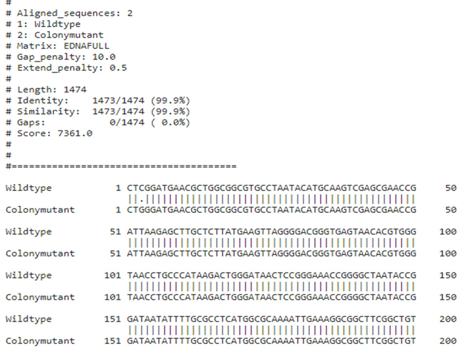
B. pseudomycoides produces rhizoidal growth on solid medium (Fig. 2a), which brings a challenge for researchers during the performance of antibacterial assays with this bacterium. In order to work on antibacterial aspects of B. pseudomycoides conveniently, it was very necessary to demolish the barrier of rhizoidal growth, therefore a colony mutant of B. pseudomycoides SB138 having no more rhizoidal growth morphology was obtained (Fig. 2b). The colony mutant strain was named as B. pseudomycoides SB138m. The 16S rRNA gene sequencing of B. pseudomycoides SB138m revealed 99% similarity between the sequences of 16S rRNA genes of wild type and the mutant strains (Fig. 3) confirming that the rhizoidal morphology of B. pseudomycoides SB138 is lost due to spontaneous mutation as reported previously .
Antibacterial activities of B. pseudomycoides strain SB138m
B. pseudomycoides strain SB138 showed antagonism against an indicator strain, S. aureus ATCC 6538 (Fig. 4a). The spectrum of the inhibitory effects of the strain was found against a range of the Bacillus spp isolated from a diverse environment and P. mirabilis isolated from a clinical sample (Fig. 4b). However, clinical isolates of E. coli, Pseudomonas spp., Klebsiella spp. were not sensitive to the B. pseudomycoides strain SB138m (Table I).
Stability of antibacterial activity of B. pseudomycoides strain SB138m
The antibacterial activity of B. pseudomycoides strain SB138m was found to be stable at high temperature and UV exposure (Fig. 5a, b). It was also observed that proteinase K enzyme demolished the antibacterial activity of B. pseudomycoides strain SB138m completely (Fig. 6). These finding suggested that B. pseudomycoides strain SB138m presumably produce bacteriocin like antibacterial substance. Furthermore, the plasmid profiling of B. pseudomycoides SB138m suggested that the strain does not contain any plasmid indicating that the gene for bacteriocin production may be genome encoded as supported by a recent study which has found the putative antimicrobial gene clusters on the genome of B. pseudomycoides .
DISCUSSION
Discovering natural antimicrobials from B. pseudomycoides remained ignored presumably due to its rhizoidal growth features on solid medium. However, a recent study reported that the B. pseudomycoides DSM 12442 produces lantibiotic namely Pseudomycoicidin, which was active against a wide range of Gram-positive bacteria only . Since Bacillus species are known to produce antibacterial substances with varied properties concurrently, it is not surprising that genes potentially involved in the synthesis of antimicrobial substances other than Pseudomycoicidin were found on the genome of B. pseudomycoides . Consequently, B. pseudomycoides BS6, an isolate of an edible oil contaminated soil was found producing biosurfactants which were lipopeptides in nature .
In the present study, B. pseudomycoides strain SB138 was found of holding strong antagonism against S. aureus and Bacillus species. Notably, inhibition of B. subtilis isolated from spoiled pulp of tomato indicated the biopreservative potential of the strain. Furthermore, the strain inhibited the growth of quinolone resistant clinical isolate of P. mirabilis. Our findings are supported by another study which identified putative antimicrobial gene clusters for 4 RPs (including a type II lantipeptide) and 3 NRPs on the genome of B. pseudomycoides .
The antibacterial activity of B. pseudomycoides SB138 was fully retained after 15 min at 75°C and it was completely destroyed by proteinase K treatment, indicating its proteinaceous nature. These finding are in contrast of previous reports which mentioned that the most of antibiotics are sensitive to the activity of proteases . However, our findings are in agreement of a recent study reporting that Pseudomycoicidin lantibiotic with four thioether rings and a disulfide bond is resistant to the activity of proteases .
CONCLUSION
The results of the present study show the antibacterial potential of B. pseudomycoides SB138 and have indicated possible use of B. pseudomycoides strain SB138m in future strategies for the development of alternative therapeutic agent or bio preservative.
ACKNOWLEDGEMENT
Authors are thankful to the Institute of Microbiology, University of Sindh for the provision of research facilities.
Funding
Higher Education Commission (HEC), Pakistan Indigenous Scholarship for PhD funded this study.
Ethical statement
This article does not contain any studies with human or animal subjects.
Statement of conflict of interest
The authors have declared no conflict of interest.
Figures
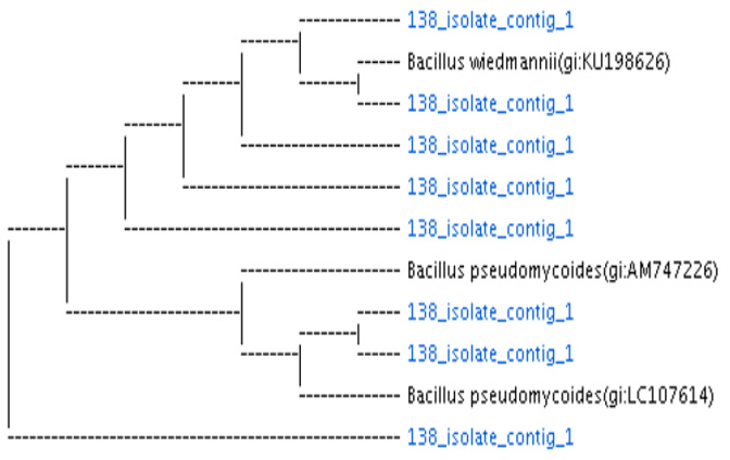
16S rRNA gene-based tree showing the phylogenetic relationship of the isolate SB 138 with type strain of B. pseudomycoides.
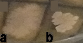
Colony characteristics of B. pseudomycoides SB138 a) the wild type B. pseudomycoides SB 138, and b) its colony mutant B. pseudomycoides SB138m.
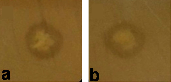
In vitro antibacterial activity of B. pseudomycoides strain SB138 against (a) indicator strain, S. aureus ATCC 6538 and (b) P. mirabilis 4S determined by stab- overlay methods.
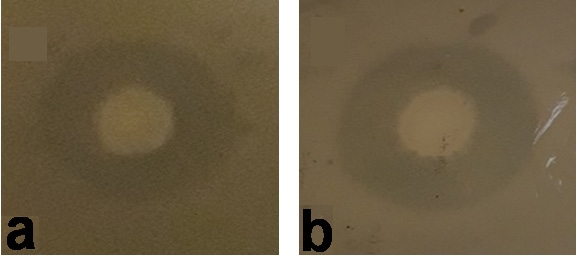
Effect of (a) heat and (b) UV on antibacterial activity of B. pseudomycoides strain SB138m determined by spot-overlay method.
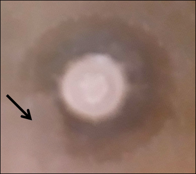
Determination of the effects of Proteinase K on antibacterial activity of B. pseudomycoides strain SB138m.
References
- Afreen, U., Bano, S., Tunio, S.A., Sharif, M. and MirJatt, A.N., 2020. Evaluation of antibacterial activity of zinc oxide nanoparticles and acrylamide composite against multidrug-resistant pathogenic bacteria. Pak. J. Anal. environ. Chem., 21: 125-131. http://dx.doi.org/10.21743/pjaec/2020.06.15[CrossRef]
- Bano, S., Tunio, S.A., Bugti, H., Khan, S. and Jatt, A.N., 2018. Exploring the rhizospheric bacterial communities of Mangifera indica. Proc. Pak. Acad. Sci., 55: 65-70.
- Bano, S., Vankemmelbeke, M., Penfold, C.N. and James, R., 2014. Detection of induced synthesis of colicin E9 using ColE9p: gfpmut2 based reporter system. World J. Microbiol. Biotechnol., 30: 2091-2099. https://doi.org/10.1007/s11274-014-1635-y[CrossRef]
- Barbour, A., Philip, K. and Muniandy, S., 2013. Enhanced production, purification, characterization and mechanism of action of salivaricin 9 lantibiotic produced by Streptococcus salivarius NU10. PLoS One, 8: e77751. https://doi.org/10.1371/journal.pone.0077751[CrossRef]
- Basi-Chipalu, S., Dischinger, J., Josten, M., Szekat, C., Zweynert, A., Sahl, H.-G. and Bierbaum, G., 2015. Pseudomycoicidin, a class II lantibiotic from Bacillus pseudomycoides. Appl. environ. Microbiol., 81: 3419-3429. https://doi.org/10.1128/AEM.00299-15[CrossRef]
- Claus, D., 1992. A standardized gram staining procedure. World J. Microbiol. Biotechnol., 8: 451-452. https://doi.org/10.1007/BF01198764[CrossRef]
- Di Franco, C., Beccari, E., Santini, T., Pisaneschi, G. and Tecce, G., 2002. Colony shape as a genetic trait in the pattern-forming Bacillus mycoides. BMC Microbiol., 2: 33. https://doi.org/10.1186/1471-2180-2-33[CrossRef]
- Díez-Méndez, A., Rivas, R., Mateos, P.F., Martínez-Molina, E., Santín, P.J., Sánchez-Rodríguez, J.A. and Velázquez, E., 2017. Bacillus terrae sp. nov. isolated from Cistus ladanifer rhizosphere soil. Int. J. Syst. Evol. Microbiol., 67: 1478-1481. https://doi.org/10.1099/ijsem.0.001742[CrossRef]
- Huang, T., Zhang, X., Pan, J., Su, X., Jin, X. and Guan, X., 2016. Purification and characterization of a novel cold shock protein-like bacteriocin synthesized by Bacillus thuringiensis. Sci. Rep., 6: 35560. https://doi.org/10.1038/srep35560[CrossRef]
- Li, J., Deng, M., Wang, Y. and Chen, W., 2016. Production and characteristics of biosurfactant produced by Bacillus pseudomycoides BS6 utilizing soybean oil waste. Int. Biodeterior. Biodegrad., 112: 72-79. https://doi.org/10.1016/j.ibiod.2016.05.002[CrossRef]
- Liu, B., Liu, G.H., Sengonca, C., Schumann, P., Che, J.M., Zhu, Y.J. and Wang, J.P., 2015. Bacillus wuyishanensis sp. nov., isolated from rhizosphere soil of a medical plant, Prunella vulgaris. Int. J. Syst. Evol. Microbiol., 65: 2030-2035. https://doi.org/10.1099/ijs.0.000215[CrossRef]
- Nakamura, L., 1998. Bacillus pseudomycoides sp. nov. Int. J. Syst. Evol. Microbiol., 48: 1031-1035. https://doi.org/10.1099/00207713-48-3-1031[CrossRef]
- Nicholson, W., 2002. Roles of Bacillus endospores in the environment. Cell Mol. Life Sci., 59: 410-416. https://doi.org/10.1007/s00018-002-8433-7[CrossRef]
- Parret, A.H., Schoofs, G., Proost, P. and De Mot, R., 2003. Plant lectin-like bacteriocin from a rhizosphere-colonizing Pseudomonas isolate. J. Bact., 185: 897-908. https://doi.org/10.1128/JB.185.3.897-908.2003[CrossRef]
- Pramanik, P., Goswami, A., Ghosh, S. and Kalita, C., 2019. An indigenous strain of potassium‐solubilizing bacteria Bacillus pseudomycoides enhanced potassium uptake in tea plants by increasing potassium availability in the mica waste-treated soil of North-east India. J. appl. Microbiol., 126: 215-222. https://doi.org/10.1111/jam.14130[CrossRef]
- Schaeffer, A.B. and Fulton, M.D., 1933. A simplified method of staining endospores. Science, 77: 194-194. https://doi.org/10.1126/science.77.1990.194[CrossRef]
- Solmaz, K.B., Ozcan, Y., Mercan Dogan, N., Bozkaya, O. and Ide, S., 2018. Characterization and production of extracellular polysaccharides (EPS) by Bacillus pseudomycoides U10. Environments, 5: 63. https://doi.org/10.3390/environments5060063[CrossRef]
- Sumi, C.D., Yang, B.W., Yeo, I.C. and Hahm, Y.T., 2014. Antimicrobial peptides of the genus Bacillus: A new era for antibiotics. Can. J. Microbiol., 61: 93-103. https://doi.org/10.1139/cjm-2014-0613[CrossRef]
- Sun, L., Lu, Z., Bie, X., Lu, F. and Yang, S., 2006. Isolation and characterization of a co-producer of fengycins and surfactins, endophytic Bacillus amyloliquefaciens ES-2, from Scutellaria baicalensis georgi. World J. Microbiol. Biotechnol., 22: 1259-1266. https://doi.org/10.1007/s11274-006-9170-0[CrossRef]
- Zhao, X. and Kuipers, O.P., 2016. Identification and classification of known and putative antimicrobial compounds produced by a wide variety of Bacillales species. BMC Genom., 17: 882. https://doi.org/10.1186/s12864-016-3224-y[CrossRef]
Services
Article usage statistics
Loading…