
Neuroinflammation is the underlying cause of a number of neurodegenerative disorders. While the current understanding of its molecular mechanisms is continually increasing, the identification and consequently the roles of transcription factors (TFs) in the respective molecular events under neuroinflammatory conditions in distinct brain regions is still lacking. To address the said gap in knowledge, first we have identified the TFs specific to IL-1β a key player in mediating inflammatory process using online databases. Next, we identified the interacting partners of the significantly differentially expressed TFs out of the selected ones, and the mRNA expression changes were studied for both the differentially expressed TF along with its interacting partners. Finally, we explored the expression of these genes from some online available RNA-seq datasets for brain tissue, neurons, microglia and astrocytes. The results showed significant upregulation of Etv5 TF in male mouse brain hippocampus along with a significant downregulation of three of its predicted interacting partners, i.e., Cop1, Det1 and Bcl6b. Besides presenting new targets for neuroinflammation studies, the discrepancies of expression between different brain regions as well as among the genders suggest that more data with respect to intricate brain region resolution along with single cell data could reflect subtle changes in brain microenvironments and individual cells, which can lead to a more precise pathological understanding of the neuroinflammatory process.
Research Article · Volume 55, Issue 5, 27 July 2023 · Open Access
ETV5 is a Putative Differentially Expressed Transcription Factor of Interleukin 1β Under Neuroinflammatory Conditions in Murine Cerebellum
·
Affiliations & Notes ⌄
- 1School of Biological Sciences, University of the Punjab, Quaid-e-Azam Campus, Lahore 54590, Pakistan * Corresponding author: arshakoori.sbs@pu.edu.pk, arshaksbs@yahoo.com
Article Info ⌄
- Journal:
- Pakistan Journal of Zoology
- Published:
- 27 July 2023

From the article
- In the male cerebellum, the only genes significantly differentially expressed were Bcl6b and Dusp6 (downregulation), and in female hippocampi and cerebellum only Bcl6b showed significant downregulation.
- We observed a downregulation of Cop1 gene in hippocampi of both the genders and in male cerebellum although significant results were those of male hippocampus only.
- In the current study, we have presented the expression profiles IL-1B associated transcription factor Etv5 along with some of its interacting partners as molecular targets of neuroinflammatory process.
- Moreover, the identification of significantly differentially expressed genes present interesting target genes for further studies on neuroinflammation.
Selected from the article's own text.
Abstract
INTRODUCTION
The inflammation of brain and spinal cord, together constituting our central nervous system (CNS), is known as neuroinflammation. Being implicated in a variety of somatic and neurological illnesses, the most common cues of its initiation known till date are toxicity to various metabolites, traumatic brain and spinal cord injuries, viral and/or bacterial toxicity, and autoimmune mediated neuroinflammation ; . Apart from being central to the mutual pathology of a number of acute and chronic neurological disorders, neuroinflammation has also been found to be a major cause and aggravating factor for the development of neurodegenerative diseases. Moreover, its role in the early development of some chronic conditions, e.g., dementia, has also been reported ; ; .
Being pleiotropic mediators and effectors of cells’ and tissues’ inflammatory processes and of various autoimmune disorders, interleukin-1 α (IL-1α) and interleukin-1 β (IL-1β) are archetypal proinflammatory cytokines. Of these, IL-1β also performs crucial homeostatic functions in a healthy organism. These include the regulation of body temperature, sleep, and feeding , 2004). Consequently, its overproduction is associated with various pathophysiological anomalies such as inflammatory bowel disease, rheumatoid arthritis, osteoarthritis, vascular diseases, neuropathy, Alzheimer’s disease and multiple sclerosis .
Some of the common documented events during the process of neuroinflammation include: Increased activation of brain’s resident immune cells (glial cells), altered permeability of blood brain barrier (BBB), leukocyte invasion, and increased concentration of the pro-inflammatory cytokines (Rejo et al., 2008; Guzyman-Martinez, 2019). IL-1 being upregulated in several of the neurodegenerative disorders such as Parkinson’s disease (PD) ; , Alzheimer’s disease , 1994), multiple sclerosis and amyotrophic lateral sclerosis , also makes IL-1β a key player in the process of neuroinflammation too, along with other inflammatory processes of the body. The pathways activated by overproduction of IL-1β include NFkB-dependent transcription of some other pro-inflammatory cytokines such as TNF-α, interferons and IL-6; activation of neutrophil recruiting chemokines; and stimulation of synaptophysin and Tau proteins’ production in neurons ; ; .
The regulatory elements of IL-1B include a TATA box within the promoter, a CRE (cAMP responsive element), an AP-1 (activator protein 1) promoter site, an SP-1/PU.1 (the ETS family of transcription factor) binding site, and an NF-κB (nuclear factor kappa B) binding site ; ; ; . The induction of IL-1B gene expression can be brought about via various stimuli including both microbial and non-microbial types. The initial translation product is a propeptide (31 kD), the pro-IL-1β, which is cleaved to the mature and biologically active form of IL-1β by the action of cysteine protease caspase-1 ; ; .
The presence of both neuroinflammation and tissue damage in post mortem brain tissues makes it difficult to determine whether upregulation of IL-1B is caused by, or a consequence of neuroinflammation . This also urged the need to explore IL-1B along with its potential mediators to get a better understanding of its precise role in the process of neuroinflammation. This study was therefore aimed at identification of the transcription factors (TF) specific to IL-1B promoter/enhancer region and then study their differential expression under neuroinflammatory conditions using in vivo LPS mouse models.
MATERIALS AND METHODS
Identification and Screening of TFs for the promoter/enhancer region of IL-1B
Various online available databases were utilized for the identification of the promoter region of Il-1B gene. These include GeneCards (https://www.genecards.org/), the eukaryotic promoter database (https://epd.epfl.ch//index.php), and super-enhancer database (http://www.licpathway.net/sedb/). These databases also identified the TFs having the binding sites for the promoter of Il-1B and among them, only those TFs were preferred having the highest scores from the online databases. The resulting TFs were further screened on the basis of their tissue specific high or low expression profiles so as to select only those relevant to the tissue being used in the study i.e., brain.
Neuroinflammation murine models
Male and female BALB/c mice, approximately 6 weeks of age, were housed in groups of 4 in cages containing rodent bedding. Food and water were available ad libitum and temperature was maintained at 25°C. All experiments were carried out on mice divided into two groups, experimental drug group and vehicle control group receiving physiological saline injections only.
BALB/c mice 6-8 weeks of age were administered with a single dose of 5 mg/kg of lipopolysaccharide (LPS) solution (Sigma Cat # L2880-25MG) dissolved in 0.9% saline solution via intraperitoneal route. The controls received injections of saline solution only. The sacrifice of animals was carried out after 24 h of LPS treatment.
Animal killing and dissections
The mice were sacrificed by cervical dislocation. Briefly, the backside of a knife was placed on the dorsal side of the neck at the junction between skull and the vertebrae. Then a little pressure was applied through the knife while pulling the animal from its tail so as to dislocate the head from the rest of the body in a single jerk to minimize animal discomfort. The bioethical clearance certificate was obtained from the Bioethics Committee of the School of Biological Sciences, University of the Punjab.
The brains were further dissected to get two regions i.e., hippocampus and cerebellum. These two regions are shown in the Supplementary Figure S1 which represents the regional map of murine brain. The protocol for dissection of these regions was followed from Li (2011). The cerebellum is located on the dorsal side of the brain and on the lower side as can be seen in the Figure 1. First, the portion of the brain containing the cerebellum was separated from the cerebral cortices by making a vertical cut. Then the tissue underneath the cerebellum consisting of midbrain, pons and medulla was separated from it very carefully with the help of fine curved forceps.
The dissection of hippocampi is a bit difficult than the cerebellum as they are located on the inside of the cerebral cortices and are covered with tissue. Therefore, the dissection procedure of hippocampus is explained a little more as in the Figure 1B, C since the crescent shaped hippocampi are placed on the dorsal side of the brain covered with a fatty tissue the removal of which makes the hippocampus visible.
qRT PCR of IL-1β TFs
For total cell RNA extraction, the tissues were homogenized in TRIzol reagent via trituration and total RNA was extracted according to the manufacturer’s protocol. The RNA was used for complementary cDNA synthesis after DNase treatment using Revert Aid First Strand cDNA synthesis kit (Thermofisher Scientific, Cat # K1622).
Extracted RNA was quantified using pico200 spectrophotometer at A260/280. For DNase treatment of RNA sample, Invitrogen kit (Cat # 18068015) was used for this procedure as per instructions of the kit. The cDNA was synthesized using Maxima H Minus First Strand cDNA Synthesis Kit (Cat # K1681).
The quantitative expression analysis of genes was done by real time PCR analysis. Maxima SYBR Green/ROX qPCR master mix (2X; Thermofisher Scientific, Cat # K0222), cDNA, and 5 µM forward and reverse primers’ mix were used for RT-PCR (the sequences of the primers are provided in Table I. Two-step thermal cycle protocol with optimized annealing temperatures (Table I) of each primer pair was used for amplification and quantification of amplicons on PikoReal Real-Time PCR system (Thermofischer Scientific). Melting curve data with every 0.2°C increment was taken to determine the uniformity of PCR product. HMBS was used as a reference gene in the study. Significance analysis for fold differences of biological replicates was done using t-test for means.
Table I. Sequences of primers used for RT-PCR along with their optimized annealing temperatures.
| Gene name | Primer | Sequence (5’-3’) | Optimized primer annealing temperature | Product length (bp) |
| HMBS | Forward | CACTGGCTTCCCGTTCACTA | 58-61 | 160 |
| Reverse | GCCAGGGGAGGTGAGAAAC | |||
| ETV5 | Forward | CAAGGTCAAGAAAACCCGCC | 60 | 70 |
| Reverse | CGCTGAGGGACCTTAATCGG | |||
| KLF16 | Forward | GTGGCCCTAATCAACACTCCT | 60 | 186 |
| Reverse | AGATGGTGCAGCTACCAAGAC | |||
| COP1 | Forward | ATGCAGCCATGAATGTGTGT | 59 | 233 |
| Reverse | TGTCCACTTGCTGTTTGCTC | |||
| DET1 | Forward | GTCTTGTACCCGTGGTGCTT | 60 | 172 |
| Reverse | AAAACCAGGACGCCTACCTT | |||
| GDNF | Forward | GCCAGCCAGAGAGAAAACAC | 59 | 234 |
| Reverse | AAGGCATTAGCTTTGGAGCA | |||
| BCL6B | Forward | ACAGCTGGGTTTGAATGTCC | 60 | 246 |
| Reverse | AAGGCAGCCCTCTTTTCTTC | |||
| DUSP6 | Forward | GAATAATCCGGCGAGAAACA | 60 | 168 |
| Reverse | AATGAAGGTGCCCAGTTTTG |
Cross-examination of differentially expressed genes via expression atlas
The genes, for which mRNA expression was significantly differential, were further cross examined with respect to their protein expression at basal level using Expression Atlas database . This database is maintained by the European Bioinformatics Institute that is a hub of information for RNA and protein expression for different studies, organisms, and experimental conditions. Since mRNA of Etv5 was being differentially expressed in the cerebellum of both the genders, its basal level protein was checked using this database.
Functional protein-protein association networks
For studying the functional protein association networks of Etv5, STRINGS (Search Tool for Retrieval of Interacting Genes and protein, Version 11.0, https://string-db.org/), BioGRID (https://thebiogrid.org/), Database of Interacting Proteins (DIP, https://dip.doe-mbi.ucla.edu/dip/Main.cgi) and IntAct (https://www.ebi.ac.uk/intact/home) databases were used to find not only the established biological interacting partners from experiments and curated databases but also the predicted interactions from gene neighborhood, fusions, co-occurrence, co-expression, homology and text-mining techniques.
The re-analysis of RNA-seq datasets taken from Gene Expression Omnibus (GEO) database was done . In short, fastq files of LPS versus control treated mouse brain (accession number GSE153369), neurons, astrocytes and microglia (accession number GSE75246) were analysed using different packages from R Bioconductor such as limma, edgeR, gplots, org.Hs.eg.db, RcolorBrewer and Glimma ; ; ; . and the average expression along their p values were noted for significance analyses.
RESULTS
Putative TFs of IL-1B
The online search resulted in the identification of a number of TFs interacting with IL-1B gene regulatory elements. Further sorting of these TFs on the basis of their differential expression in different tissues resulted in the identification of Klf16 and Etv5 exhibiting the highest expression in brain as compared to other tissues (Supplementary Table SI).
mRNA expression analysis of TFs under neuroinflammatory conditions
mRNA expression analysis revealed that Klf16 did not show any significant difference in expression in both the brain regions of male and female mice (Fig. 2). However, in case of Etv5, significant (p < 0.005) upregulation of 1.25 folds was observed only in male hippocampus (Fig. 3A). Quite interestingly, its expression was found to be downregulated in male cerebellum (Fig. 3B) as well as in female hippocampus (Fig. 3C) and cerebellum (Fig. 3D), although these fold differences were found to be non-significant.
After getting significant expression difference in Etv5 transcript, we further searched for the interacting protein partners of Etv5 of both human and mouse origin of which the mRNA expression of Cop1, GDNF, Det1, Bcl6b and Dusp6 was further analyzed using RT-PCR. The protein partners for mouse and human Etv5 protein from STRING database are given in Figure 4 in the form of node diagrams. Supplementary Table SII shows the interacting protein partners of Etv5 from all the databases for mouse and human, respectively.
In line with the significant upregulation of Etv5 gene, three of its interacting partners, i.e., Cop1, Det1 and Bcl6b showed significant downregulation, while non-significant upregulation was observed for GDNF and Dusp6 in male hippocampus. In male cerebellum, significant downregulation of Bcl6b and Dusp6 was seen. In female mice, only Bcl6b was found to be showing significant downregulation in both the hippocampus and cerebellum.
When the protein expression of Etv5 was checked as a validation to see its baseline conditions under normal conditions via Human Protein Atlas project , it was found that in the cerebellum of both male and female subjects, its expression was moderate (Fig. 5).
Expression of ETV5 from RNA seq data sets
When the expression of Etv5 and its selected interacting partners was checked from RNA-seq data sets for brain tissue, neurons, microglia and astrocytes, only Det1 significant difference in average expression with a value of -1.729 (p < 0.005) (Table II).
Table II. Average expression of Etv5 from LPS treated neuroinflammation RNA-seq datasets. * (p < 0.05), ** (p < 0.005), NS (p > 0.05 = non-significant).
| Genes | Average expression | |||
| Brain | Neurons | Microglia | Astrocytes | |
| Etv5 | -0.424ns | 6.352ns | N/A | N/A |
| Cop1 | N/A | 0.73ns | N/A | -1.310ns |
| GDNF | -0.913ns | -0.619ns | N/A | N/A |
| Det1 | N/A | N/A | N/A | -1.729** |
| Bcl6b | 0.524NS | 0.351NS | N/A | N/A |
| Dusp6 | -0.160ns | 0.624 | N/A | N/A |
DISCUSSION
This study was focused on the identification of IL-1B specific transcription factors of brain as IL-1β serves a pivotal role in the mediation of inflammatory responses. Consistent with the significant differential expression of Etv5 mRNA only in male hippocampus, most of its interacting partners (Cop1, Det1 and Bcl6b) also showed significant differential expression (downregulation) only in male hippocampus. An interesting difference to note was that while Etv5 was being upregulated, the interacting partners were being downregulated in male hippocampi. In the male cerebellum, the only genes significantly differentially expressed were Bcl6b and Dusp6 (downregulation), and in female hippocampi and cerebellum only Bcl6b showed significant downregulation. Further exploration of the genes under study from the RNA-seq data sets of LPS treated brain tissue as well as from neurons, microglia and astrocytes revealed significant downregulation of Det1 in astrocytes (Table II). Among all the genes, Bcl6b was found to be common among the two brain regions in both the genders, all exhibiting downregulation of it. Also, the downregulation of Det1 in male hippocampus was corroborated with its down regulation in astrocytic RNA-seq data.
Being a member of the PEA3 family of TFs, Etv5 binds to a conserved DNA motif having sequence GGAA/T (Oh et al., 2010). Some of the important known roles of Etv5 include: Coordination of limb development; regulation of epithelial-to-mesenchymal transition in some cancer types; control of gene expression in spermatogonial stem cells; inhibition of sonic hedgehog gene expression; and positive regulation of some miRNAs important in spermatogonial stem cells renewal ; 2010; ; . Besides these, although still poorly understood, the roles of Etv5 in immune responses include an increased interferon gamma (IFN-γ) production from TH1 cells ; , increased IL-17 production in TH17 cells (thus playing a role in allergic inflammation development) , and suppression of the activation of adipose tissue resident macrophages . Since activation of these macrophages contributes in the development of chronic inflammation, and Etv5 being identified as a major suppressor of the process, the upregulation of Etv5 transcript in male hippocampus in the current study presents an interesting target for future studies of Etv5 on brain resident macrophages i.e., microglia. Corroborated with the previous studies indicating a regulatory role of Etv5 on transcriptional activation of IL-10, IL-17 and IL-9 expression ; , 2017), the precise mechanism of its role in macrophage activation regulation is yet to be unveiled.
We observed a downregulation of Cop1 gene in hippocampi of both the genders and in male cerebellum although significant results were those of male hippocampus only. Previous studies have found a neuroprotective role of Cop1 in neurodegenerative disorders , 2008). Corroborating the neuroprotective role, recent knockout studies of Cop1 have shown an increased pro-inflammatory response in brain resident macrophages, i.e., microglial cells (Ndojaa et al., 2020). Since Det1 is a partner of Cop1 E3 ubiquitin ligase complex, its downregulation is in concert with Cop1 downregulation (which has been found to be almost double in magnitude to that of Det1 (Fig. 3B). Previous roles of Det1 have been found in the mediation of cellular differentiation and proliferation in multicellular organisms , however, no known roles could be found of Det1 in the process of neuroinflammation. Similarly, although previous roles of Bcl6b and Dusp6 have been found in the regulation of inflammatory responses and endothelial inflammation , respectively, their precise roles in the process of neuroinflammation are yet to be studied. These findings present important genes for further detailed studies with respect to the modulation of neuroinflammatory process in hippocampus tissue. Also, the brain region along with gender differences are also evident from the current study highlighting the need to focus on individual brain regions of each gender.
CONCLUSIONS AND RECOMMENDATIONS
In the current study, we have presented the expression profiles IL-1B associated transcription factor Etv5 along with some of its interacting partners as molecular targets of neuroinflammatory process. Also, the current study presents the dissection of these TFs with respect to not only the two important brain regions hippocampus and cerebellum due to their functional implications in memory and motor function, respectively, but also with respect to gender differences. Moreover, the identification of significantly differentially expressed genes present interesting target genes for further studies on neuroinflammation. Furthermore, future research on the extensive molecular characterization of all brain regions as well as individual cell types could provide a complete landscape not only for the recognition of each of these brain regions and cells in the process of neuroinflammation, but also would guide the development of more targeted therapeutic interventions.
ACKNOWLEDGEMENT
Funds for this study were provided by the University of the Punjab.
DECLARATIONS
Ethical statement
The study was approved by the Institutional Review Board (IRB) and Institutional Animal Care and Use Committee (IACUC) of University of the Punjab
Data availability statement
All the data has been included in this article.
Supplementary material
There is supplementary material associated with this article. Access the material online at: https://dx.doi.org/10.17582/journal.pjz/20220612100622
Statement of conflict of interest
The authors have declared no conflict of interest.
Figures
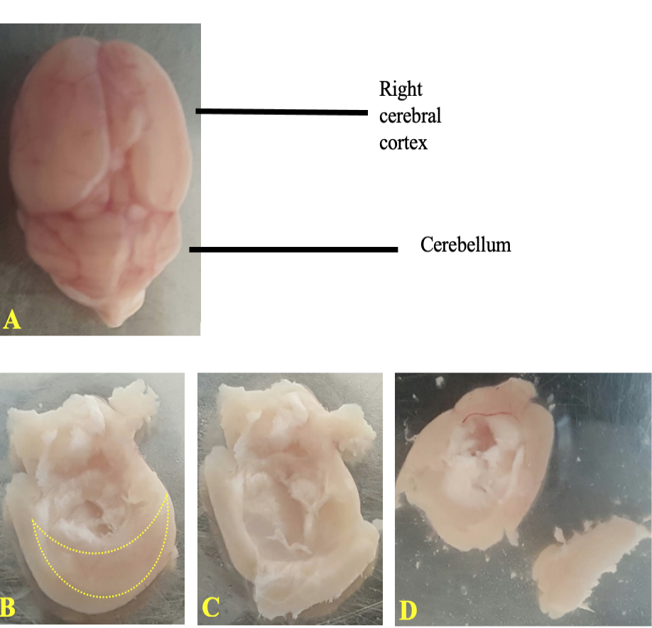
Dissection of murine brain to separate out hippocampus. A, Dorsal view of the mouse brain. On the lower side is the cerebellum and on the upper side are the cerebral cortices. Each cerebral cortex contains hippocampus embedded in its ventral inner side. B, right cerebral cortex is placed on its dorsal side with the crescent shaped hippocampus clearly visible. C, hippocampus flipped inside out; and D, hippocampus separated from the rest of the tissue.
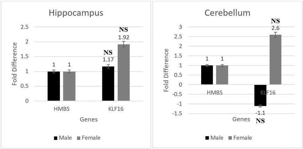
mRNA expression of the transcription factor Klf16 represented in terms of fold difference up/ down regulation in hippocampus and cerebellum. NS - (p > 0.05 = non-significant)
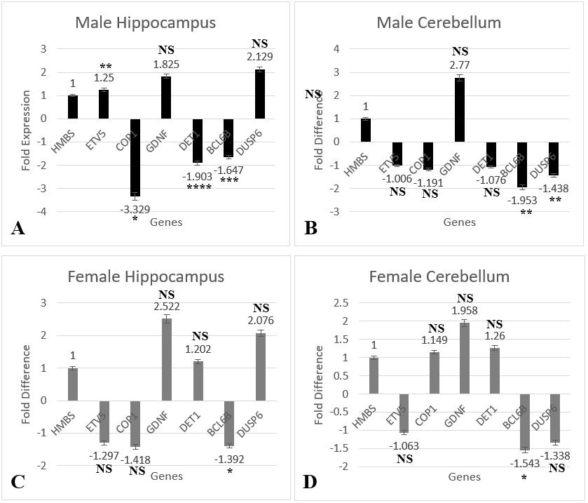
mRNA expression of the transcription factor Etv5 along with its interacting partners Cop1, GDNF, Det1, Bcl6b, and Dusp6, represented in terms of fold difference up/down regulation. A, male hippocampus; B, male cerebellum; C, female hippocampus; and D, female cerebellum. * (p < 0.05), ** (p < 0.005), *** (p < 0.0005), **** (p < 0.00005), and NS (p > 0.05 = non-significant).
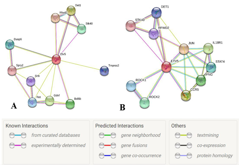
Node graph of the interacting partners of Etv5. A, Mus musculus and B, Homo sapiens, with proteins represented by network nodes. The node in red color is the query protein while other nodes are the proteins having first shell of interaction with the query protein (STRINGS, ETV5 Homo sapiens).
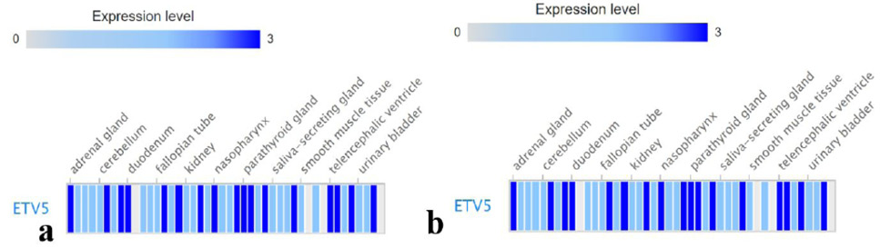
Protein expression levels of Etv5 in different tissues of human subjects. A, male; and B, female.
References
- Acuner-Ozbabacan, S.E., Gursoy, A., Nussinov, R., and Keskin, O., 2014. The structural pathway of interleukin 1 (IL-1) initiated signaling reveals mechanisms of oncogenic mutations and SNPs in inflammation and cancer. PLoS Computat. Biol., 10: e1003470. https://doi.org/10.1371/journal.pcbi.1003470[CrossRef]
- Braddock, M., and Quinn, A., 2004. Targeting IL-1 in inflammatory disease: New opportunities for therapeutic intervention. Nat. Rev. Drug Discov., 3: 330-340. https://doi.org/10.1038/nrd1342[CrossRef]
- Bunting, K.L., and Melnick, A.M., 2013. New effector functions and regulatory mechanisms of BCL6 in normal and malignant lymphocytes. Curr. Opin. Immunol., 25: 339-346. https://doi.org/10.7554/eLife.37892[CrossRef]
- Cacabelos, R., Alvarez, X.A., Fernandez-Novoa, L., Franco, A., Mangues, R., Pellicer, A., and NIshimura, T., 1994. Brain interleukin-1 beta in Alzheimer’s disease and vascular dementia. Meth. Find. exp. clin. Pharmacol., 16: 141-151.
- Cacabelos, R., Franco-Maside, A., and Alvarez, X.A., 1991. Interleukin-1 in Alzheimer’s disease and multi-infarct dementia: neuropsychological correlations. Meth. Find. exp. clin. Pharmacol., 13: 703-708.
- Dinarello, C.A., 1996. Biologic basis for interleukin-1 in disease. Blood, 87: 2095-2147. https://doi.org/10.1111/jnc.13607[CrossRef]
- Dinarello, C.A., 2004. Therapeutic strategies to reduce IL-1 activity in treating local and systemic inflammation. Curr. Opin. Pharmacol., 4: 378-385. https://doi.org/10.1016/j.coph.2004.03.010[CrossRef]
- DiSabato, D.J., Quan, N., and Godbout, J.P., 2016. Neuroinflammation: The devil is in the details. J. Neurochem., 139(S2): 136-153. https://doi.org/10.1111/jnc.13607[CrossRef]
- Eder, C., 2009. Mechanisms of interleukin-1beta release. Immunobiology, 214: 543-553. https://doi.org/10.1016/j.imbio.2008.11.007[CrossRef]
- Expression Atlas, 2020. Human immunochemistry data on 83 different normal cell types from 44 tissue types from the human protein atlas project. [cited 2020 21 Aug]; Available from https://www.ebi.ac.uk/gxa/experiments/E-PROT-3/Results?geneQuery=%5B%7B%22value%22%3A%22ensg00000244405%22%7D%5D https://www.surveymonkey.co.uk/r/EAsurvey22
- Fagerberg, L., Hallstroem, B.M., Oksvold, P., Kampf, C., Djureinovic, D., Odeberg, J., Habuka, M., Tahmasebpoor, S., Danielsson, A., Edlund, K., Asplund, A., Sjoestedt, E., Lundgerg, E., Szigyarto, C.A.K., Skogs, M., Takanen, J.O., Berling, H., Tegel, H., Mulder, J., Nilsson, P., Schwenk, J.M., Lindskog, C., Danielsson,F., Mardinoglu, A., Sivertsson, A., von Feilitzen, K., Forsberg, M., Zwahlen, M., Olsson, I., Navani, S., Huss, M. Nielson, J., Ponten, F. and Uhlen, M., 2014. Analysis of the human tissue-specific expression by genome-wide integration of transcriptomics and antibody-based proteomics. Mol. Cell. Proteomics, 13: 397-406. https://doi.org/10.1074/mcp.M113.035600[CrossRef]
- Fan, Z., Soderr, S., Oehler, S., Fundel, K. and Aigner, T., 2007. Activation of interleukin-1 signaling cascades in normal and osteoarthritic articular cartilage. Am. J. Pathol., 171: 938-946. https://doi.org/10.2353/ajpath.2007.061083[CrossRef]
- Feinstein, D.L., Kalinin, S. and Braun, D., 2016. Causes, consequences, and cures for neuroinflammation mediated via the locus coeruleus: Noradrenergic signaling system. J. Neurochem., 139(S2): 154-178. https://doi.org/10.1111/jnc.13447[CrossRef]
- Gorantla, S., Liu, J., Sneller, H., Dou, H., Holguin, A., Smith, L., Ikezu, T., Volsky, D.J., Poluektova, L., and Gendelman, H.E., 2007. Copolymer-1 induces adaptive immune anti-inflammatory glial and neuroprotective responses in a murine model. https://doi.org/10.4049/jimmunol.179.7.4345[CrossRef]
- Gorantla, S., Liu, J., Wang, T., Holguin, A., Sneller, H.M., Dou, H., Kipnis, J., Poluektova, L., Gendelman, H.E., 2008. Modulation of innate immunity by copolymer-1 leads to neuroprotection in murine HIV-1 encephalitis. Glia, 56: 223-232. https://doi.org/10.1002/glia.20607[CrossRef]
- Guzman-Martinez, L., Maccioni, R.B., Andrade, V., Navarrete, L.P., Pastor, M.G. and Ramos-Escobar, N., 2019. Neuroinflammation as a common feature of neurodegenerative disorders. Front. Pharmacol., 10: 1008. https://doi.org/10.3389/fphar.2019.01008[CrossRef]
- Herzog, C., Kaushal, G.P. and Haun, R.S., 2005. Generation of biologically active interleukin-1beta by meprin B. Cytokine, 31: 394-403. https://doi.org/10.1016/j.cyto.2005.06.012[CrossRef]
- Hewett, S.J., Jackman, N.A. and Claycomb, R,J., 2012. Interleukin-1β in central nervous system injury and repair. Eur. J. Neurodegr. Dis., 1: 195-211.
- Hsu, S.F., Lee, Y-B., Lee, Y-C., Chung, A-L., Apaya, M.K., Shyur, L-F., Cheng, C-F., Ho, F-M., and Merng, T-C., 2018. Dual specificity phosphatase DUSP6 promotes endothelial inflammation through inducible expression of ICAM-1. FEBS J., 285: 1593-1610. https://doi.org/10.1111/febs.14425[CrossRef]
- Hu, R.D., Zhang, W., Li, L., Zuo, Z-Q., Ma, M., Ma, J-F., Yin, T-T., Gao, C.Y., Yang, S-H., Zhao, Z-B., Li, Z-J., Qiao, G-B., Lian, Z-X. and Qu, K., 2021. Chromatin accessibility analysis identifies the transcription factor ETV5 as a suppressor of adipose tissue macrophage activation in obesity. Cell Death Dis., 12: 1023. https://doi.org/10.1038/s41419-021-04308-0[CrossRef]
- Khan, A., Khalid, Z., and Shakoori, A.R., 2022. Identification of transcription factors differentially expressed under neuroinflammatory conditions: Analysis of rna-seq datasets combined with an unconventional exploratory approach. Crit. Rev. Eukary. Gene Expr., https://doi.org/10.1615/CritRevEukaryotGeneExpr.2022044272[CrossRef]
- Koh, B., Hufford, M.W., Pham, D., Olson, M.R., Wu, T., HJaberen, R., Sun, X. and Kaplan, M.H., 2016. The ETS family transcription factors Etv5 and PU.1 function in parallel to promote Th9 cell development. J. Immunol., 197: 2465-2472. https://doi.org/10.4049/jimmunol.1502383[CrossRef]
- Koh, B., Hufford, M.W., Sun, X., and Kaplan, M.H., 2017. Etv5 regulates IL-10 Production in Th cells. J. Immunol., 198: 2165-2171. https://doi.org/10.4049/jimmunol.1600801[CrossRef]
- Kwon, H.S. and Koh, S-H., 2020. Neuroinflammation in neurodegenerative disorders: The roles of microglia and astrocytes. Transl. Neurodegener., 9: 42. https://doi.org/10.1186/s40035-020-00221-2[CrossRef]
- Lahiri, T., Moore, P.E., Baraldo, S., Whitehead, T.R., Mckenna, M.D., Panettieri Jr., R.A. and Shore, S.A., 2002. Effect of IL-1beta on CRE-dependent gene expression in human airway smooth muscle cells. Am. J. Physiol. Lung Cell. mol. Physiol., 283: L1239-1246. https://doi.org/10.1152/ajplung.00231.2001[CrossRef]
- Leal, M.C., Casabona, J.C., Puntel, M. and Pitossi, F.J., 2013. Interleukin-1β and tumor necrosis factor-α: Reliable targets for protective therapies in Parkinson’s Disease? Front. Cell. Neurosci.,7:1-10. https://doi.org/10.3389/fncel.2013.00053[CrossRef]
- Li, K.W., 2011. Springer protocols: Neuroproteomics (Neuromethods, 57). Humana Press, Springer, New York.
- Lin, C.C. and Edelson, B.T., 2017. New Insights into the role of IL-1β in experimental autoimmune encephalomyelitis and multiple sclerosis. J. Immunol., 198: 4553-4560. https://doi.org/10.4049/jimmunol.1700263[CrossRef]
- Lyman, M., Lloyd, D.G., Ji, X., Vizcaychipi, M.P., and Ma, D., 2014. Neuroinflammation: The role and consequences. Neurosci. Res., 79: 1-12. https://doi.org/10.1016/j.neures.2013.10.004[CrossRef]
- McCarthy, D.J., Chen, Y. and Smyth, G.K., 2012. Differential expression analysis of multifactor RNA-Seq experiments with respect to biological variation. Nucl. Acid Res., 40: 4288-4297. https://doi.org/10.1093/nar/gks042[CrossRef]
- Meissner, F., Molawi, K., and Zychlinsky, A., 2010. Mutant superoxide dismutase 1-induced IL-1beta accelerates ALS pathogenesis. Proc. natl. Acad. Sci. USA, 107: 13046-13050. https://doi.org/10.1073/pnas.1002396107[CrossRef]
- Nassrallah, A., Rougee, M., Bourbousse, C., Drevensek., S., Fonseca, S., Iniesto, E., Ait Mohamed, O., Deton-Cabanillas, A-F., Zebulon, G., Ahmed, I., Stroebel, D., Masson, V., Lombard, B., Eeckhout, D., Gevaert, K., Loew, D., Genovesio, A., Breyton, C., De Jaeger, G. Bowler, C., Rubio, V., and Barneche, F., 2018. DET1-mediated degradation of a SAGA-like deubiquitination module controls H2Bub homeostasis. Elife, 7: e37892. https://doi.org/10.7554/eLife.37892[CrossRef]
- Ndoja, A., Reja, R., Lee, S-H., Wenbster, J.D., Ngu, H., Rose, C.M., Kirkpatrick, D.S., Modrusan, Z., Chen, Y-J. J., Dugger, D.L., Gandham, V., Xie, L., Newton, K. and Dixit, V.M., 2020. Ubiquitin ligase COP1 suppresses neuroinflammation by degrading C/EBPβ in microglia. Cell, 182: 1156-1169.e12. https://doi.org/10.1016/j.cell.2020.07.011[CrossRef]
- Niu, Z., Goodyear, S.M., Rao, S., Wu, X., Tobias, J.W., Avarbock, M.R., and Brinster, R.L., 2011. MicroRNA-21 regulates the self-renewal of mouse spermatogonial stem cells. Proc. natl. Acad. Sci. USA, 108: 12740-12745. https://doi.org/10.1073/pnas.1109987108[CrossRef]
- Oh, S., Shin, S. and Janknecht, R., 2010. ETV1, 4 and 5: An oncogenic subfamily of ETS transcription factors. Biochim. biophy. Acta, 1826: 1-12. https://doi.org/10.1016/j.bbcan.2012.02.002[CrossRef]
- Ouyang, W., Jacobson, N.G., Bhattacharya, D., Gorham, J.D., Fenoglio, D., Sha, W.C., Murphy, T.L., and Murphy, K.M., 1999. The Ets transcription factor ERM is Th1-specific and induced by IL-12 through a Stat4-dependent pathway. Proc. natl. Acad. Sci. USA, 96: 3888-3893. https://doi.org/10.1073/pnas.96.7.3888[CrossRef]
- Perez, R.L., Ritzenthaler, J.D., and Roman, J., 1999. Transcriptional regulation of the interleukin-1beta promoter via fibrinogen engagement of the CD18 integrin receptor. Am. J. Respir. cell. mol. Biol., 20: 1059-1066. https://doi.org/10.1165/ajrcmb.20.5.3281[CrossRef]
- Pham, D., Sehra, S., Sun, X. and Kaplan, M.H., 2014. Transcription factor Etv5 controls TH17 cell development and allergic airway inflammation. J. Allergy clin. Immunol.,134: 204-214. https://doi.org/10.1016/j.jaci.2013.12.021[CrossRef]
- Pyrillou, K., Burzynski, L.C., and Clarke, M.C.H., 2020. Alternative pathways of IL-1 activation, and its role in health and disease. Front. Immunol., 11: 613170. https://doi.org/10.3389/fimmu.2020.613170[CrossRef]
- Ritchie, M.E., Phipson, B., Wu, D., Hu, Y., Law, C.W., Shi, W., and Smyth, G.K., 2015. limma powers differential expression analyses for RNA-sequencing and microarray studies. Nucl. Acid Res., 43: e47-e47. https://doi.org/10.1093/nar/gkv007[CrossRef]
- Rojo, L.E., Fernwandez, J.A., Maccioni, A.A., Jimenez, J.M., and Maccioni, R.B., 2008. Neuroinflammation: Implications for the pathogenesis and molecular diagnosis of Alzheimer’s disease. Arch. med. Res., 39: 1-16. https://doi.org/10.1016/j.arcmed.2007.10.001[CrossRef]
- Shirakawa, F., Saito, K., Bonagura, C.A., Galson, D.L., Fenton, M.J., Webb, A.C., and Auron, P.E., 1993. The human prointerleukin 1 beta gene requires DNA sequences both proximal and distal to the transcription start site for tissue-specific induction. Mol. cell. Biol., 13: 1332-1344. https://doi.org/10.1128/mcb.13.3.1332-1344.1993[CrossRef]
- Stojakovic, A., Paz-Filho, G., Arcos-Burgos, M., Licinio, J., Wong, M-L. and Mastronardi, C.A., 2017. Role of the IL-1p pathway in dopaminergic neurodegeneration and decreased voluntary movement. Mol. Neurobiol., 54: 4486-4495. https://doi.org/10.1007/s12035-016-9988-x[CrossRef]
- Su, S., Law, C.W., Ah-Cann, C., Asselin-Labat, M-L., Blewitt, M.E. and Ritchie, M.E., 2017. Glimma: Interactive graphics for gene expression analysis. Bioinformatics, 33: 2050-2052. https://doi.org/10.1093/bioinformatics/btx094[CrossRef]
- Szklarczyk, D., Gable, A.L., Lyon, D., Junge, A., Wyder, S., Huerta-Cepas, J., Simonovic, M., Doncheva, N.T., Morris, J.H., Bork, P. Jensen, L.J., and von Mering, C., 2018. STRING v11: Protein–protein association networks with increased coverage, supporting functional discovery in genome-wide experimental datasets. Nucl. Acids Res., 47: D607-D613. https://doi.org/10.1093/nar/gky1131[CrossRef]
- Thieu, V.T., Yu, Q., Chang, H-C., Yeh, N., Nguyen, E.T., Sehra, S., and Kaplan, M.H., 2008. Signal transducer and activator of transcription 4 is required for the transcription factor T-bet to promote T helper 1 cell-fate determination. Immunity, 29: 679-690. https://doi.org/10.1016/j.immuni.2008.08.017[CrossRef]
- Tjitro, R., Campbell, L.E., Basova, L., Johnson, J., Najera J.A., Lindsey, A., Marcondes, M.C.G., 2018. Modeling the function of TATA box binding protein in transcriptional changes induced by HIV-1 Tat in innate immune cells and the effect of methamphetamine exposure. Front. Immunol., 9: 3110. https://doi.org/10.3389/fimmu.2018.03110[CrossRef]
- Warnes, G., Bolker, B., Bonebakker, L., Gentleman, R., Huber, W., Liaw, A., Lumley, T., Maechler, M., Magnusson, A., and Moeller, S., 2005. Gplots: Various R programming tools for plotting data. 2. https://cran.r-project.org/package=gplots
- Wu, X., Goodyear, S.M., Tobias, J.W., Avarbock, M.R. and Brinster, R.L., 2011. Spermatogonial stem cell self-renewal requires ETV5-mediated downstream activation of brachyury in mice. Biol. Reprod., 85: 1114-1123. https://doi.org/10.1095/biolreprod.111.091793[CrossRef]
- Zepter, K., Haffner, A., DeLuca, D., Tang, H., Fisher, P., Chavinson, J., and Elmets, C., 1997. Induction of biologically active IL-1 beta-converting enzyme and mature IL-1 beta in human keratinocytes by inflammatory and immunologic stimuli. J. Immunol., 159: 6203-6208.
- Zhang, Z., Sui, P., Dong, A., Hassell, J., Cserjesi, P., Chen, Y-T., Behringer, R.R. and Sun, X., 2010. Preaxial polydactyly: interactions among ETV, TWIST1 and HAND2 control anterior-posterior patterning of the limb. Development, 137: 3417-3426. https://doi.org/10.1242/dev.051789[CrossRef]
- Zhang, Z., Verheyden, J.M., Hassell, J.A., and Sun, X., 2009. FGF-regulated Etv genes are essential for repressing Shh expression in mouse limb buds. Dev. Cell, 16: 607-613. https://doi.org/10.1016/j.devcel.2009.02.008[CrossRef]
Supplementary material
Services
Article usage statistics
Loading…