
Heat stress exert a significant toll on global poultry industry in the context of climate change. While extensive research has delved into the impact on poultry gut health and their productivity caused by heat stress, the fundamental mechanisms associated with heat stress responses and intestinal barrier function, especially in the large intestine, remain inadequately defined. This study aimed to determine histomorphology change and tight junction genes expression in the cecum of chicken exposing to heat stress. Fifty broiler chickens at 14 days old were randomly allocated to the thermoneutral control (TC) treatment and the high ambient temperature (HT) treatment (5 cages of 5 chickens per treatment). The broilers of the TC treatment remained under a constant 26 ± 1°C (mean ± standard deviation), while those of the HT treatment were maintained under a constant high temperature of 35 ± 1°C. At 24 days of age, all chickens were sacrificed, and their cecum were collected for histomorphological observation by HE staining and checking the gene expression of tight junction genes by RT-qPCR. Results showed that chickens in the HT group tended to reduce mucosal length and higher lesion score in cecum (p=0.057). The mRNA expression levels of tight junction genes in the cecum revealed claudin 3 expression increased in the HS treatment (p<0.05), while there was no difference in claudin 1, claudin 2, E-cadherin and ZO-1 expression (p>0.05). Overall, high temperature exposure upregulated claudins gene expression thus reduced intestinal integrity. This caused an increased inflammation and mucosal damage in the cecum of chickens.
Research Article · Volume 12, Issue 1, 21 December 2023 · Open Access
Effects of Heat Stress on Histomorphology and Tight Junction Genes Expression in the Cecum of Broiler Chickens
· · · · · · ·
Affiliations & Notes ⌄
- 1Faculty of Animal Sciences and Veterinary Medicine, University of Agriculture and Forestry, Hue University, Hue City, Vietnam. *Correspondence | Pham Hoang Son Hung, Faculty of Animal Sciences and Veterinary Medicine, University of Agriculture and Forestry, Hue University, Hue City, Vietnam
- 2Email: phamhoangsonhung@hueuni.edu.vn
Article Info ⌄
- Journal:
- Advances in Animal and Veterinary Sciences
- Published:
- 21 December 2023

From the article
- Overall, high temperature exposure upregulated claudins gene expression thus reduced intestinal integrity.
- This caused an increased inflammation and mucosal damage in the cecum of chickens.
- Therefore, it is important to maintain the thermoneutral zone of chicken flocks to protect the cecum, thereby promoting overall health and ensuring the efficient production of chickens.
Selected from the article's own text.
Abstract
Introduction
Recently, our health and wellbeing have been adversely affected by the abnormal increase in global temperature caused by climate change. Animal husbandry is therefore also influenced in numerous ways. This can be seen in the case of broiler chickens, which are particularly susceptible to heat stress because of their elevated metabolic activity and the absence of sweat glands. The adverse consequences of heat stress for broilers, such as impaired growth, diminished feed consumption, reduced feed efficiency, alterations in yield and quality of meat, heightened mortality, and compromised welfare, are extensively reported in the literature ; , 2012a). Emerging research provides evidence of heat stress impact on the morphological characteristics of the gastrointestinal tract, leading to reduced crypt depth, mucosal area, and villus height, with subsequent implications for nutrient and toxic substance absorption. The severity of these effects is contingent upon the frequency and duration of heat exposure . Prolonged heat exposure induces substantial physiological changes in animals, resulting in systemic damages ; . Conversely, brief episodes of heat exposure result in tissue damages and the rapid synthesis of heat shock proteins , b; ; .
The gastrointestinal tract functions as a physical barrier, safeguarding the internal environment against harmful agents. This barrier comprises of an outer mucus layer, the cells lining the intestine, and tight junctions that connect these cells. Tight junctions play a crucial role in forming an uninterrupted intercellular barrier among epithelial cells. This function is essential for maintaining the separation of tissue compartments and regulating the selective passage of solutes through the epithelial layer . These tight junctions consist of cytoplasmic scaffolding proteins and transmembrane proteins, including occludin and claudins. Claudins play a pivotal role within tight junctions by controlling the permeability of the paracellular barrier, while a cytoplasmic scaffolding protein, ZO-1, interacts with both transmembrane and cytoskeletal proteins .
Preserving the integrity of the gut barrier is crucial for promoting overall health and ensuring the efficient production of chickens. Numerous studies have documented that heat stress can lead to significant damages to the intestinal morphology of chickens. Exposure to heat stress conditions for a 24-hour period resulted in a notable reduction in both the ratio of villus height to crypt depth and an increase in the concentration of plasma endotoxin ; . Additionally, heat stress has the potential to impact gut permeability by disrupting tight junction proteins. Previous research has shown that the expression of claudins and ZO-1 in the small intestine was upregulated under heat stress condition ; . Most studies investigating the effects of heat stress on the integrity of gastrointestinal tract have primarily focused on the small intestine. In contrast, the research focus on the large intestine, critical for water absorption and carbohydrate fermentation, has been relatively limited. The objective of the present study was therefore to investigate the histopathological alterations and the expression profile of tight junction genes in the ceca of broilers upon exposing to heat stress.
Materials and Methods
Animals
One-day-old broiler chickens (MD02) were purchased from Minh Du company (Binh Dinh, Vietnam). These chickens were vaccinated against Marek’s disease at the hatchery, and were fed ad libitum with commercial feed containing 22% crude protein and 3100 Kcal ME of energy. The incubation was carried out at environmental temperature adhering to the recent recommendations for broiler production (0-7 days old: 35oC; 7-14 days old: 30oC). Approval for the ethical aspects of the experiment was granted by the Animal Ethics Committee of Hue University (HUVN0022).
Experimental design
Fifty 14-day-old chickens with a similar body weight of 116±2 g (mean±SD) was randomly allocated to two treatments: thermoneutral control – TC (constant temperature of 26 ± 1°C, mean±SD) and high ambient temperature – HT (constant high temperature of 35 ± 1°C, mean±SD). Each treatment had 5 replicates, each replicate consists of 5 chickens per battery cage (60 cm wide and 65 cm deep with a height that goes from 40 cm at the front of the cage to 35 cm at the back). Heat in the HS treatment was produced by brooding lamps connected with automatic temperature controller and heat sensor clocks (future world XH-W3001 10A/220V) to control the temperature in the room. The humidity of 50-60% was controlled in both treatments using dehumidifiers (Sharp DW-D12A-W). The temperature was recorded by a humidity and temperature recorder (LogTag 1178X33, Thomas Scientific). All chickens were sacrificed when they reached 24 days old. One cecum was collected from each chicken and fixed in formalin 10% for further morphological observation by hematoxylin and eosin staining. Meanwhile, the other cecum was immediately stored under -80oC for checking the gene expression of tight junction genes using quantitative RT-qPCR.
Histopathological observation
To observe the histopathological changes in broilers’ ceca, the middle part of tissues fixed in formalin were separated and then embedded in paraffin. The cecum tissues embedded in paraffin were subsequently sliced at a thickness of 6 μm and underwent de-paraffinization before applying the hematoxylin-eosin (HE) stain. Three specimens with 200 μm intervals were produced for each chicken. The mucosal thickness was evaluated at eight random fields per specimens using light microscopy (Optika, Italy) and Optikam B1 digital camera. A 0 to +4 scoring system was used to evaluate the inflammation levels of ceca ; .
Gene expression analysis
The cecum tissues (50 mg) were subjected to total RNA extraction using Freezol Reagent (Nanjing Vazyme Biotech Co., Ltd). A nanodrop lite spectrophotometer
Table 1: The sequence of primer pairs used for amplification of target genes.
| Gene name | Primer sequence (5’ to 3’) | Accession No. | |
| Forward | Reverse | ||
| Claudin-1 | AAGGTGTACGACTCGCTGCT | CAGCAACAAACACACCAACC | NM_001013611.2 |
| Claudin-2 | CCTGCTCACCCTCATTGGAG | GCTGAACTCACTCTTGGGCT | NM_001277622.1 |
| Claudin-3 | GCCAAGATCACCATCGTCTC | CACCAGCGGGTTGTAGAAAT | NM_204202.1 |
| ZO-1 | AAGTGGGAAGAATGCCAAAA | GGTCCTTGGATCCCGTATCT | XM_015278981.2 |
| E-cadherin | TCACGGGCAGATTTCTAT | CACGGAGTTCGGAGTTTA | NM_001039258.2 |
(Thermo scientific) was used to quantify RNA concentrations. The complementary DNA (cDNA) was reverse transcripted from mRNA of the extracted total RNA by using the FIRE Script RT cDNA Synthesis MIX with oligo (dT) and random primers (Solid Biodyne, Estonia). The products of cDNA preparations were then preserved at −20°C for further amplification. The Quant StudioTM 5 Real-Time PCR System (Thermo Fisher Scientific) was used to specifically amplify intersted genes from the prepared cDNA using real-time reverse-transcriptase quantitative polymerase chain reaction (RT-qPCR). The reactions were performed using the HOT FIREPol® EvaGreen® qPCR Supermix Kit (Solid Biodyne, Estonia). Primers used for cDNA amplification are presented in Table 1. Thermocycler conditions included initial denaturation at 95°C for 1 min; followed by 40 cycles of 95°C for 15 seconds and 60°C for 60 seconds. Twenty-five independent samples from each treatment were analyzed to identify the level of gene expression, with a triplicated repetition for each sample on the same plate. Finally, 2−ΔΔCt technique was employed to detect the relative mRNA level .
Statistical analysis
Statistical analysis was conducted by the Kyplot 5.0 software, and the statistically significant differences was considered at p < 0.05 using the unpaired t-test. Means and standard errors (SE) were also reported.
Results and Discussion
Histological observation
The morphometrical analysis revealed the reduction of the mucosal thickness in the HS treatment (179 vs 199 µm, respectively for HS and TC treatments) (Figures 1 and 2). However, there was no statistically significant difference of the mucosal length between two treatments (p>0.05) (Figure 2). The lesion score was higher in HS supposed chickens compared to control characterized by inflammatory cells infiltrate to mucosa and submucosa (p=0.057, Figures 1 and 2).
Tight junction genes expression
The expression analysis of tight junction gene using quantitative RT-PCR indicated that the expression of claudin-3 in cecum of chickens was remarkably up-regulated by heat stress. Claudin-1, E-cadherin and ZO-1 expression level was down-regulated after 10 days exposed to heat stress compared to chickens in the TC treatment, however no statistical significance had been found (p>0.05). In contrast, claudin-2 tended to express at higher level in the HS compared to the CT treatment.
The morphometrical analysis in the present study showed that chickens tended to reduce the mucosal thickness in the HS treatment, compared to the TC treatment, which are comparable with previous studies. Indeed, histopathological and morphometric changes in the gastrointestinal tracts have been reported in chickens exposed to the heat stress. Numerous publications have documented that heat stress results in a decrease in villi height and an increase in crypt depth, leading to a reduced villi-to-crypt ratio ; . Additionally, previously published studies have described a decrease in the area of epithelial cells and an expansion in the width of villi within the mucosa of the duodenum, jejunum, and ileum . Mucosal damages and morphological changes of chickens exposing to the heat stress is due to the decreased feed intake, and the diminished blood supply which thereby cause reduced availability of oxygen and nutrients.
The intestinal epithelial barrier, a thin monolayer of cells, line the gastrointestinal tract and consists of various types of epithelial cells. Among their crucial functions, these cells closely link to the intestinal barrier integrity. This integrity is vital as it permits the selective permeation of essential water, ions, and nutrients, while simultaneously prevents chickens from pathogen infections . The effective maintenance is supported by tight connections between neighboring epithelial cells. Several proteins, including claudins, occludin, and ZO, play key roles in sealing the paracellular pathway and regulating gate and fence functions. The preservation of intestinal barrier integrity is obviously of paramount significance in broiler production. When this integrity is compromised, a condition known as intestinal barrier dysfunction occurs, thus leading to increased intestinal permeability . The increase of intestinal permeability frequently cause “leaky gut” which is the localized and potentially systemic inflammatory reactions ; ; ; . Hyperthermia, induced by high temperatures, causes changes in tight junction proteins. These changes often result in increased permeability of tight junctions due to alterations in the expression of tight junction genes. In this study, it was found that prolonged exposure to heat stress led to an upregulation of claudin-2 and claudin-3 expression in the ceca of chickens. Claudins, with multiple claudin isoforms, regulate the paracellular pathway, with each potentially fulfills a distinct function. Preserving a delicate equilibrium among these isoforms is essential for upholding the integrity of paracellular pathways, as fluctuations in their abundance can result in various impacts on the intestinal barrier . Our findings align with previous research in pigs examining the adverse effect of heat stress on the expression of tight junction genes in the ileum, in which the increase of claudin-3 gene expression has been observed in heat-exposed animals, however no significant difference in claudin-1 gene expression were noted . Additionally, Barekatain et al. (2019) noted a correlation between enhanced paracellular transport of FITC-d through the intestinal epithelium and increased claudin-3 gene expression in the ileum of chickens. Considering the fact that intestinal epithelial cells and the tight junction proteins are dynamic and subjected to a rapid turnover , the upregulation of claudin-3 expression may represent an adaptive response to restore the compromised intestinal barrier upon heat stress. Furthermore, other studies have also suggested that claudin-2, a tight junction protein involving in the formation of paracellular water channels, was highly expressed in leaky epithelial tissues, associated with inflammation ; ; .
One of the predominant factors contributing to the suboptimal performance of chickens exposed to the heat stress is the provocation of inflammatory processes within the chicken’s gut . Our findings corroborate this, as we observed a higher lesion score in chickens subjected to HS, compared to the control treatment. This elevated score was characterized by the infiltration of inflammatory cells into the mucosal and submucosal layers of the intestine. Certainly, previous investigations , 2012a, b) have also indicated that heat stress has detrimental effects on the integrity of the intestinal barrier. This disruption leads to the elevation of intestinal permeability and the emergence of localized inflammation in poultry. Consequently, there is a noticeable increase in lymphoplasmacytic inflammatory infiltrates observed throughout the entire extent of the intestine.
Conclusions and Recommendations
Overall, high temperature exposure upregulated claudins gene expression thus reduced intestinal integrity. This caused an increased inflammation and mucosal damage in the cecum of chickens. Therefore, it is important to maintain the thermoneutral zone of chicken flocks to protect the cecum, thereby promoting overall health and ensuring the efficient production of chickens.
Acknowledgments
This work was supported/partially supported by University of Agriculture and Forestry, Hue University under the Strategic Research Program, Grant No. NCM.DHNL.2022.02. Ho Thi Dung was funded by the Postdoctoral Scholarship Programme of Vingroup Innovation Foundation (VINIF) code VINIF.2022.STS.33.
Novelty Statement
To the best of our knowledge, this is the first study investigated the effect of heat stress on tight junction genes expression in the cecum of chickens. The research findings indicate that chickens exposed to heat stress experience increased damage and significantly up-regulated the expression of claudin-3 in their cecum.
Author’s Contribution
HTD, PHSH: Conceptualization and design the experiment, investigation, supervision, editing and finalization.
HTD, PHSH, NTT, LDP, NTH, TTN, NVC, TNT: Investigation, methodology, formal analyses, manuscript preparation.
Conflict of interest
The authors have declared no conflict of interest.
Figures

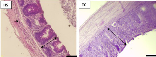
The histological observations in ceca of chickens shown by HE stains. HE-stained specimens were observed under light microscopy. The arrow indicates inflammatory cells infiltrate to mucosa and submucosa. The double-headed arrows indicate the mucosal length. Bar scale is 50 µm.
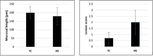
Mucosal length and lesion score in ceca of broiler chickens exposed with heat stress. The mucosal thickness and lesion score was evaluated under light microscopy in the TC (thermoneutral control) and HS (heat stress). All data are represented as the mean ± SE.
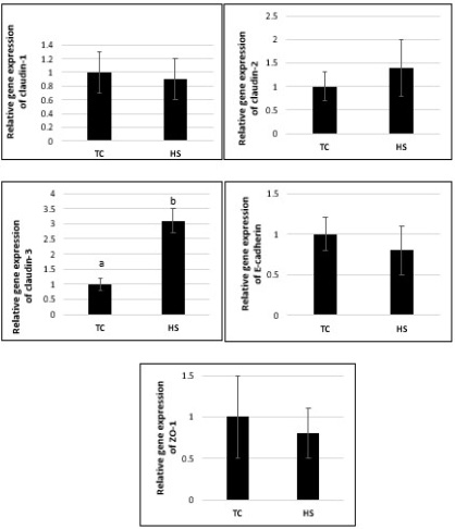
mRNA expression levels of tight junction genes in the ceca of chickens. Amplifications were performed on twenty-five independent samples with triplicate reactions carried out for each sample. The relative mRNA level was calculated using the 2−ΔΔCt method. All data are represented as the mean ± SE. Different letters in the upper of columns represent significantly different (p < 0.05) by unpaired t-test.
References
- Ahmad R, Chaturvedi R, Olivares-Villagómez D, Habib T, Asim M, Shivesh P, Polk DB, Wilson KT, Washington MK, Van KL, Dhawan P, Singh AB (2014). Targeted colonic claudin-2 expression renders resistance to epithelial injury, induces immune suppression, and protects from colitis. Mucosal Immunol., 7(6): 1340-1353. https://doi.org/10.1038/mi.2014.21[CrossRef]
- Alan SF, Brian JJ, Lynne A, Jesaitis JMA (1998). The tight junction protein ZO-1 establishes a link between the transmembrane protein occludin and the actin cytoskeleton. J. Biol. Chem., 273(45): 29745-29753. https://doi.org/10.1074/jbc.273.45.29745[CrossRef]
- Anderson JM, Van ICM (1995). Tight junctions and the molecular basis for regulation of paracellular permeability. Am. J. Physiol., 269: 467–475. https://doi.org/10.1152/ajpgi.1995.269.4.G467[CrossRef]
- Barekatain R, Chrystal PV, Howarth GS, McLaughlan CJ, Gilani S, Nattrass GS (2019). Performance, intestinal permeability, and gene expression of selected tight junction proteins in broiler chickens fed reduced protein diets supplemented with arginine, glutamine, and glycine subjected to a leaky gut model. Poult. Sci., 98(12): 6761-6771. https://doi.org/10.3382/ps/pez393[CrossRef]
- Burkholder KM, Thompson KL, Einstein ME, Applegate TJ, Patterson JA (2008). Influence of stressors on normal intestinal microbiota, intestinal morphology, and susceptibility to Salmonella enteritidis colonization in broilers. Poult. Sci., 87: 1734–1741. https://doi.org/10.3382/ps.2008-00107[CrossRef]
- Chelakkot C, Ghim J, Ryu SH (2018). Mechanisms regulating intestinal barrier integrity and its pathological implications. Exp. Mol. Med., 50(103). https://doi.org/10.1038/s12276-018-0126-x[CrossRef]
- Deng W, Dong XF, Tong JM, Zhang Q (2012). The probiotic Bacillus licheniformis ameliorates heat stress-induced impairment of egg production, gut morphology, and intestinal mucosal immunity in laying hens. Poult. Sci., 91: 575–582. https://doi.org/10.3382/ps.2010-01293[CrossRef]
- Erben U, Loddenkemper C, Doerfel K, Spieckermann S, Haller D, Heimesaat MM, Zeitz M, Siegmund B, Kühl AA (2014). A guide to histomorphological evaluation of intestinal inflammation in mouse models. Int. J. Clin. Exp. Pathol., 7: 4557-4576.
- Findley MK, Koval M (2009). Regulation and roles for claudin-family tight junction proteins. Int. Union Biochem. Mol. Biol. Life, 61: 431–437. https://doi.org/10.1002/iub.175[CrossRef]
- Gunzel D, Yu AS (2013). Claudins and the modulation of tight junction permeability. Physiol. Rev., 93: 525–569. https://doi.org/10.1152/physrev.00019.2012[CrossRef]
- Ho DT, Pham HHS, Aota W, Matsubayashi M, Tsuji N, Hatabu T (2021). Reduction of macrophages by carrageenan decreases oocyst output and modifies local immune reaction in chick cecum with Eimeria tenella. Res. Vet. Sci., 139: 59-66. https://doi.org/10.1016/j.rvsc.2021.07.003[CrossRef]
- Lambert GP (2009). Stress-induced gastrointestinal barrier dysfunction and its inflammatory effects. J. Anim. Sci., 87(14): 101–108. https://doi.org/10.2527/jas.2008-1339[CrossRef]
- Horowitz M (2002). From molecular and cellular to integrative heat defense during exposure to chronic heat. Comp. Biochem. Physiol. A Mol. Integr. Physiol., 131(3): 475–483.
- Livak KJ, Schmittgen TD (2001). Analysis of relative gene expression data using real-time quantitative PCR and the 2-ΔΔCT method. Methods, 25: 402-408. https://doi.org/10.1016/S1095-6433(01)00500-1[CrossRef]
- Mazzoni M, Zampiga M, Clavenzani P, Lattanzio G, Tagliavia C, Sirri F (2022). Effect of chronic heat stress on gastrointestinal histology and expression of feed intake regulatory hormones in broiler chickens. Animal, 16(8). https://doi.org/10.1016/j.animal.2022.100600[CrossRef]
- Mu Q, Kirby J, Reilly CM, Luo XM (2017). Leaky gut as a danger signal for autoimmune diseases. Front. Immunol., 8: 598. https://doi.org/10.3389/fimmu.2017.00598[CrossRef]
- Nanto-Hara F, Kikusato M, Ohwada S, Toyomizu M (2020). Heat stress directly affects intestinal integrity in broiler chickens. J. Poult. Sci., 57(4): 284-290. https://doi.org/10.2141/jpsa.0190004[CrossRef]
- Pearce SC, Mani V, Boddicker RL, Johnson JS, Weber TE, Ross JW (2013). Heat stress reduces intestinal barrier integrity and favors intestinal glucose transport in growing pigs. PLoS One, 8(8). https://doi.org/10.1371/journal.pone.0070215[CrossRef]
- Pham HHS, Matsubayashi M, Tsuji N, Hatabu T (2021). Relationship between Eimeria tenella associated-early clinical signs and molecular changes in the intestinal barrier function. Vet. Immunol. Immunopathol., 240: 110321. https://doi.org/10.1016/j.vetimm.2021.110321[CrossRef]
- Quigley EMM (2016). Leaky gut concept or clinical entity? Curr. Opin. Gastroenterol., 32: 74–79. https://doi.org/10.1097/MOG.0000000000000243[CrossRef]
- Quinteiro-Filho WM, Gomes AV, Pinheiro ML, Ribeiro A, Ferraz-de-Paula V, Astolfi-Ferreira CS, Ferreira AJ, Palermo-Neto J (2012a). Heat stress impairs performance and induces intestinal inflammation in broiler chickens infected with Salmonella enteritidis. Avian Pathol., 41: 421–427. https://doi.org/10.1080/03079457.2012.709315[CrossRef]
- Quinteiro-Filho WM, Ribeiro A, Ferraz-de-Paula V, Pinheiro ML, Sakai M, Sa LR, Ferreira AJ, Palermo-Neto J (2010). Heat stress impairs performance parameters, induces intestinal injury, and decreases macrophage activity in broiler chickens. Poult. Sci., 89: 1905–1914. https://doi.org/10.3382/ps.2010-00812[CrossRef]
- Quinteiro-Filho WM, Rodrigues MV, Ribeiro A, Ferraz-de-Paula V, Pinheiro ML, Sa LRM, Ferreira AJP, Palermo-Neto J (2012b). Acute heat stress impairs performance parameters and induces mild intestinal enteritis in broiler chickens: role of acute hypothalamic-pituitary-adrenal axis activation. J. Anim. Sci., 90: 1986–1994. https://doi.org/10.2527/jas.2011-3949[CrossRef]
- Rescigno M (2011). The intestinal epithelial barrier in the control of homeostasis and immunity. Trends Immunol., 32: 256–264. https://doi.org/10.1016/j.it.2011.04.003[CrossRef]
- Santos RR, Awati A, Roubos-van den Hil PJ, Tersteeg-Zijderveld MH, Koolmees PA, Fink-Gremmels J (2015). Quantitative histo-morphometric analysis of heat-stress-related damage in the small intestines of broiler chickens. Avian Pathol., 44: 19–22. https://doi.org/10.1080/03079457.2014.988122[CrossRef]
- Tabler TW, Greene ES, Orlowski SK, Hiltz JZ, Anthony NB, Dridi S (2020). Intestinal barrier integrity in heat-stressed modern broilers and their ancestor wild jungle fowl. Front. Vet. Sci., 7: 249. https://doi.org/10.3389/fvets.2020.00249[CrossRef]
- Uerlings J, Song ZG, Hu XY, Wang SK, Lin H, Johan B, Everaert N (2018). Heat exposure affectes jejunal tight junction remodeling independently of adenosine monophosphate-activated protein kinase in 9-day-old broiler chicks. Poult. Sci., 97(10): 3681-3690. https://doi.org/10.3382/ps/pey229[CrossRef]
- Xie J, Tang L, Lu L, Zhang L, Xi L, Liu HC, Odle J, Luo X (2014). Differential expression of heat shock transcription factors and heat shock proteins after acute and chronic heat stress in laying chickens (Gallus gallus). PLoS One, 9(7). https://doi.org/10.1371/journal.pone.0102204[CrossRef]
- Zeissig S, Bürgel N, Günzel D, Richter J, Mankertz J, Wahnschaffe U, Kroesen AJ, Zeitz M, Fromm M, Schulzke JD (2007). Changes in expression and distribution of claudin 2, 5 and 8 lead to discontinuous tight junctions and barrier dysfunction in active Crohn’s disease. Gut, 2007; 56(1): 61-72. https://doi.org/10.1136/gut.2006.094375[CrossRef]
Services
Article usage statistics
Loading…