
Creatine kinase (CK) and lactate dehydrogenase (LDH) are myosin heavy chain associated molecules which do not have the capacity to cross muscle cell membrane under normal conditions. Their increase in serum concentration have been used as indicators for muscle damage. The aim of the current study was to determine the effect of Lactobacillus rhamnosus administration on CK and LDH levels in the serum of mice following extensor digitorium longus (EDL) muscle grafting. Blood from the intact control as well as experimental and EDL muscle grafted mice was secured on day 3, 5, 7 and 14 post-transplantations. CK activity of control and EDL muscle grafted mice with probiotics showed 157% and 130% increase, respectively on day 5, whereas on 14th days post-transplantation CK values showed 45.6% and 4%, respectively increase compared with the values of intact control. The LDH activity increase 52.27% and 19.46%, respectively on day 7 when compared with intact control. On 14th days post-transplantation corresponding values for control and probiotics were 23.0% and 1.7 %, respectively increase when compared with the value of intact control. Current study revealed that probiotic administration in orthotropic-transplanted EDL muscle reduce CK and LDH activities in mice, which is positive indication for increase in muscle regeneration.
Short Communication · Volume 56, Issue 2, 21 February 2024 · Open Access
Effect of Lactobacillus rhamnosus (NR_113332.1) on Creatine Kinase and Lactate Dehydrogenase Levels in Orthrotopically Transplanted EDL Muscle of Mice
·
Affiliations & Notes ⌄
- 1Microbial Biotechnology Laboratory, Institute of Zoology, University of the Punjab, Lahore, Pakistan. * Corresponding author: qazi.zool@pu.edu.pk
Article Info ⌄
- Journal:
- Pakistan Journal of Zoology
- Published:
- 21 February 2024

From the article
- Significant increases in CK during the regenerative phases of control mice over the probiotic treated group were recorded.
- The control mice group had CK activity of 625.00 ± 184.68 and 354.67 ± 13.92 U/L on day 5 and 14 days of post grafting.
- This study showed that the probiotics supplementation after EDL muscle transplantation helped to reduce the level of CK and LDH than that of control group.
- The study also revealed that treatment with probiotics caused CK and LDH increases during early stages of regeneration with comparatively lower values than control group.
Selected from the article's own text.
Abstract
Materials and methods
Male albino mice were purchased and housed 15 days before the experiment at 25±5 oC with a roughly light and dark cycle of 12:12h. They were provided free access to water and pelleted food to minimise the cage effect. After adopting the local environmental condition animals were randomly divided into three groups (Intact control, Control, Probiotic), each containing 6 animals. EDL muscle were orthrotransplant in right legs of the mice in groups 2 and 3 under ether anesthesia.
After surgeries, probiotic (Lactobacillus rhamnosus NR_113332.1) dose was administered orally via a gavage method on daily basis. Each animal in control group was given a dose of 200µL of saline on daily basis for 3, 5, 7 and 14 days. Whereas probiotic group was given a dose of 1x10⁸ C.F.U. of the probiotic (NR_113332.1)/200ul/animal/day for 3, 5, 7 and 14 days after surgeries.
For preparation of probiotic dose ten autoclaved vials containing 9 mL of sterile MRS broth (Biolife, Italiana), and added 1 mL of cultural broth of Lactobacillus rhamnosus in vial label as one, and then serially diluted to vial labelled as ten. Then 1mL was taken from each serially diluted broth vials to measure its optical density and 0.1mL was taken from each vial and spread over MRS agar plates. Lactobacillus rhamnosus showed 1 × 10⁸ C.F.U. mL−1 at 0.5 optical density when measured at 600nm using spectrophotometer (V-M5). Every time culture broth was diluted with un-inoculated broth until obtained 0.5 optical density. Diluted broth with optical density 0.5 was centrifuged to have C.F.U. 1× 10⁸ mL−1 following the procedure by Hasan et al. (2022).
For collection of blood and tissue samples, animals were killed with overdose of ether. Grafted as well as non-grafted EDL muscles were recovered. Blood was collected from each mouse by direct puncturing of the heart.
Serum was seperated from the blood and used for the estimation of CK and LDH activities with the help of commercially available bio-kits (Merck, Pvt, Ltd) in automatic bioanalyzer (Erba Chem 5 V3, Germany).
The data were analyzed by two-way analysis of variance (ANOVA) using MINITAB-16 software. The mean was compared by using Tukey multiple range test. Differences between means ± SEM were considered significant at P < 0.05.
Results and discussion
Following the grafting of EDL muscles there were no signs of sickness, diarrhea and aggressive behavior for both in the control as well as probiotic supplemented groups throughout the experimental period. All the experimental animals remained alive during the observational period. All animals were recovered after 3, 5, 7 and 14 days after surgeries, whereas intact control group was recovered at 0 day. At the time of recovery there was no apparent difference in size and morphology of visceral organs of probiotic treated and control mice were noticed.
Figure 1 shows the effect of probiotic on the level of CK and LDH in regenerating EDL muscle of mice. Significant increases in CK during the regenerative phases of control mice over the probiotic treated group were recorded. The control mice group had CK activity of 625.00 ± 184.68 and 354.67 ± 13.92 U/L on day 5 and 14 days of post grafting. Whereas the corresponding value in the probiotics treated group of mice were 558.67 ± 125.24 and 255.33 ± 12.88 (U/L), respectively (P<0.001). The intact control group showed 243.33 ± 19.10 U/L of CK activity peaked on day 5, which was 157.2% for control and 129.6% for probiotic treated group. Whereas, day 14 post-transplantation corresponding values for control and probiotics were 45.6% and 4.9%, respectively. Muscle grafting increased serum CK level despite from the fact that probiotic treatment tended to minimize the injury effect.
LDH level in blood increases with response to increase in muscle damage. Value of control EDL grafted mice group at 7 and 14 days of regeneration were 258.3 ± 42.1 and 208.7 ± 11.6 (mg/dl), respectively. Whereas probiotic group had the corresponding values of 230.00 ± 65.6 and 172.7 ± 26.00 (mg/dl) (P<0.016). Percentage increase was calculated from intact control which had value 169.67 ± 9.74 mg/dl. Control group represent highest increase in lactate level which was observed at day 7 of regeneration which was 52.27% but in probiotic supplemented group the peak 35.5% was observed at day 5 of post-transplantation. Whereas, at 14-days post-transplantation corresponding values for control and probiotics were 23.0% and 1.7 %, Results showed positive effect of probiotic supplementation reducing the serum lactate level as compared to control group (Fig. 1).
Probiotic treatment tended to minimize the injury effect in terms of creatine kinase at first as well as last sampling period. Presence of CK and LDH in blood stream of an organism is indicator of tissue damage . Creatine kinase is normally found in the muscles tissues but this enzyme can leak and be present in the bloodstream owing to the injury to the epimysium membrane surrounding the muscles. That’s why elevated CK levels is suggestive of muscular injury and is considered a marker of muscle damage. It is well documented that probiotics reduce levels of CK, LDH and exhibit a therapeutic impact compared with protective impact. Many authors have reported that different Lactobacillus species significantly reduce CK activity ; ; . Highest % increase in LDH level in control group was observed at day 7 day of regeneration which was 52.27% but in probiotic supplemented group the peak 35.5% was observed at day 5 of post-transplantation when compared with the value of intact control. The shifting of the peak of LDH level at an early phase and its regression in the later phase, is a good indicator of early occurring of degeneration and regeneration processes with EDL muscle grafts of the Lactobacillus rhamnosus (NR_113332.1) supplemented mice than the control grafted mice.
Delay in maximal elevations of CK and LDH contents may be caused by increasing membrane permeability due to secondary or delayed onset damage as a result of increasing Ca2+ leakage into the muscle, and thus increased calpain activity and further reductions in membrane integrity . Furthermore, the exercise-induced muscular activity release of predominantly cytoplasmic CK can be due to final death of the muscle fibre which result in temporary muscle fibre damage accompanied by membrane leakage , this process does not necessarily reflect myofibrillar disruption. Indeed, any correlation between leakage of muscle enzymes and force production is a complex process, as it is well established that the greatest elevation in plasma enzyme levels occurs in the days following injury at a time when force has considerably recovered ; . However, Friden and Lieber (2001) suggested that increase in serum CK levels may provide a gross indication that skeletal muscle injury has occurred, differences between serum CK levels do not necessarily provide information regarding the extent of muscle damage.
Conclusion
This study showed that the probiotics supplementation after EDL muscle transplantation helped to reduce the level of CK and LDH than that of control group. The study also revealed that treatment with probiotics caused CK and LDH increases during early stages of regeneration with comparatively lower values than control group.
Funding
There study received no external funds.
IRB approval
Study was approved by the Punjab University Institutional Ethics Review Board vide letter No.D/68/FIMS.
Ethical statement
The current study was approved by the Punjab University Ethics Review Board.
Statement of conflict of interest
The authors have declared no conflict of interest.
Figures
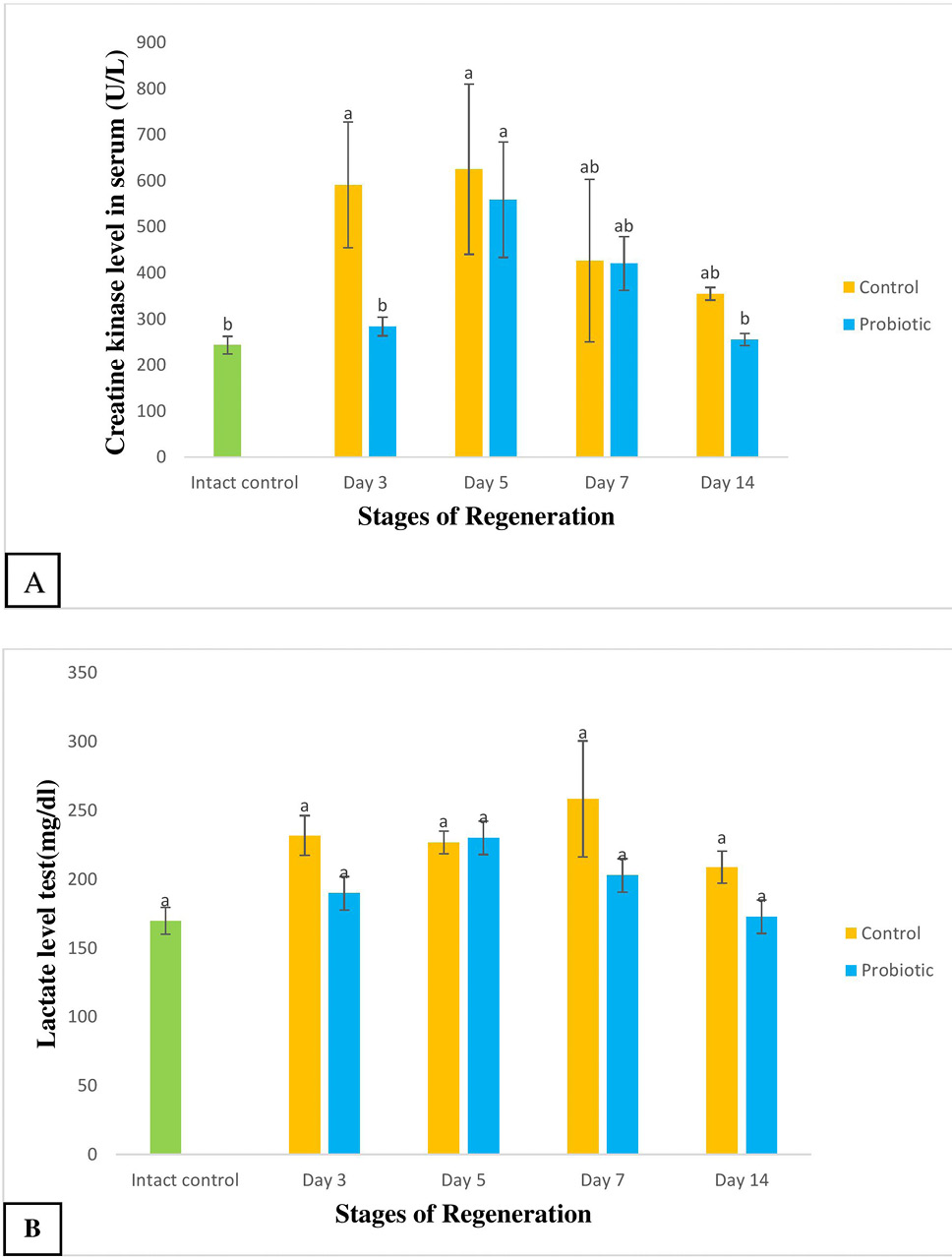
Creatine kinase (A) and lactate dehydrogenase (B) levels of intact mice, the EDL muscle grafted control and probiotic supplemented mice at different days of post-transplantation. The data were analyzed statistically by using two-way ANOVA, means that do not share same alphabets in the same column were significantly different from each other. The effect were declared highly significant at p<0.05.
References
- Al-Orf, N., El-Ansary, A., Bjørklund, G., Moubayed, N., Bhat, R.S. and Bacha, A.B., 2018. Metab. Brain Dis., 33: 1811-1820. https://doi.org/10.1007/s11011-018-0284-5[CrossRef]
- Al-Osaimi, M., El-Ansary, A., Al-Daihan, S., Bhat, R.S. and Ben-Bacha, A., 2018. J. mol. Neurosci., 65: 327-335. https://doi.org/10.1007/s12031-018-1107-1[CrossRef]
- Atherton, P.J. and Smith, K., 2012. J. Physiol., 590: 1049-1057. https://doi.org/10.1113/jphysiol.2011.225003[CrossRef]
- Baird, M.F., Graham, S.M., Baker, J.S. and Bickerstaff, G.F. 2012. J. Nutr. Metab., 1: 960363. https://doi.org/10.1155/2012/960363[CrossRef]
- Beaton, D., Bombardier, C., Guillemin, F. and Ferraz, M.B., 2002. J. Am. Acad. Orthop. Surg., 12: 1-9.
- Brown, S.J., Child, R.B., Day, S.H. and Donnelly, A.E., 1997. J. Sports Sci., 15: 215-222. https://doi.org/10.1080/026404197367498[CrossRef]
- Cezar, C.A. and Mooney, D.J., 2015. Adv. Drug Deliv. Rev., 84: 188-197. https://doi.org/10.1016/j.addr.2014.09.008[CrossRef]
- Charge, S.B. and Rudnicki, M.A., 2004. Physiol. Rev., 84: 209-238. https://doi.org/10.1152/physrev.00019.2003[CrossRef]
- Chen, T.C. and Hsieh, S.S., 2001. Med. Sci. Sports Exerc., 33: 1732-1738. https://doi.org/10.1097/00005768-200110000-00018[CrossRef]
- Chen, Y.M., Wei, L., Chiu, Y.S., Hsu, Y.J., Tsung, Y.T., Wang, M.F. and Huang, C.C., 2016. Nutrients, 8: 205-220. https://doi.org/10.3390/nu8040205[CrossRef]
- Foschini, D. and Prestes, J., 2007. Fit. Perform J., 6: 38-44. https://doi.org/10.3900/fpj.6.1.38.e[CrossRef]
- Friden, J. and Lieber, R.L., 2001. Acta Physiol. Scand., 171: 321-326. https://doi.org/10.1046/j.1365-201x.2001.00834.x[CrossRef]
- Gissel, H. and Clausen, T., 2001. Acta Physiol. Scand., 171: 327-334. https://doi.org/10.1046/j.1365-201x.2001.00835.x[CrossRef]
- Hasan, A., Qazi, J.I., Muzaffer, N., Jabeen, S. and Hussain, A., 2022. Pakistan J. Zool., 54: 2577-2583. https://doi.org/10.17582/journal.pjz/20210803100802[Full text][CrossRef]
- Jarvinen, T.A. and Jarvinen, T.L., 2005. Am. J. Sports Med., 33: 745-764. https://doi.org/10.1177/0363546505274714[CrossRef]
- Liu, J., Saul, D., Boker, K.O., Ernst, J., Lehman, W. and Schilling, A.F., 2018. Biol. Med. Res. Int., 218: 11-21. https://doi.org/10.1155/2018/1984879[CrossRef]
- Mach, N. and Fuster-Botella, D., 2017. J. Sport Hlth. Sci., 6:179-197. https://doi.org/10.1016/j.jshs.2016.05.001[CrossRef]
- McNeil, P.L. and Khakee, R., 1992. Am. J. clin. Pathol., 140: 1097.
- Philp, A., Hargreaves, M. and Baar, K., 2012. Am. J. Physiol. Endocrinol. Metab., 302: E1343-E1351. https://doi.org/10.1152/ajpendo.00004.2012[CrossRef]
- Przewłocka, K., Folwarski, M., Kazmierczak-Siedlecka, K., Skonieczna-Zydecka, K. and Kaczor, J.J., 2020. Nutrients, 12: 1451. https://doi.org/10.3390/nu12051451[CrossRef]
- Shreiner, A.B., Kao, J.Y. and Young, V.B., 2015. Curr. Opin. Gastroenterol., 31: 69. https://doi.org/10.1097/MOG.0000000000000139[CrossRef]
- Spriet, L.L., 2014. Sports Med., 44: 87-96. https://doi.org/10.1007/s40279-014-0154-1[CrossRef]
- Willoughby, D.S., McFarlin, B. and Bois, C., 2003. Int. J. Sports Med., 24: 15-21. https://doi.org/10.1055/s-2003-37197[CrossRef]
- Wozniak, A.C. and Kong, J., 2005. Muscle Nerve, 31: 283-300. https://doi.org/10.1002/mus.20263[CrossRef]
Services
Article usage statistics
Loading…