
This experiment was done on twelve cross breed male goats of age (2-3 months) having 8-9 kg body weight. After 15 days of adaptation period, they were divided into two groups viz. control (C) and selenium yeast supplemented (SY) (n=6), respectively. Basal diet given to the animals consist of roughage and concentrate at the ratio of 65:35. In group C, the goats received only basal diet while in group SY the goats consumed basal diet along with SY 0.3 mg/kg.BW for 8 weeks. The results revealed that testicular weight, thickness, and circumference increased in SY as compared to C. The body weight of goats was also increased in SY as compared to C. while, the Gonado-somatic index in goats of SY was not different compared to C. The histomorphological studies revealed that the height of the germinal epithelium was significantly higher in SY as compared to C. Although the diameter of seminiferous tubule and thickness of the basal membrane were not significantly different in both groups. The serum glutathione peroxidase (GPx) activity as well the testicular GPx activity was significantly increased in SY as compared to C. Furthermore, the serum T-SOD activity was significantly elevated in SY as compared to C. Moreover, the T-SOD activity in testicular tissues was also significantly increased in SY as compared to C. The results revealed that the SY supplementation has positive effects on gross morphological parameters of testis by increasing its weight, thickness, and circumference. Besides this, the SY also improved the height of germinal epithelium of Seminiferous tubule and improved antioxidant status by enhancing the activities of GPx and T-SOD in both serum and testicular tissues.
Research Article · Volume 51, Issue 3, 24 March 2019 · Open Access
Effect of Dietary Selenium Yeast Supplementation on Morphology and Antioxidant Status in Testes of Young Goat
· · · · · · · · ·
Affiliations & Notes ⌄
- 1Department Veterinary Physiology and Biochemistry, SBBUVAS, Sakrand, 67210 Pakistan 2Department Veterinary Physiology and Biochemistry, Sindh Agricultural University, 70060 Tandojam, Pakistan 3Department Animal Reproduction, Sindh Agricultural University, 70060 Tandojam, Pakistan 4Key Laboratory of Agricultural Animal Genetics, Breeding, and Reproduction, Education Ministry of China, Huazhong Agricultural University, Wuhan, Hubei 430070, People’s Republic of China 5Department Veterinary Microbio
Article Info ⌄
- Journal:
- Pakistan Journal of Zoology
- Published:
- 24 March 2019

From the article
- In present investigation, we found amplified activities of the GPx and T-SOD in testis as well as in the serum of goats who consumed SY supplemented diet as compared to the C group.
- Increased GPx and T-SOD levels in both serum as well as within the testicles indicated that SY supplementation improved protection against lipid peroxidation and oxidative stress.
- The SY has positive effects on Gross morphological parameters of testis by increasing weight, thickness and circumference of testicles.
- Further, it improved diameter of lumen of seminiferous tubule, hence it possess positive implications on testicular histology.
Selected from the article's own text.
Abstract
Introduction
The selenium (Se) is an important trace element composed of some selenoproteins and enzyme glutathione peroxides, which possess antioxidant characteristics. It also plays substantial role in the maintenance of reproductive health of animals therefore, minute fluctuations of its level in diet has many significant impacts on animal’s health, performance, and reproduction ; . The Se also applies various activities upon the cancer prevention agents named as antioxidants ; , key factor of immune system , acts as regenerative agent and has implications on endocrine system of animal body . The testis is a primary reproductive organ in the male goat which produces spermatozoa and the male sex hormone. The sperms are synthesized insides Seminiferous tubules (SFT), which are beneficial for the continuation of subsequent generation . The male reproductive organs, including both the testis and epididymis require Se to regulate the synthesis of selenoproteins .
The spermatogenesis is well disciplined and highly organized process of germ cell maturation along with the proliferation of a diploid cell known Spermatogonia into a mature haploid cell known as Spermatozoa. The entire procedure comprises of the mitotic division of Spermatozoids and the meiotic division of Spermatocytes, constituting of both growth as well as differentiation of the Spermatids. Moreover, any disturbance during Spermatogenesis can lead to infertility or lowered production of Spermatozoa. For elimination of unwanted cells, in a germ cell, the procedure of apoptosis occupies a key task in maintaining stability during the procedure of sperm formation . The covering of spermatozoa in mammals exhibits an elevated content of polyunsaturated fatty acids (PUFA), which are extremely prone to the reaction called lipid peroxidation (LPO) produced due to over creation of reactive oxygen species (ROS). The ROS possess very short half-life time and are instable compounds in nature. They can affect adversely certain cellular metabolic procedures when present inside the cells in higher ranges as compared to normal concentrations required for physiological procedures. Generally, they are produced at inner mitochondrial sheath due to aerobic metabolism and respiratory oxidation. As protons are released during these procedures, which eventually lead to the formation of the ROS. The substrates for the ROS production are present in all subcellular organelles of the cell . However, to prevent occurrence of LPO of sperm cell, there are several defense mechanisms including water-soluble antioxidants, fat-soluble antioxidants, and some enzymatic reactions have established inside testicular cells to maintain sperm viability and sperm count . The Se has outstanding effects on the enzymes of the male reproductive system . Because it serves as a catalytic hub in the active sites of antioxidant enzymes, for instance, it contains glutathione peroxidase (GSH-Px) along with the selenoprotein .
The Se is acknowledged to manipulate both gross as well as the histomorphology of the testis. The morphometric investigation of the testis of several breeds is essential to assess and estimate the qualitative variations in testicular machinery and spermatogenic functions . The utilization of male with superior testicular development and consequently with high fecundation capability is significant to ensure the good reproductive competence of the flock. Moreover, the increase in the size of the testes may result in better fertility . The Se has been reported to have an influence on the structural architecture of the testes within male goats . The spermatozoa of boars utilized Se deficient diet showed abnormalities in their midpiece, plasma membrane, tail and in mitochondrial gaps (Marin-Guzman et al., 2000). The Se deficiency has been revealed to result in bilateral atrophy of the testes of rats. Additionally, some studies revealed that due to Se deficiency the diameter of the SFT was condensed, sheathed by sertoli cells or a small number of amount of stem cells, in the company of osseous metaplasia, and partial or compact spermatogenic action was experiential .
The GSH-Px (Glutathione Peroxidase) besides Glutathione reductase (GR) are the chief reducing mediators in the living cells and is regarded as hunting antioxidants in male reproductive tract especially at the region of epididymis and testicles . Their alteration of the spermatozoa sheath consults fortification on the lipid ingredients, consequently protecting the motility and viability of sperms . The earlier studies performed in vitro have revealed that Gpx conserves the tail-beat consistency, decreases LPO, and progresses the sperm casing characteristics. Another important antioxidant is the Superoxide dismutase (SOD), which strolls both extracellular as well as intracellular superoxide anion and prohibits LPO of the plasma casing . The proper research data on the possessions of the Se supplementation on histomorphology and testicular biometry of male goat is scanty, particularly in Sindh Pakistan. Consequently, the aim of present study was to observe the effects of SY added diet on the antioxidative status and the histomorphology of testes in goats.
Materials and methods
Twelve male crossbred young goats of nearly 3 month of age having 8-9 kg body weight (BW) were used for this experiment. All animals were purchased from nearby goat farm and brought at Livestock Experimental Station, Faculty of Animal Husbandry and Veterinary Sciences, Sindh Agriculture University Tandojam. All animals were housed in individual pens of 2.5x4 ft2 area per individual pen. The animals were given a duration of 15 days for adaptation to experimental surroundings. All animals were properly examined physically to ensure their health status and were ear tagged for identification. Following acclimatization, the goats were arbitrarily divided into two groups (n=6 in each group) and fed basal diet without any supplementation i.e. Control (C) or supplemented with Selenium Yeast (SY). The basal diet consisted of concentrate and roughage (65:35) and Water was provided ad-libitum. The source of Se was (Selemax™, Lencois, Paulista, São Paulo, Biorigin®, Brazil) and mixed in the diet having quantity of 0.3 mg/kg/diet. The entire trial persisted for 8 weeks.
Collection of blood for analysis of enzymatic activity
The blood samples for determination of oxidative stress parameters were collected after completion of adaptation period till the slaughtering of animals between 8 and 9 AM, to avoid diurnal effects. During the blood sample collection, the goats were restrained properly and blood sample was taken from the jugular vein by using sterilized needle. From each animal, approximately 5 ml blood was taken weekly. Following collection, the samples were carried to the Post Graduate Research Laboratory of Physiology and Biochemistry, Faculty of Animal Husbandry and Veterinary Sciences. The serum was obtained via centrifugation of blood at 3000 rpm for 15-20 min. The serum was stored in cryovials of 5ml at 4°C for analysis of enzymatic activity.
Slaughtering of goats and data collection
After completion of experiment the live mass of all goats was calculated at around 0800 hours (not below 12 h afterward contact to the previous feeds) then the goats were slaughtered by “Muslim Halal Method” and the testicles including and the epididymis, were removed. These testicles were possessing bilateral symmetry and lacking any sort of superficial lesions, except fluctuating sizes and weights. They were kept on ice packets and then were immediately transported to the Research laboratory of Physiology and Biochemistry, Faculty of Animal Husbandry and Veterinary Sciences. The epididymis was separated from testicles and weight of each testicle was recorded to measure the gonadosomatic index.
Determination of gonadosomatic index (GSI, g/Kg)
The gonadosomatic index (GSI) is the determination of the mass of gonad as a percentage of total body mass. It is signified by means of the formula: GSI= weight of gonad / Entire tissue weight or else entire body weight x 100. It is a tool for calculating the sexual maturity of animals in relation to the growth and progress of reproductive organs. Generally, the GSI signifies proportion of body mass that links to the testes. Each testicle was weighed to determine GSI index. The Weigh was done by putting the testes upon the triple beam weighing machine.
Testicular biometry / gross morphology of testes
The gross features of the fresh testis were examined and recorded. Testicular volume was measured by a method so-called water displacement. In brief, this was done through filling up a measuring cylinder (100 ml) with normal saline up to an identified initial range for instance 70 ml, interpreting the level of normal saline at the upper most of the meniscus. After that the testis was smoothly released into the measuring cylinder and the concluding range of the normal saline within the cylinder was examined e.g. 84 ml. The final volume of the normal saline displaced from the testis was estimated via subtracting the last range from the initial range of normal saline (i.e. 84 ml – 70ml = 14 ml). Finally, this volume in ml was renewed in to cm3, as 1ml is equal to 1 cm3. The photographs of the testes were captured by using a digital camera (Samsung ES95, 16.2 megapixel. The length (cm) and width (cm) and circumference (cm) were measured by using measuring tape, respectively. The entire length of the testes was measured from the anterior to the posterior border and the testicular circumference was measured by encircling each testis at the mid-portion with a thread which was then straightened and measured on a meter rule and by encircling measuring tape directly upon the surface of testicles. The width was determined by measuring tape after dividing testicles into two equal halves. All measurements were done thrice to avoid any error and results were noticed.
Histomorphometric analysis
After performing gross morphological studies the testes were divided into small pieces. These pieces were then immediately fixed by complete immersion in 10% neutral buffered formalin, labeled and kept for two days, after 48 h the sample was washed with distilled water to remove salts and ions. Then the sample was fixed in the Bouin’s solution, for approximately 18 h. After fixation in Bouin’s solution the sample was washed in 70 % alcohol in order to remove picric acid in tissues. The histological slides were prepared and were stained by hematoxylin and eosin. Five slides of each group were arbitrarily selected and were observed for by using the compound microscope by means of low power (10x) as well as high power (40x) magnifications, respectively. The micrographs were conducted by using a light microscope linked to a video based, computer related system (Tuscen CMOS Camera: IS500, resolution: 5.0 megapixels) was programmed to take micrograph. The histological assemblies of the testes were noticed by using light microscope using both low 4x as well as high 40x magnifications. The diameter of SFT, lumen and thickness of BM was measured by using Image J 1.50i software. The snapshots of each sample were taken for better estimation of the results.
Determination of enzyme activities in serum and testicular tissues
The activity of GPx and Total-SOD (T-SOD) was determined by means of using commercially available kits including Cat. No. A005 for the GPx enzyme assay and the Cat. No A001 for the SOD assay, respectively. The enzyme activity was observed in both serum as well as in testicular tissues. The optical density (OD) of samples was recorded by using spectrophotometer model Vis 721 spectrophotometer model at 412 nm wave length for Gpx and 550 nm wave length for SOD determination per the protocol provided by manufacturer.
Results
The effects of dietary supplementation of SY on gross morphology of goat testis of both C and SY is shown in Table I. The testicular weight (3.5 ± 0.54 g vs 2.15 ± 0.39 g, p < 0.05), thickness (2.95 ± 0.12 cm vs 2.15 ± 0.32 cm, p < 0.05) and circumference (5.3 ± 0.23 cm vs 3.9 ± 0.44 cm, p < 0.05) increased in SY as compared to C. While the length, width and volume of testicles in SY (3.25 ± 0.38 cm, 2.07 ± 0.39 cm and 2.02 ± 0.05 cm) were not different (p > 0.05) compared to C (2.58 ± 0.25 cm, 1.60 ± 0.29 cm and 2.06 ± 0.05).
Table I.- Effect of dietary selenium yeast (SY) supplementation on testicular morphology of goat.
| Items | Groups | P-Value | |
| C | SY | ||
| Weight (g) | 2.15 ± 0.39 | 3.5 ± 0.54* | 0.044 |
| Length (cm) | 2.58 ± 0.25 | 3.25 ± 0.38 | 0.094 |
| Width (cm) | 1.60 ± 0.29 | 2.07 ± 0.39 | 0.183 |
| Thickness (cm) | 2.15 ± 0.32 | 2.95 ± 0.12* | 0.031 |
| Circumference (cm) | 3.9 ± 0.44 | 5.3 ± 0.23* | 0.016 |
| Volume (cm) | 2.06 ± 0.05 | 2.02 ± 0.05 | 0.2982 |
* values (mean ± SE) differ at P < 0.05 between two groups.
Table II.- Effects of dietary SY supplementation on GSI of goat.
| Items | Groups | P-Value | |
| C | SY | ||
| Final BW (kg) | 9.12 ± 0.427 | 10.88 ± 0.427* | 0.013 |
| Testicles weight (g) | 2.15 ± 0.393 | 3.5 ± 0.54* | 0.044 |
| GSI (%) | 24.25 ± 5.618 | 32.46 ± 5.209 | 0.162 |
GSI, Gonado-somatic Index; BW, body weight. * values (mean ± SE) differ at P < 0.05 between two groups.
The impacts of diet supplemented with SY and diet without any supplementation of SY on the gonado-somatic index (GSI) of goat is shown in Table II. The body weight (10.88 ± 0.427 kg vs 9.12 ± 0.427 kg, p < 0.05) and testicular weight (3.5 ± 0.54g vs 2.15 ± 0.393, p < 0.05) increased in SY as compared to the C. Moreover, the GSI in goats of SY was not different (32.46 ± 5.209 %, p > 0.05) compared to goats of C (24.25 ± 5.618 %, p > 0.05).
The micro-anatomical features of the testis were observed in the present study. Histologically, the testicles of goat comprised of three coatings, the outer most tunica vaginalis, the internal and the densest tunica albuginea and the innermost tunica vasculosa. The internal portion of the capsule was occupied by the SFT comprising of elongated, oval, comma, straight and elliptical shapes. The SFT was surrounded by Basal membrane (BM). The lumen was absent in SFT so they were termed as sex cords. The innermost area of sex cords was comprised of Germinal epithelium (GE) which was bulkier in SY goats as compared to C. The cells within region of GE were not properly differentiated and were distributed virgoursly in both groups comprising of both Sertoli cells and Gonocytes. The sertoli cells were somewhat polygonal in shape and their cytoplasm was homogenous. The Gonocytes were huge and round and were holding globular nucleus. While the leydig cells were present within interstitial spaces. The density of SFT was higher in SY compared to C (Fig. 1A, B). The SFT were closely arranged by a thin basement membrane (BM) which was more thick and prominent in the testis of goats fed SY supplemented diet as compared to C group (Fig. 1C, D). The morphometry of SFT of goat testis is presented in Table III and Figure 2. The diameter of GE was significantly higher (p < 0.05) in SY (25.55 ± 0.346 µm) compared to C (17.78 ± 0.424 µm). Although the diameter of SFT and thickness of BM in SY (28.15 ± 1.194 µm and 1.52 ± 0.143 µm) were not significantly different (p < 0.05) compared to C (26.50 ± 2.209 µm and 1.53 ± 0.143 µm).
Table III.- The effects of dietary SY supplementation on morphometry of seminiferous tubules (SFT) in the testis of goat.
| Items | Groups | P-Value | |
| C | SY | ||
| Diameter of SFT (µm) | 26.50±2.209 | 28.15±1.194 | 0.267 |
| Diameter GE (µm) | 17.78±0.424 | 25.55±0.34* | 0.000 |
| Thickness of BM (µm) | 1.53±0.143 | 1.52±0.143 | 0.497 |
SFT, seminiferous tubules; GE, Germianl epithelium; BM, basement membrane. * values (mean ± SE) differ at P < 0.05 between two groups.
The effects of diet without SY supplementation upon GPx activity in the serum of goat is expressed in Figure 3A. The GPx activity was significantly increased (p< 0.05) in SY (22.23 ± 0.23) as compared to C (17.80 ± 0.495). The effects of diet without SY supplementation upon GPx activity in the testicular tissues of goat is expressed in Figure 3B. The GPx activity was significantly increased (p< 0.05) in SY (113.25 ± 2.5) as compared to C (90 ± 5.33).
The impacts of diet treated with SY on SOD enzyme activity in serum of goats is shown in Figure 3C. The serum SOD activity was significantly elevated (p < 0.05) in SY (36.00 ± 0.741) as compared to C (30.13 ± 2.791). The impacts of diet treated with SY on T-SOD activity in testis of goats is shown in Figure 3D. The testicular T-SOD activity was significantly elevated (p< 0.05) in SY (302 ± 13.44) as compared with C (215 ± 4.04).
Discussion
The morphometric study of the testis is crucial to evaluate the qualitative distinctions in testicular machinery and its functions. The utilization of male with superior testicular development and consequently with high fecundation capability is significant to ensure the good reproductive competence of the flock. The mammalian gonads including testicles need a constant supply of trace minerals including Se. according to some studies, it has been proved that the morphology of testicular organ powerfully rest on on suitable amount of Se, either in organic or inorganic states. Thus, the diets which are deficient in Se affect the gross as well as histological morphology of testicles in various species including buck .
The current study assessed the influences of dietary SY supplementation on both gross as well as histomorphology of testicular tissues in addition calculation of Gonado-somatic index (GSI) of goats. The results revealed that SY consumption did not altered the volume, width and length of testicles, however, the thickness, weight and circumference of testicular tissues was significantly increased in SY goats as compared to C. Consistent with our results, the previous studies have shown that testicular functions and morphology is affected by Se deficiency in diet . Conferring to an investigation it was observed that the male progeny of mice consuming little grades of dietary Se (2–7g/kg in feed) revealed late testicular enlargement and maturation . Conversely, this outcome was minute in contrast with that in the second and third progeny in which Se shortage affected the morphology of testes to a greater degree. While in the fourth generation, testis size being a minor quantity than partially of offspring of those mice consuming adequate Se (250-300 g/kg sodium selenite), and testis were observed to be bilaterally shrunken lacking mitotic action in spermatogonia .
Some reviews have revealed that the declined weight of the testicular tissues designates extensive diffuse or damage of the epithelial cells of SFT and the testicles having superior quantity of sustenocytes were weightier and fashioned additional spermatozoa than testes possessing rarer sustenocytes . Consistent with our results it can be suggested that sophisticated testicular weight in our research indicates that the testes could comprise additional interstitial endocrine cells, sustenocytes and SFT. Furthermore, a research showed that there was a positive association among the testicular weight, testicular diameter and circumference . Thus, due to improved weight of testicles, the thickness of testicles also improves along with its circumference. Hence, Se supplementation improved gross morphological parameters of testicles. The GSI characterizes fraction of body form that matches to the gonad of animal. It is meant for determining the sexual adulthood of animal in association to ovary and testes growth. In current research, there was no significant difference in GSI of both groups. It is suggested that non-significant modification in GSI is possibly since of the minor rise of body weight in animals .
The microphotographs of testicles were examined in present study and result showed that the diameter of lumen of SFT was larger in SY as compared to the C. However, there was no any significant modification between the diameter of entire SFT and the thickness of BM in both groups. Some authors revealed that Se shortage resulted in reduction of diameter of SFT in rat. Moreover, the varying consequences in animals may be due to the species modifications and ratio of Se in diet . Additionally, it also depends on the different stages of bio activity of Se such as the organic form of Se like SY has an increased rate of bioactivity as compared to the inorganic enhancements. Moreover, the nutrients usually act on the endocrine instead of the spermatogenic task of the testis . A previous study reveals that there was no significant difference among the spermatozoa number in both Se treated bulls and control group despite of increased levels of Se level in testicles of Se treated bulls as compared to control animals. Thus, it has been proved that increased concentration of Se in testis is not associated with the spermatogenic factor of testicles . The current study showed no momentous modifications in thickness of BM and diameter of SFT as the diet nourished to the goats in present experiment was already comprising satisfactory Se and consequently, additional Se in diet did not express noteworthy consequences. Though, our study shows that 0.3mg SY supplementation augmented the diameter of lumen in the SFT in goats.
The ROS are extremely active oxidizing factors consisting of single or multiple unpaired types of an electron in their structure belonging to the set of free radicals. The majority of these possess forceful implications on the reproductive physiology which include H2O2, O2-, ROO- and OH- radicals which are very reactive in nature. The ROS normally have advantageous impacts upon the functions of spermatozoa, depending on the nature and the amount. During normal physiological state, the spermatozoa manufacture a very little quantity of ROS that are required in support of the sperm capacitation and acrosomal reaction. The production free radicals should be inactivated constantly to maintain normal homeostatic environment of the cell. The plasma of semen is gifted with a range of antioxidant molecules to shield the sperm cells from effects of oxidants. Generally, antioxidants are the substances which suppress the synthesis of free radical molecules or resist their actions. There are many antioxidant compounds including catalase, GPx and SOD.
The SY is an important mineral consisting of variety of enzymes possessing defensive mechanisms against oxidative stress . Due to this reason, currently the research on Se has attracted incredible attention. The Se contains GPx enzyme which is regarded as a basic factor for fortification against LPO and OS. Hence the normal levels of GPx enzyme are very much imperative for maintenance of OS conditions which rest on utilization of diet supplemented with Se, since the Se controls wide zones of cellular occupations including the antioxidant activities. Meanwhile many isoforms of GPx are only Se dependent hence, it is thinkable to measure their status in the provision of testicular job by investigating the influence of Se shortage upon male reproductive traits. In present investigation, we found amplified activities of the GPx and T-SOD in testis as well as in the serum of goats who consumed SY supplemented diet as compared to the C group. According to some investigations the serum and testicular GPx level is regarded as an indicator of the Se ratio in the animal body because of the high association found among the intake of dietary SY and the actions of Gpx within both plasma and red blood cells of animal . Likewise, it has been proved that addition of SY in diet of ruminants improves antioxidant status . Furthermore, the animals consumed a Se scarce diet displayed a substantial decrease of GPx action and an additional damage of germ cells within the testes .
Some authors revealed that the Se supplementation in the regime of livestock surges the action of SOD . Likewise, the consumption of Se rises antioxidant bulk in sows by the elevation of SOD, GPx, levels . These results were inveterate similarly in our results. Though, one more study defines that an absenteeism of Se chiefs to the imperative drop of GPx in blood serum of guinea pigs . These results revealed that addition of Se in diet can overcome from damages cause by OS and enhance protection by enhancing oxidants in blood.
Conclusions
Increased GPx and T-SOD levels in both serum as well as within the testicles indicated that SY supplementation improved protection against lipid peroxidation and oxidative stress. The SY has positive effects on Gross morphological parameters of testis by increasing weight, thickness and circumference of testicles. Further, it improved diameter of lumen of seminiferous tubule, hence it possess positive implications on testicular histology.
Statement of conflict of interest
The authors declare no conflict of interest.
Figures
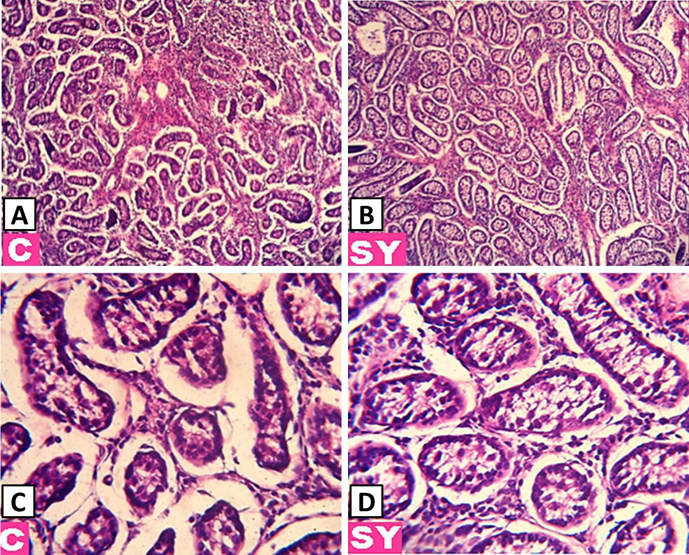
Effect of dietary selenium yeast (SY) on the histological structure of goat testes. Magnification: A, B, 10X; C, D, 40X. Stain: H&E.
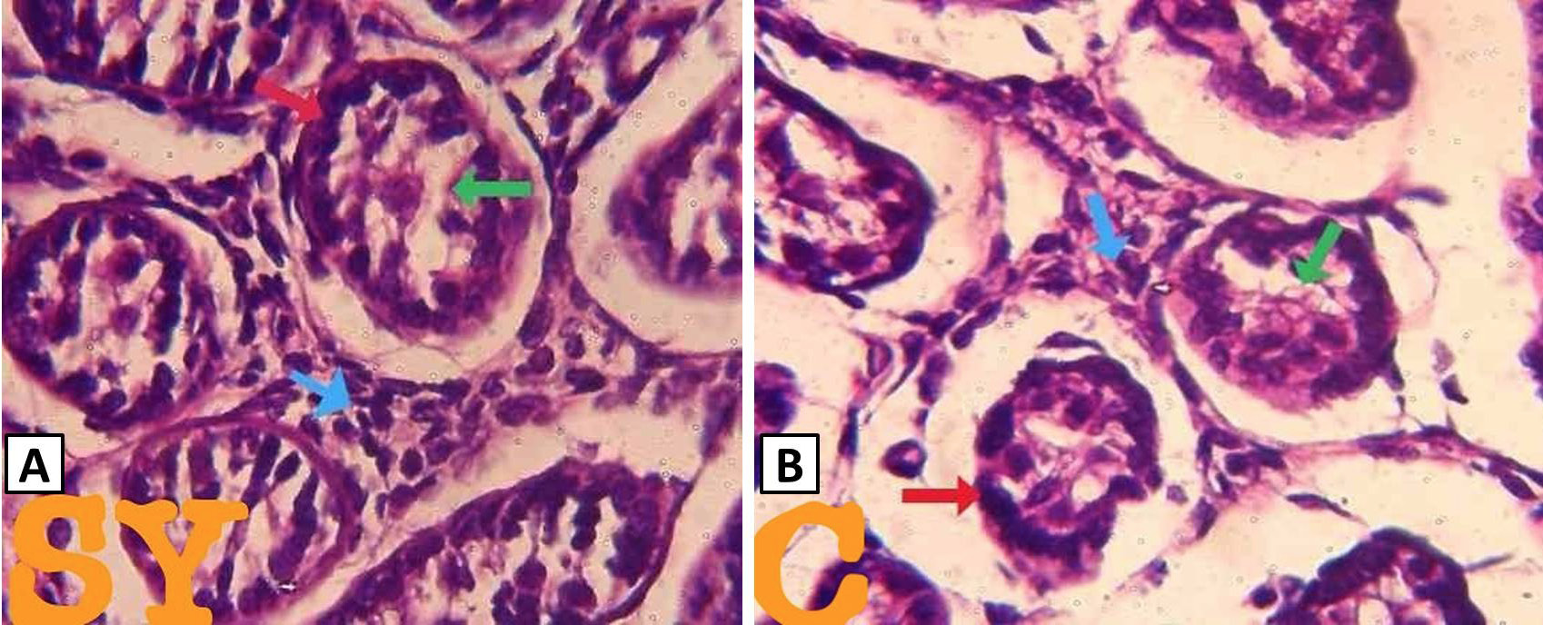
Histological structure of selenium yeast (SY) and control (C) demonstrating assembly of SFT including the BM (red arrow), germinal epithelium comprising of gonocytes and sertoli cells (green arrow) interstitial cells (blue arrow). Stain: H&E. Magnification: 100X.
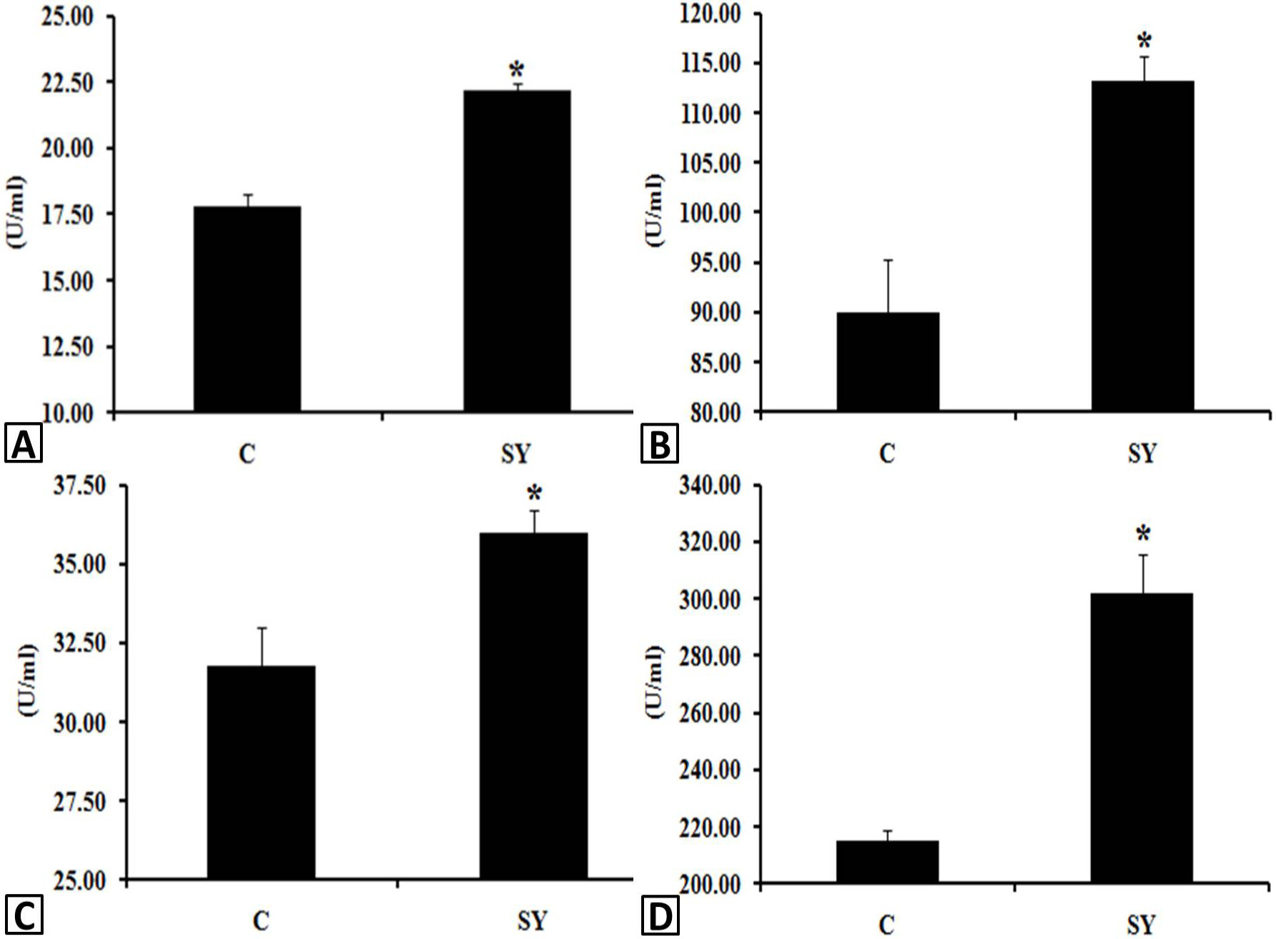
The effect of dietary SY supplementation on glutathione peroxidase (GPx) and super oxide dismutase (SOD) activity (U/mL) in serum (A, B) and testis (C, D) of goat. * values (mean± SE) differ at p < 0.05 between the groups. The goats in SY were fed 0.3 mg/kg SY while the goats in C did not consumed SY in diet.
References
- Adaramoye, O.A., Akanni, O.O., Adewumi, O.M. and Owumi, S.E., 2015. Lopinavir / ritonavir, an antretroviral drug, lowers sperm quality and induces testicular oxidative damage in rats. Tokai J. Exp. Clin. Med., 40: 51-57.
- Agarwal, A. and Majzoub, A., 2017. Free radicals in andrology. In: Antioxidants in andrology. Springer International Publishing, pp. 1-21. https://doi.org/10.1007/978-3-319-41749-3_1[CrossRef]
- Ahsan, U., Kamran, Z., Raza, I., Ahmad, S., Babar, W., Riaz, M.H. and Iqbal, Z., 2014. The role of selenium in the male reproduction- A review. Anim. Reprod. Sci., 146: 55-62. https://doi.org/10.1016/j.anireprosci.2014.01.009[CrossRef]
- Andres, S., Jimenez, A. and Mañé, M.C., 1997. Relationships between some soil parameters and the blood glutathione peroxidase activity of grazing sheep. Vet. Rec., 141: 267-268. https://doi.org/10.1136/vr.141.11.267[CrossRef]
- Arthur, J.R., 2001. The glutathione peroxidases. Cell. Mol. Life Sci., 57: 1825-1835. https://doi.org/10.1007/PL00000664[CrossRef]
- Beckett, G.J. and Arthur, J.R., 2005. Selenium and endocrine systems. J. Endocrinol., 184: 455-465. https://doi.org/10.1677/joe.1.05971[CrossRef]
- Behne, D., Weiler, H. and Kyriakopoulos, A., 1996. Effects of selenium deficiency on testicular morphology and function in rats. J. Reprod. Fertil., 106: 291-297. https://doi.org/10.1530/jrf.0.1060291[CrossRef]
- Brenneisen, P., Steinbrenner, H. and Sies, H., 2005. Selenium, oxidative stress, and health aspects. Mol. Aspects Med., 26: 256-267. https://doi.org/10.1016/j.mam.2005.07.004[CrossRef]
- Dhamsaniya, H.B., Parmar, S.C., Jadav, S.J., Bhatti, I.M. and Patel, V.K., 2016. Plasma minerals profile in delayed pubertal Surti buffalo heifers treated with GnRH alone and with phosphorus. J. Livest. Sci., 7: 157-161.
- Foresta, C., Flohé, L., Garolla, A., Roveri, A., Ursini, F. and Maiorino, M., 2002. Male fertility is linked to the seleno-protein phospholipid hydro-peroxide glutathione peroxidase. Biol. Reprod., 67: 967-971. https://doi.org/10.1095/biolreprod.102.003822[CrossRef]
- Ganabadi, Jr. S., Halimatun, Y., Amelia, C.K., Nor, J.A. and Mohammed, H.A., 2010. Effect of selenium supplementation on spermatogenic cells of goats. Malaysian J. Nutr., 16: 187-193.
- Gofur, M.R., Khan, M.Z.I., Karim, M.R. and Islam, M.N., 2008. Histomorphology and histochemistry of testis of indigenous bull (Bos indicus) of Bangladesh. J. Vet. Med., 6: 67-74. https://doi.org/10.3329/bjvm.v6i1.1341[CrossRef]
- Griswold, M.D., 2016. Spermatogenesis: The commitment to meiosis. Physiol. Rev., 96: 1-17. https://doi.org/10.1152/physrev.00013.2015[CrossRef]
- Hall, J.A., Bobe, G., Vorachek, W.R., Kasper, K., Traber, M.G., Mosher, W.D., Pirelli, G.J. and Gamroth, M., 2014. Effect of supranutritional organic selenium supplementation on postpartum blood micronutrients, antioxidants, metabolites and inflammation biomarkers in selenium-replete dairy cows. Biol. Trace Elem. Res., 161: 272-287. https://doi.org/10.1007/s12011-014-0107-4[CrossRef]
- Hatfield, D.L., Tsuji, P.A., Carlson, B.A. and Gladyshev, V.N., 2014. Selenium and selenocysteine: Roles in cancer, health, and development. Trends Biochem. Sci., 39: 112-120. https://doi.org/10.1016/j.tibs.2013.12.007[CrossRef]
- Henkel, R.R., 2011. Leukocytes and oxidative stress: Dilemma for sperm function and male fertility. Asian J. Androl., 13: 43. https://doi.org/10.1038/aja.2010.76[CrossRef]
- Horky, P., Ruttkay-Nedecky, B., Kremplova, M., Krystofova, O., Kensova, R., Hynek, D., Babula, P., Zitka, O., Zeman, L., Adam, V. and Kizek, R., 2013. Effect of different doses of organically bound selenium on antioxidant status and levels of metal ions in postpartum sows. Int. J. Electrochem. Sci., 8: 6162-6179.
- Hu, H.J., Wang, M., Zhan, X.A., Li, X. and Zhao, R.Q., 2011. Different selenium sources on productive performance, serum and milk Se concentrations and antioxidant status of sows. Biol. Trace Elem. Res., 142: 471-480. https://doi.org/10.1007/s12011-010-8803-1[CrossRef]
- Ibrahim, H.A., Zhu, Y., Wu, C., Lu, C., Ezekwe, M.O., Liao, S.F. and Haung, K., 2012. Selenium-enriched probiotics improves murine male fertility compromised by high fat diet. Biol. Trace Elem. Res., 147: 251-260. https://doi.org/10.1007/s12011-012-9353-5[CrossRef]
- Jana, K., Samanta, P.K., Manna, I., Ghosh, P., Singh, N., Khetan, R.P. and Ray, B.R., 2008. Protective effect of sodium selenite and zinc sulfate on intensive swimming-induced the testicular gamatogenic and the steroidogenic disorders in mature male rats. Appl. Physiol. Nutr. Metab., 33: 903-914. https://doi.org/10.1139/H08-065[CrossRef]
- Jebur, A.B., Nasr, H.M. and El-Demerdash, F.M., 2014. Selenium modulates β-cyfluthrin-induced liver oxidative toxicity in rats. Environm. Toxicol., 29: 1323-1329.
- Kaur, P. and Bansal, M.P., 2004. Influence of selenium induced oxidative stress on spermatogenesis and lactate dehydrogenase-X in mice testis. Asian J. Androl., 6: 227-232.
- Luo, J., Nikolaev, A.Y., Imai, S.I., Chen, D., Su, F., Shiloh, A., Guarente, L. and Gu, W., 2001. Negative control of p53 by Sir2α promotes cell survival under stress. Cell, 107: 137-148. https://doi.org/10.1016/S0092-8674(01)00524-4[CrossRef]
- Maneesh, M. and Jayalekshmi, H., 2006. Role of reactive oxygen species and antioxidants on pathophysiology of male reproduction. Indian J. clin. Biochem., 21: 80-89. https://doi.org/10.1007/BF02912918[CrossRef]
- Mann, T. and Lutwak-Mann, C., 2012. Male reproductive function and semen: Themes and trends in physiology, biochemistry and investigative andrology. Springer Science and Business Media.
- Marin-Guzman, J., Mahan, D.C., Chung, Y.K., Pate, J.L. and Pope, W.F., 1997. Effects of dietary selenium and vitamin E on boar performance and tissue responses, semen quality, and subsequent fertilization rates in mature gilts. J. Anim. Sci., 75: 2994-3003. https://doi.org/10.2527/1997.75112994x[CrossRef]
- McKenzie, R.C., Rafferty, T.S. and Beckett, G.J., 1998. Selenium: An essential element for immune function. Immunol. Today, 19: 342-345. https://doi.org/10.1016/S0167-5699(98)01294-8[CrossRef]
- Melo, F.C.S.A., Matt, S.L.P., Paula, T.A.R., Gomes, M.L.M. and Oliverira, L.C., 2010. The effects of Tynnanthus fasciculatus (Bignoniaceae) infusion on testicular parenchyma of adult Wistar rats. Biol. Res., 43: 445-450. https://doi.org/10.4067/S0716-97602010000400009[CrossRef]
- Omeje, V.I., 2016. Effect of dietary supplementation of organic selenium at different levels on reproductive performance of rabbit does. Ph. D. thesis, University of Nigeria, Nsukka.
- Ostovan, F., Gol, A. and Javadi, A., 2017. Investigating the effects of Citrullus colocynthis pulp on oxidative stress in testes and epididymis in streptozotocin-induced diabetic male rats. Int. J. Reprod. Med., 15: 41-48. https://doi.org/10.29252/ijrm.15.1.41[CrossRef]
- Oyeyemi, M.O., Fayomi, A.P., Adeniji, D.A. and Ojo, O.M., 2012. Testicular and epididymal parameters of sahel buck in the humid zone of Nigeria. Int. J. Morphol., 30: 489-492. https://doi.org/10.4067/S0717-95022012000200021[CrossRef]
- Potts, R.J., Notarianni, L.J. and Jefferies, T.M., 2000. Seminal plasma reduces exogenous oxidative damage to human sperm, determined by the measurement of DNA strand breaks and lipid peroxidation. Mutat. Res. Fund. Mol. Mechan. Mutag., 447: 249-256. https://doi.org/10.1016/S0027-5107(99)00215-8[CrossRef]
- Puglia, C.D. and Loeb, G.A., 1984. Influence of rat brain superoxide dismutase inhibition by diethyldithiocarbamate upon the rate of development of central nervous system oxygen toxicity. Toxicol. appl. Pharmacol., 75: 258-264. https://doi.org/10.1016/0041-008X(84)90208-4[CrossRef]
- Raji, A.Y. and Njidda, A.A., 2014. Gonadal and extra-gonodal sperm reserves of the red Sokoto goats fed Moringa oleifera supplemented diets. Int. J. Agric. Biosci., 3: 61-64.
- Riaz, M. and Mehmood, K.T., 2012. Selenium in human health and disease: A review. J. Postgrad. Med. Inst. Peshawar Pakistan, 2: 26.
- Roy Choudhury, S., Sharma, R., Sikka, S. and Agarwal, A., 2016. Diagnostic application of total antioxidant capacity in seminal plasma to assess oxidative stress in male factor infertility. J. Assist. Reprod. Genet., 33: 627-635. https://doi.org/10.1007/s10815-016-0677-5[CrossRef]
- Saleh, R.A. and Agrawal, A., 2002. Oxidative stress and male infertility: From research bench to clinical practice. J. Androl., 23: 737-752.
- Samo, S.P., Malhi, M., Gadahi, J., Lei, Y., Kaciwal, A.B. and Soomro, S.A., 2018. Effect of organic selenium supplementation in diet on gastrointestinal tract performance and meat quality of goat. Pakistan J. Zool., 50: 995-1001. http://dx.doi.org/10.17582/journal.pjz/2018.50.3.995.1001[CrossRef]
- Segerson, E.C. and Johnson, B.H., 1980. Selenium and reproductive function in yearling Angus bulls. J. Anim. Sci., 51: 395-401. https://doi.org/10.2527/jas1980.512395x[CrossRef]
- Shalini, S. and Bansal, M.P., 2007. Alterations in selenium status influences reproductive potential of male mice by modulation of transcription factor NFκB. Biometals, 20: 49-59. https://doi.org/10.1007/s10534-006-9014-2[CrossRef]
- Sirota, T.V., 2010. Effect of selenium-containing products on antioxidant enzyme activity in the kidneys, liver and blood of guinea pigs. Bull. Exp. Biol. Med., 149: 412-415. https://doi.org/10.1007/s10517-010-0958-4[CrossRef]
- Surai, P.F., 2006. Selenium in nutrition and health, Vol. 974. Nottingham University Press, Nottingham.
- Tapiero, H., Townsend, D.M. and Tew, K.D., 2003. The antioxidant role of selenium and seleno-compounds. Biomed. Pharmacother., 57: 134-144. https://doi.org/10.1016/S0753-3322(03)00035-0[CrossRef]
- Tremellen, K., 2008. Oxidative stress and male infertility-a clinical perspective. Human Reprod. Update, 14: 243-258. https://doi.org/10.1093/humupd/dmn004[CrossRef]
- Ufer, C. and Wang, C.C., 2011. The roles of glutathione peroxidases during embryo development. Front. Mol. Neurosci., 28: 4-12. https://doi.org/10.3389/fnmol.2011.00012[CrossRef]
Cited by 3 articles
In journals published by ResearchersLinks.
- Dietary Selenium Supplementation Reduces Oxidative Stress and Attenuates Apoptotic Gene Expression in Testes of Pre-Pubertal Male GoatPakistan Journal of Zoology · 2025
- Dietary Organic Selenium Supplementation Improved Some Histo-Morphological Features and Stimulated G1 Cell Cycle Progression in Testis of Pre-Pubertal Male GoatPakistan Journal of Zoology · 2025
- Study on the Therapeutic Effect of Modified Yujin Powder on E. coli in the Procapra przewalskiiPakistan Journal of Zoology · 2022
Services
Article usage statistics
Loading…