After prolonged storage in low-temperature conditions, colonies that were not formed during 3-month incubation at 37°C were detected. The growth of the culture of the first generation (Figure 1) was characterized by a “incrustation”, the colonies separate and different in size, shape and appearance, slightly orange, in the second - the continuous intensive growth of orange colonies with a smooth and rough surface (Figure 2).
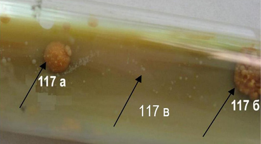
Figure 1: Colonies M. bovis of the first generation, cultivated at a temperature of 3°C (117a, 117b, 117c).
The consistency of the colonies was fragile and dry, transparent - matte, and emulsification - moderate.
By means of microscopy was established (Figure 3), that all cells of M. bovis variant 117-c of the first-generation were kept the paint of fuchsin (acid-resistant), 117-a variant - partially (in some events also had been found non-acid resistant cells), 117-b - all non-acid resistant variants (forms) of the microorganism. At the same time in the second generation of the colonies formed: only non-acid-resistant forms (117-b, c); non-acid resistant and acid-resistant (single) mycobacteria (117-a generation). In the following subcultures, beginning with the third, only non-acid-resistant mycobacteria were detected.
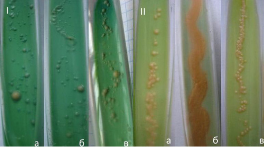
Figure 2: Culture M. bovis of dissociative forms a passage 117th, that has been formed at a temperature of 3°С: I-two weeks, II-two to four weeks, the second generation (a, b, c).
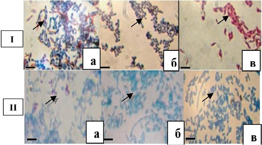
Figure 3: Dissociative forms M. bovis 117th subculture of the first (1) and the second (II) generations. Bar = 10 microns (a, b, c).
All microorganisms of the being studied subcultures, both first and second generations, had the shape of straight rod-shaped bacteria with rounded ends. The length of the microorganisms did not reveal any significant difference: short mycobacteria were dominated. This also applies to the thickness: in the vast majority, thin, sometimes thin and thick forms of mycobacteria mostly appeared in the first generation. However, mycobacteria of the first generation turned out to be longer than the traditional in the general background of short coconut like forms, which were placed by aggregations. In the second generation it is noted in the majority of single and aggregate short forms.
By studying the subcultures of the third generation, different growth rates were established on the dense nutrient medium at temperatures of 3°C and 37°C of cultivation. Almost twice as fast colonies were formed at 3°C (3-4 day) than at 37°C (5-10 days). The surface of subcultures was smooth and moist. However, they all formed a pigment: at 3°C – cream-coloured, 37°C – orange. Under the immersion of the microscope, mycobacteria from the colonies, which were obtained at 3°C and 37°C, were non-acid-resistant, short, thin, straight, with rounded edges and granular. However, in the curtain suspensions of mycobacteria, prepared from cultures that were obtained at 3°C have been revealed non-acid-resistant elementary small bodies.
Dissociative forms (117-a, b, c) cultivated and accumulated at 3°C and 37°C did not cause at guinea pigs the pathological and anatomical changes of typical tuberculosis and allergic reactions to PPD-tuberculin for mammals. The weight of the body of the experimental animals corresponded to the physiological norm. There were no ulcers in the area of introduction of mycobacteria suspension.
M. bovis 100th reinoculation in guinea pigs (control) stimulated the development of the infectious process, weight loss, allergic condition and ulcers in the inoculation zone of mycobacteria curvature. The death of animals from tuberculosis was marked on the 32nd and 42nd day of the experiment. At 10th reinoculation of mycobacteria the rate of formation of subcultures (colonies) at both temperatures of cultivation was the same – 4-7 days. Colonies of all dissociative variants are classified into S-shapes, pigmented for both 3°C and 37°C cultivation. M. bovis of dissociative forms were long and short, with the exception of the mycobacteria of the 117-a subculture of the 37°C cultivation temperature, where short, filiform forms with dark grains were established along the entire length of the cell. The thickness of mycobacteria was the same: thick and thin.
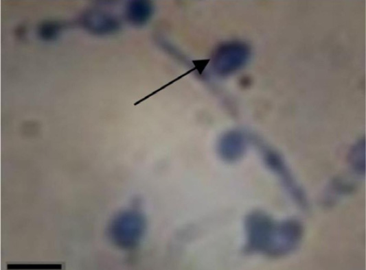
Figure 4: M. bovis of dissociative forms 117-b (10th generation), cultivated at temperature 37°C. Bar=10 microns.
However, M. bovis 117-c (at 37°C) and 117-b, c (at 3°C and 37°C) variants differed (Figure 4): thick, slightly oval rod-shaped bacteria, filiform shapes, in which dark blue grainy formations were contained. The latter, in some cases, pushed out from such cells, from which later formed typical L-shapes (ovals with different optical density of the surface).
In the form of mycobacteria – curved, straight, granular (almost equal in proportion), their ends are rounded. All cultures contained elementary small bodies (non acid-resistant), but only this was not the case for cultures that were cultivated at 37°C. L-forms of mycobacteria (protoplasts/ spheroplasts) were found in 117-b, c variants that were cultivated at 37°C and in subculture 117-c variant at 3°C.
The contamination of guinea pigs, rabbits, and chickens with mycobacteria of dissociative forms of 10th subculture was accompanied by sensitization of the macroorganism in relation to PPD-tuberculin for mammals in 50% of experimental animals, in particular guinea pigs. Some of them (for No 14) responded to the 30th and 60th day of the experiment. At the same time, none of the experimental animals showed pathological anatomical changes characteristic for tuberculosis. Guinea pigs and rabbits that are infected with the pathogenic strain M. bovis responded to tuberculin (Table 1).
The death is noted: guinea pigs for 35-48 days; rabbits 60-72 days; hens did not get sick. Only six cultures (50.0%) were isolated from bacteriological investigations of biological material from each separately tested guinea pig. Only three cultures cultivated at 3˚C, only one (16.7%) cultivated at 37°C, in cultivation simultaneously at 3°C and 37°C were two (33.3%) cultures. Morphologically, mycobacteria under the immersion system were characterized, as a rule, by grains, non-acid-resistant rod-shaped bacteria (117-a, No. No. 3-4, 117-b, No. No. 13-14), L-forms of protoplast type (117-c, No. 25), and only L-forms (117c, No. 28).
Two cultures were obtained at 3°C and 37°C simultaneously from mammals infected with mycobacteria (No. 4, 14) accumulated at 3°C and 37°C, respectively. Three cultures were obtained only at 3°C (No. No. 3, 13, 28), of which 117-a, c cultures were accumulated at 37°C, one – 117-b at 3°C. One culture (117-c) was isolated at 37°C from guinea pig (number 25) infected with mycobacteria, which was accumulated at 3°C. At the same time, as can be seen from the table, the dependency of the morphology of isolated microbial cells and the temperature of their cultivation were not detected.
The rabbits and chickens infected by M. bovis did not react to tuberculin tuberculosis with dissociative forms, and bacteriological investigations turned out as negative. Mycobacteria of the 20th subculture compared with the 10th changed the rate of reproduction (formation of colonies), especially at 37°C. At this temperature, all variants of the studied subcultures form miserable, but macroscopically visible colonies in the form of continuous growth along the line of inoculation of suspension on the second day of cultivation, while the growth of the culture of the tenth generation was marked for 4-7 days. The same subcultures were formed at the temperature 3°C, but only on the fourth day of cultivation. A week after the two temperature regimes, the subculture was formed in the form of thick continuous growth. The surface of subcultures was smooth, though, after 2-4 weeks of cultivation, individual rough colonies climbed over continuous growth.
On the 2-4 days of cultivation of dissociative forms of 20th generation at both temperatures the cream pigment was formed at 21 days – at 37°C it remained creamy, and at 3°C it acquired orange color.
In the morphology and tinctorial properties of M. bovis 20 subculture, a certain dependence on the temperature of the cultivation was observed: 1) non-acid-resistant M. bovis dissociative forms were usually generated at a low positive temperature in the dynamics of the reinoculations, as a rule, generated (with the exception of 117-a and b variants); 2) elementary bodies were formed at 37°C cultivation (except for the 117-a variant of mycobacteria, where the bodies were generated at 3°C); 3) L-forms of the protoplast type with different optical density of the surface more stably generated at 37°C with the simultaneous formation of elementary acid-resistant bodies, at 3°C against the background of a certain change in the morphology of L-forms, non-acid resistant, short and long rod-shaped bacteria (elementary small bodies are absent).
At the same time, studying the possibility of multiplying in mycobacteria of the dissociative forms of generation 25 on simple nutrient media and on the Lövenstein-Jensen medium containing 0.5% and 1% by volume of salicylic acid, it was noted that all the studied mycobacteria (117-a, b, c) formed colonies at 3°C and 37°C for 2-4 days. Pathogenic mycobacteria (maternal strain) did not show growth in the medium except for such Lövenstein-Jensen medium (without sodium salicylate), in which colonies were formed on the 14th day of incubation.
Under imersium, in smears prepared from weekly cultures obtained on agar, in the broth (film) and on Lövenstein-Jensen medium with sodium salicylate have been revealed differences in morphology and tinctorial properties of mycobacteria forms.
In the culture of the variant 117-a on the FPA at 37°C, only acid-resistant elementary small bodies and L-forms of the protoplast type were observed, in the culture on the FPB, at the same temperature, only acid and non-acid resistant grains and L-forms, while at temperatures of 3°C in the culture on the FPA registered non-acid resistant rod-shaped bacteria and acid-resistant grains, and in the FPB - non-acid resistant rod-shaped bacteria and non-acid-resistant grains.
Non-acid resistant rod-shaped bacteria and acid-resistant grains were found in 117-b subculture at FPA and FPB at temperature of 3°C, at 37°C in FPA - non-acid resistant rod-shaped bacteria, acid-resistant elementary small bodies, non acid-resistant grains and L-shapes, and only non-acid-resistant grains on FPB. Non-acid-resistant rod-shaped bacteria of different shapes and sizes and non-acid-resistant grains were found in 117-c subculture obtained at FPA at 3°C, and at 37°C there were filamentous forms and L-forms of two variants: a) large oval formations with different optical density of the surface; b) in two to three times smaller, similar in morphology to the previous formations. The latter, as shown in Figure 5, are pushed out of filiform shapes. Grain sticks and acid and non-acid-resistant grains were found on the FPB at 3°C; at 37°C only acid and non-acid-resistant grains were found.
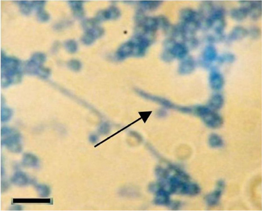
Figure 5: M. bovis 117-c variant of dissociative forms cultivated at 37°C on flesh-peptone agar (5 generation of weekly culture). Bar = 10 microns.
Consequently, in cultures on simple nutrient media at 37°C, dissociative forms were predominantly L-shaped, while at 3°C – sticks of different shapes and grains (although there were also L-forms).
Having obtained such results, we conducted repeated studies to find out the influence of the temperature of cultivation on the morphology of the dissociative forms of M. bovis, with reinoculation them to the same dense Lövenstein-Jensen nutrient medium 20 times.
The reinoculation from 3 to 23 generation confirmed the influence (Figure 6) of the temperature on the morphology of mycobacteria: in all three variants of M. bovis at 3°C the morphology remained practically stable grains, short rod-shaped bacteria were constantly generated during 20 reinoculations, whereas at 37°C sticks, grains were replaced by L-shapes and elementary small bodies.
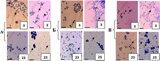
Figure 6: Morphological signs and tinctorial properties of M. bovis variants 117-a, b, c of dissociative forms, passaged through dense nutrient medium, cultivated for: left column 3°C; rights - 37°C. Bar: 10 microns.
Under such conditions, the pattern of the culture changed over time. In particular, with the advent of elementary small bodies in culture after 4-5 days of incubation, the film of continuous growth thinned, through 2-4 weeks the medium was thinned, while at 3°C the culture in the form of continuous growth along the line of inoculation of the suspension of mycobacteria intensively increased in time.
Investigating the morphogenesis of dissociative forms of mycobacteria (variant 117-c), isolated from the biomaterial of experimentally infected guinea pigs, it was established that colonies at 3ºC cultivation formed (on the 10th day of colony growth) only oval-shaped forms (Figure 7-1) did not contain grains and elementary small bodies around them. Seven days later (Figure 7-2), after repeated microscopy, spheroplasts (L-forms) acquired different optical density of the surface with clearly visible grains inside, a significant number of elementary small bodies near and around them.
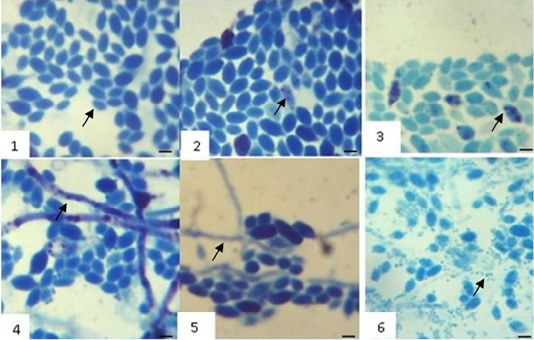
Figure 7: Initial and subsequent stages of separation of nuclear substance in M. bovis spheroplasts and formation of morphological forms: 1 - on 7; 2 - on 14; 3 - on 21; 4 - on 28; 5 - on 35; 6 – on 240 day of colony growth (117-v variant, 11 subculture isolated from guinea pig biomaterial). Bar: 10 μm.
After 21 days (Figure 7-3), the intensity of separation the nuclear material in spheroplasts (grains, elementary small bodies) significantly increased. This may indicate the persistence in the macroorganism of single mycobacteria in the form of spheroplasts that are capable of cultivating in the future on the dense nutrient medium at 3ºC with the separation of nuclear material (elementary small bodies).
After studying the morphology of the transformed M. bovis of the same colony for the next 14 days, it was revealed (Figure 7-4, 5) law-based dynamic changes that were characterized, along with the morphological forms previously given, an increase in the number of elementary small bodies, the formation of filiform variants, in which the grains (large and small) and oval-shaped formations with the same optical density of the surface that were freed from them and were clearly observed. On the 240th day of cultivation, the fluffy threads with the contents of grains (Figure 7-b), which were pushed out and transformed into L-forms (oval formations with different optical density of the surface), were established, and a sharp increase in the number of submicroscopic and microscopic, as in previous studies, non-acid-resistant elementary small bodies.
These data confirm once again that, under the condition of investigation, in the dynamics of growth of one colony, which was formed from one L-form of the spheroplastic type, the formation of spheroplasts occurs precisely from the filiform variants of M. bovis, although, in addition, in these structures, grains (elementary small bodies). At the same time, our investigations have not revealed a clear mechanism for the formation of filiform variants of M. bovis, that is, from which morphological forms they are formed. Most likely, one can only assume that from the elementary small bodies, since other formations in the investigated colony were not detected, but they, elementary small bodies (nuclear matter) were separated in spheroplasts: the threads were not observed until the appearance of elementary small bodies in the colony.
Generally, phenotypic changes in mycobacteria are closely related to the chemical composition of the cell, which, depending on the medium, varied by defining morphology, tinctorial properties, biochemical activity that contributed to adaptation to survival. At the same time, Table 2 shows that the content of total lipids in investigated mycobacteria strains was significantly different.
Their highest content was found in M. bovis of the pathogenic epizootic strain of the second subculture, however, it was lower by almost two and three times in the reference strain
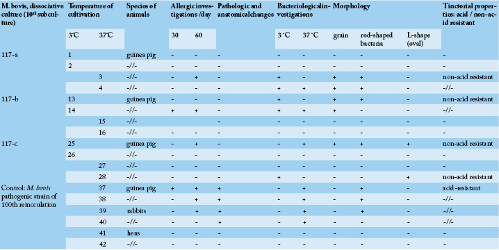
Table 1: Biological effect in experimental animals depending on infection with dissociative forms M. bovis.
Table 2: Total lipids of mycobacteria, % by weighed portion.

